Periostin在肾细胞癌中的表达及其临床意义
2015-04-13江伟东廖国龙曾志宇刘志龙
江伟东,朱 黎,廖国龙,曾志宇,刘志龙
(珠海市第二人民医院泌尿外科,广东 珠海 519020)
Periostin在肾细胞癌中的表达及其临床意义
江伟东,朱 黎,廖国龙,曾志宇,刘志龙
(珠海市第二人民医院泌尿外科,广东 珠海 519020)
目的 研究骨细胞特异性因子2(Periostin)在肾细胞癌组织中的表达,探讨其与肾细胞癌临床病理因素和预后的关系及意义。方法应用Western blot和Real-time PCR检测90例肾细胞癌组织及相对应的癌旁肾脏组织中Periostin表达情况,并与临床病理资料及预后进行相关性分析。结果76.67%(69/90)的肾癌组织的Periostin表达高于相对应的癌旁组织,65.56%(59/90)肾癌组织的Periostin mRNA表达高于相对应的癌旁组织,肾细胞癌组织Periostin高表达与肿瘤大小、肾盂侵犯、癌栓、淋巴转移相关(P<0.05),肾细胞癌组织Periostin高表达的患者术后生存时间显著短于Periostin低表达者(P<0.01)。结论Periostin表达升高与肾细胞癌的增殖、转移、浸润与生存时间密切相关,Periostin可作为预测肾细胞癌转移和判断预后的指标。
肾细胞癌;Periostin;转移;预后
肾细胞癌是泌尿系统中恶性度较高的肿瘤,起源于肾实质泌尿小管上皮系统的恶性肿瘤,占肾恶性肿瘤的80%~90%[1-2]。判断肾细胞癌预后的主要依据是临床分期和病理分级,寻求敏感、早期预测肾细胞癌发生发展及预后的分子学标记物尤为重要。骨细胞特异性因子2(Periostin)在多种肿瘤组织中存在过表达,与多种肿瘤发生、发展密切相关[3]。本研究通过Western blot和Real-time PCR的方法检测肾细胞癌组织和相对应的癌旁肾脏组织中Periostin的蛋白以及mRNA表达,以探讨其与肾细胞癌临床病理因素和预后的关系。
1 资料与方法
1.1 标本来源 选用2004年12月至2010年1月手术切除临床病理资料完整的肾细胞癌及相对应的癌旁组织石蜡标本90例。所有病例术前均未行放、化疗。所有病例均重新切片,确认其病理类型、组织学分级、包膜和肾盂浸润、有无肾静脉癌栓及肾门淋巴结转移等。术后均随访5年以上,随访截止日期为2014年12月。
1.2 Western blot检测Periostin 吸去培养基,以细胞裂解液RIPA 100 μl于冰上裂解细胞30 min,高速离心10 min,测定蛋白浓度。加上样缓冲液,煮沸变性,经过聚丙烯酰胺凝胶电泳(SDS-PAGE)、转膜,10%BSA封闭1 h,加入兔抗人GAPDH和Periostin一抗(1:500),在4℃过夜孵育;辣根过氧化酶标记的山羊抗兔二抗(1:5 000),在室温下孵育1 h;ECL孵育3 min,显色,以抗GAPDH作为内参。
1.3 Real-time PCR检测Periostin的mRNA表达 (1)RNA提取:肾癌组织及肾癌旁组织均加入Trizol裂解液,经研磨后,提取细胞总RNA。(2)逆转录:采用逆转录试剂盒(Takara),所有操作按照试剂盒说明书进行操作。(3)Real-time PCR:反应体系采用20 μl体系,cDNA 1 μl,上下游引物共1 μl,2×SYBR Green染料10 μl,ROX 0.4 μl,水7.6 μl。反应条件为95℃30 s,95℃5 s,60℃30 s,40个循环。用β-actin mRNA为内参。引物序列如下:β-Actin引物:上游5'-CAGGATCGTTAAGGAGATTA-3',下游5'-CAGTC TCGCCAGGATAG-3';Periostin引物:上游5'-TGTCCC AAACTGGGACGATA-3',下游5'-GGATCTTTACGGA TGTCAGCA-3'。
1.4 统计学方法 应用SPSS19.0软件进行统计学处理,计数资料组间比较采用χ2检验,相关性检验采用Pearson秩相关分析,生存分析采用Kaplan-Meier法,行Log-rank检验。以P<0.05表示差异有统计学意义。
2 结 果
2.1 Periostin蛋白在肾癌组织及相对应肾癌旁组织中的表达 76.7%(69/90)的肾癌组织的Periostin表达高于相对应癌旁组织(χ2=19.521,P<0.0001),见图1。

图1 Periostin蛋白在肾癌及癌旁组织中的表达
2.2 PeriostinmRNA在肾癌组织及相对应肾癌旁组织中的表达 65.56%(59/90)的肾癌组织的Periostin mRNA表达高于相对应癌旁组织(χ2=17.780,P<0.0001),见图2。
2.3 Periostin蛋白水平和mRNA水平的相关性 肾癌组织Periostin mRNA表达水平和Periostin蛋白表达的灰度值有显著的相关性(r2=0.783,P<0.0001),见图3。
2.4 Periostin在肾癌中表达与临床病理参数的关系 肾细胞癌组织Periostin高表达阳性率与患者性别、年龄、血尿比较差异均无统计学意义(P>0.05),而与肿瘤大小、肾盂侵犯、癌栓、淋巴转移比较差异均有统计学意义(P<0.05),见表1。
2.5 Periostin在肾癌中表达与预后的关系 Periostin的表达水平与预后显著相关(P<0.000 1),肾细胞癌组织Periostin高表达的患者预后差,见图4。

图2 Periostin mRNA在肾癌组织及相对应肾癌旁组织中的表达

图3 Periostin蛋白水平和mRNA水平的相关性

图4 Periostin在肾癌中表达与预后的生存曲线
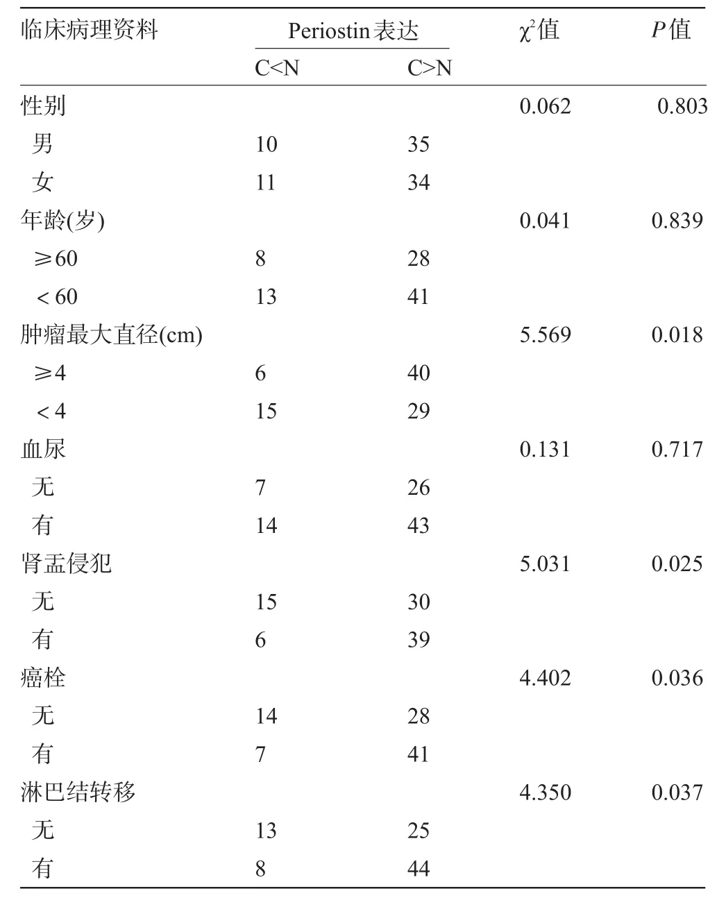
表1 Periostin的表达与肾细胞癌临床病理因素的关系(例)
3 讨 论
Periostin在多种恶性肿瘤组织中高表达,包括乳腺癌、结肠癌、胰腺癌、卵巢癌、前列腺癌、肺癌和肝癌等[4-7]。Periostin能促进表皮生长因子受体(EGFR)表达和激活Akt-FAK介导的信号传导途径,促进血管形成,抵抗低氧环境下诱导的细胞死亡,促进肿瘤细胞的侵袭性、转移能力[8-10];Periostin能够诱导上皮间质转化,促进肿瘤细胞的转移和定植[11];此外,Periostin的C末端区域能与细胞外基质相互作用,作用于细胞外基质纤维生成,影响肿瘤的生长[12]。Periostin本身属于一种细胞外基质分泌蛋白,可能通过改变细胞外基质,参与肿瘤微环境的变化,影响肿瘤的浸润性、侵袭性[13-14]。本研究显示Periostin在肾癌组织中高表达,提示肾癌细胞中Periostin的过表达可能促进肿瘤细胞生长以及转移。
本研究显示Periostin mRNA和蛋白水平呈显著相关性,说明Periostin的过表达主要是由Periostin mRNA的过表达所致,提示Periostin的过表达主要受转录水平的调控,而不是蛋白翻译后的调控,开发特异性的针对结合Periostin基因启动子活性的特异性小分子药物,可能是治疗肾癌的一个新的方向。
本组资料显示Periostin高表达阳性率与患者性别、年龄、血尿的差异均无统计学意义,与肿瘤大小、肾盂侵犯、癌栓、淋巴转移的差异均有统计学意义,且与患者的预后生存相关。提示了Periostin的表达与肾细胞癌的生长和转移能力呈正相关,Periostin的升高可能会导致肿瘤细胞侵袭力的增强,从而促进肿瘤的转移浸润。Periostin作为一种分泌形式的蛋白,从血清检测Periostin的水平,可能可以作为预测肾细胞癌转移和预后的一个潜在指标。
[1]么恩亮,张秀虹.肾细胞癌分期与复发的CT诊断[J].海南医学, 2007,18(10):39-41.
[2]Kyriakopoulos CE,Chittoria N,Choueiri TK,et al.Outcome of patients with metastatic sarcomatoid renal cell carcinoma:results from the International Metastatic Renal Cell Carcinoma Database Consortium[J].Clin Genitourin Cancer,2014,58(14):1010-1016.
[3]Kapoor S.Periostin and its emerging role in systemic carcinogenesis[J].Osteoporos Int,2014,25(4):1423-1424.
[4]Sirica AE,Almenara JA,Li C.Periostin in intrahepatic cholangiocarcinoma:Pathobiological insights and clinical implications[J]. Exp Mol Pathol,2014,97(3):515-524.
[5]Romanos GE,Asnani KP,Hingorani D,et al.Periostin:role in formation and maintenance of dental tissues[J].J Cell Physiol,2014, 229(1):1-5.
[6]Lv Y,Wang W,Jia WD,et al.High-level expression of periostin is closely related to metastatic potential and poor prognosis of hepatocellular carcinoma[J].Med Oncol,2013,30(1):385-389.
[7]Wu G,Wang X,Zhang X.Clinical implications of periostin in the liver metastasis of colorectal cancer[J].Cancer Biother Radiopharm,2013,28(4):298-302.
[8]Utispan K,Sonongbua J,Thuwajit P,et al.Periostin activates integrin alpha5beta1 through a PI3K/AKTdependent pathway in invasion of cholangiocarcinoma[J].Int J Oncol,2012,41(3):1110-1118.
[9]Watanabe T,Yasue A,Fujihara S,et al.PERIOSTIN regulates MMP-2 expression via the alphavbeta3 integrin/ERK pathway in human periodontal ligament cells[J].Arch Oral Biol,2012,57(1):52-59.
[10]Wong GS,Lee JS,Park YY,et al.Periostin cooperates with mutant p53 to mediate invasion through the induction of STAT1 signaling in the esophageal tumor microenvironment[J].Oncogenesis,2013, 2:59-64.
[11]Watanabe T,Yasue A,Tanaka E.Hypoxia-inducible factor-1alpha is required for transforming growth factor-beta1-induced type I collagen,periostin and alpha-smooth muscle actin expression in human periodontal ligament cells[J].Arch Oral Biol,2014,59(6):595-600.
[12]Lu Y,Liu X,Jiao Y,et al.Periostin promotes liver steatosis and hypertriglyceridemia through downregulation of PPARalpha[J].J Clin Invest,2014,124(8):3501-3513.
[13]Yamaguchi Y,Ono J,Masuoka M,et al.Serum periostin levels are correlated with progressive skin sclerosis in patients with systemic sclerosis[J].Br J Dermatol,2013,168(4):717-725.
[14]Hu F,Wang W,Zhou HC,et al.High expression of periostin is dramatically associated with metastatic potential and poor prognosis of patients with osteosarcoma[J].World J Surg Oncol,2014,12:287-292.
Expression and clinical significance of Periostin in renal cell carcinoma.
JIANG Wei-dong,ZHU Li,LIAO Guo-long,ZENG Zhi-yu,LIU Zhi-long.Department of Urology,the Second People's Hospital of Zhuhai,Zhuhai 519020,Guangdong,CHINA
Objective To evaluate the mRNA and preotein levels of Periostin in renal cell carcinoma(RCC), and to analyze the relationships between its protein level and the clinical pathological features,prognosis of RCC.MethodsWestern blot and real-time PCR were applied to detect the expression levels of Periostin in 90 cases of RCC and the adjacent tissues.The correlations between expression of Periostin and pathological features,prognosis were analyzed.Results76.67%(69/90)and 65.56%(59/90)patients showed significant higher protein and mRNA levels in RCC than adjacent tissues,respectively.The abnormal expression of Periostin correlated with tumor size,renal pelvis invasion,tumor thrombus and lymph metastasis(P<0.05).Patients with elevated expression of Periostin in RCC had significantly shorter postoperative survival time than those with low expression of Periostin(P<0.01).ConclusionThe high expression of Periostin correlates with proliferation,invasion,metastasis and patient prognosis.Our results suggest that Periostin may be a potential marker for determining the degree of malignancy and prognosis.
Renal cell carcinoma;Periostin;Metastasis;Prognosis
R737.11
A
1003—6350(2015)11—1578—03
10.3969/j.issn.1003-6350.2015.11.0565
2015-01-02)
广东省中医药局课题立项(编号:20131049)
江伟东。E-mail:jweidong1965@126.com
