雌激素受体α对血小板Aβ所诱导的血管内皮细胞损伤的保护作用
2015-03-13罗秀梅王继申龙大宏
罗秀梅 王继申 石 纯 龙大宏
(广州医医科大学基础学院人体解剖学教研室,广东 广州 510182)
·论著·
雌激素受体α对血小板Aβ所诱导的血管内皮细胞损伤的保护作用
罗秀梅 王继申 石 纯*龙大宏
(广州医医科大学基础学院人体解剖学教研室,广东 广州 510182)
目的:探讨血小板β淀粉样蛋白(Aβ)在动脉粥样硬化(AS)发生中的作用机制以及雌激素受体α(ERα)对血小板淀粉样前体蛋白(APP)代谢的调节作用。方法:分别提取12只6月龄APP/PS1鼠和6只同月龄野生型小鼠的血小板。将所获得的血小板分为3组:正常对照组、APP/PS1组和APP/PS1+ ERα激动剂(PPT)组。在APP/PS1+PPT组的血小板悬液中加入PPT溶液;在正常对照组和APP/PS1组的血小板悬液中加入等量生理盐水。ELISA法检测各组血小板释放Aβ的量以及α、β内分泌酶活性;离心提取各组血小板悬液的上清,并将其加入主动脉内皮细胞的培养液中,用流式细胞仪JC-1法检测主动脉内皮细胞线粒体膜电位。结果:与正常对照组相比,APP/PS1组血小板释放Aβ的量明显增加 (P<0.01);APP/PS1+PPT组血小板释放Aβ的量与APP/PS1组血小板相比明显减少(P<0.01);APP/PS1+PPT组血小板而α内分泌酶活性较APP/PS1组增强(P<0.01);与对照组血小板的上清相比,APP/PS1组血小板上清可增强血管内皮细胞线粒体膜电位的去极化(P<0.01)。相比之下,APP/PS1+PPT组血小板的上清对血管内皮细胞线粒体膜电位的作用较APP/PS1组血小板的上清明显减弱(P<0.01)。结论:血小板Aβ的过度生成可导致内皮细胞凋亡,这可能是血小板Aβ促进AS形成的机制之一。激活ERα可通过增强α分泌酶介导的血小板APP代谢减少血小板Aβ的生成,为AS的防治提供了新思路和新途径。
雌激素受体α;血小板;β淀粉样蛋白;动脉粥样硬化;小鼠
Aβ是一种由39~43个氨基酸残基组成的多肽。有研究结果显示,血液中Aβ与AS的发生有关[1-3];血浆Aβ水平与AS的发生及其严重程度呈正相关[1],且动脉粥样硬化斑块中存在大量淀粉样前体蛋白(APP)和Aβ[2]。动物实验表明,APP转基因小鼠早期即可发生AS,且其严重程度与血液中Aβ的水平呈正相关[3]。已知血液中超过90%的Aβ由血小板APP代谢而来[4]。但血小板Aβ在AS发生中的作用机制还不清楚。本组的前期研究结果表明,一些植物雌激素,如人参皂甙Rg1可通过雌激素受体(estragen receptor ol,ERα)调节APP代谢,抑制Aβ的生成[5]。那么调节血小板ERα能否抑制血小板Aβ生成,进而防止AS形成?目前还不清楚。APP/PS1是小鼠表达突变的人类早老素(DeltaE9)和淀粉样前体蛋白(APPswe)的融合体。有研究结果表明,6~7月龄的APP/PS1小鼠脑内会形成Aβ沉淀[6,7]。本研究采用APP/PS1鼠作为动物模型,对血小板Aβ在AS发生中的作用以及ERα激动剂(PPT)的保护作用进行探讨。
1 材料与方法
1.1 动物
12只6月龄APP/PS1鼠和6只同月龄正常对照小鼠(同品系正常小鼠)购自广东省实验动物中心,雌雄各半。饲养温度(24±1)℃,普通饮食,自由饮食、饮水,自然明暗周期。
1.2 血小板提取、药物处理及主动脉内皮细胞的培养
在10%水合氯醛(400 mg/kg)腹腔注射麻醉下,对APP/PS1及其对照小鼠进行右心房穿刺,抽取血液1.0~1.5 mL。5 μmol/L EDTA抗凝,150 g 离心20 min。上清再以400 g 离心15 min所得到的沉淀物即为血小板。将血小板重悬于改良的Tyrode液(150 mmol/L NaCl,5 mmol/L N-2-hydroxyethylpiperazine-N-2-ethanesulfonic acid,0.55 mmol/L NaH2PO4,7 mmol/L NaHCO3,2.7 mmol/L KCl,0.5 mmol/L MgCl2,5.6 mmol/L glucose,pH 7.4)。调节血小板密度为1×10⁸/mL。
将所获得的血小板悬液分为3组:对照组、APP/PS1组和APP/PS1+PPT组。在APP/PS1+PPT组中加入PPT(终浓度为10 nmol/L),37℃孵育12 h。在对照组和APP/PS1组的血小板悬液中加入生理盐水37℃孵育12 h。为保持血小板在静息状态,在各组血小板悬液中再加入10 ng/ml前列腺环素I2;为激活血小板,在血小板悬液中加入10 mg/mL I型胶原,孵育10 min。
主动脉内皮细胞培养于含LSGS的Medium 200PRF中。以104/cm2的细胞密度在培养皿中进行单层培养。将经上述药物处理后的血小板悬液以400 g 离心15 min。将离心后所得到的上清加入主动脉内皮细胞的培养基中,37℃孵育12 h。
1.3 ELISA法检测血小板释放Aβ情况
将经上述药物处理后的血小板悬液离心后收集上清。用Aβ ELISA试剂盒(美国BetaMark)检测血小板在静息和激活状态下释放到上清中Aβ含量。
1.4 ELISA法检测血小板α、β内分泌酶活性
收集经上述药物处理后的血小板。用α内分泌酶活性试剂盒(美国R&D Systems)和β内分泌酶活性检测试剂盒(美国Sigma)分别对血小板α、β内分泌酶活性进行检测。
1.5 流式细胞仪JC-1法检测主动脉内皮细胞凋亡情况
收集经上述处理后的主动脉内皮细胞,重悬于PBS中。在细胞悬液中加入JC-1(终浓度为5 μg/mL),避光的情况下孵育20 min。用流式细胞仪分析主动脉内皮细胞线粒体膜电位情况。
1.6 统计学分析

2 结 果
2.1 血小板释放Aβ情况
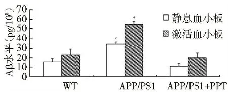
与对照组相比,APP/PS1组血小板释放的Aβ量明显增加(P<0.01);APP/PS1+PPT组血小板释放的Aβ量较APP/PS1组明显减少(P<0.01),见图1。

注:与对照组比较,*P<0.01。
图1 各组血小板释放Aβ情况
2.2 血小板α、β内分泌酶活性
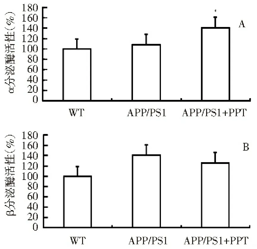
APP/PS1组血小板α、β内分泌酶活性与对照组无明显差异(P>0.05),;APP/PS1+PPT组血小板β内分泌酶活性与APP/PS1组比较无明显差异(P>0.05),而α内分泌酶活性则较APP/PS1组增强(P<0.01),见图2。
2.3 主动脉内皮细胞凋亡情况
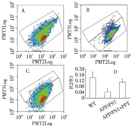
与对照组血小板上清相比,APP/PS1组血小板上清可增强血管内皮细胞线粒体膜电位的去极化(P<0.01);APP/PS1+PPT组血小板上清对血管内皮细胞线粒体膜电位的作用较APP/PS1组明显减弱(P<0.01),见图3。

注:A:血小板α分泌酶活性;B:血小板β分泌酶活性;与对照组比较,*P<0.01
图2 各组血小板α、β分泌酶活性

注:A:对照组;B:APP/PS1组;C:APP/PS1+ PPT组;D:各组主动脉内皮细胞的线粒体膜电位(以R2区细胞数/R1区细胞数表示);与APP/PSI组比较,*P<0.01
图3 各组主动脉内皮细胞的线粒体膜电位
3 讨 论
在血管细胞的培养物中加入100 nmol/L的Aβ即可诱发血管内皮细胞功能失调,表现为凋亡、诱导性一氧化氮合酶的表达增加以及一氧化氮的生物利用度下降[8]。已知血液中Aβ主要来源于血小板,那么血小板Aβ过表达能否导致血管内皮的损伤呢?为检测这一假设,我们检测了APP/PS1血小板释放Aβ情况以及APP/PS1血小板的释放产物对血管内皮细胞活性的影响。本研究结果显示,相较于对照组血小板,APP/PS1血小板释放的Aβ明显增多。将APP/PS1血小板的释放产物加入主动脉内皮细胞的培养液中可促进其凋亡。这些结果提示:血小板Aβ的过度生成可导致内皮细胞凋亡,这可能是血小板Aβ促进AS形成的机制之一。
本组的前期研究结果表明,一些植物雌激素如人参皂甙Rg1可通过ERα调节APP代谢,抑制Aβ的生成[5]。那么激活血小板ERα能否抑制血小板Aβ生成?为检测这一假设,我们检测了ERα激动剂PPT对APP/PS1血小板Aβ生成的调节作用。结果发现,PPT可抑制APP/PS1血小板Aβ的生成,并防止APP/PS1血小板释放产物对血管内皮细胞的毒性作用。这结果提示:激活ERα可调节血小板APP代谢,抑制血小板Aβ的生成,进而防止血小板Aβ过生成对血管内皮细胞造成的毒性作用。已知体内APP主要经2条途经进行代谢:α-分泌酶介导的APP代谢和β-分泌酶介导的APP代谢,这两条代谢途径可相互制约[4-5]。促进α-分泌酶介导的APP代谢可减少APP经β-分泌酶途径进行代谢的机会,进而减少APP经β-分泌酶代谢所产生的Aβ[4-5]。为检测PPT调节血小板APP代谢的机制,本组进一步检测了血小板α、β分泌酶活性。本研究结果还显示,PPT可增强APP/PS1血小板α分泌酶的活性。这证实激活ERα可通过增强α分泌酶介导的血小板APP代谢减少血小板Aβ的生成。
[1] Baiguera S, Fioravanzo L, Grandi C, et al. Involvement of the receptor for advanced glycation-end products (RAGE) in beta-amyloid-induced toxic effects in rat cerebromicrovascular endothelial cells cultured in vitro[J]. Int J Mol Med, 2009, 24(3):9-15.
[2] Fujiwara Y, Takahashi M, Tanaka M, et al. Relationships between plasma beta-amyloid peptide 1-42 and atherosclerotic risk factors in commol/Lunity-based older populations[J]. Gerontology, 2003, 49(2):374-379.
[3] Li L, Cao D, Garber DW, et al. Association of aortic atherosclerosis with cerebral beta-amyloidosis and learning deficits in a mouse model of Alzheimer′s disease[J]. Am J Pathol, 2003, 163(3):2155-2164.
[4] Shi C, Na N, Zhu X, et al. Estrogenic effect of Ginsenoside Rg1 on APP processing in post-menopausal platelets[J]. Platelets, 2013, 24(4):51-62.
[5] Shi C, Zheng DD, Fang L, et al. Ginsenoside Rg1 promotes nonamyloidgenic cleavage of APP via estrogen receptor signaling to MAPK/ERK and PI3K/Akt[J]. Biochim Biophys Acta, 2012, 1820(8):453-460.
[6] Chen L, Choi JJ, Choi YJ, et al. HIV-1 Tat-induced cerebrovascular toxicity is enhanced in mice with amyloid deposits[J]. Neurobiol Aging,2012,33(6):1579-1590.
[7] Takeda S, Hashimoto T, Roe AD, et al. Brain interstitial oligomeric amyloid β increases with age and is resistant to clearance from brain in a mouse model of Alzheimer’s disease[J] FASEB J, 2013, 27(3):3239-3248.
[8] Howlett GJ, Moore KJ. Untangling the role of amyloid in atherosclerosis. Curr Opin Lipidol, 2006, 17(5):541-547.
(本文编辑:郑颖)
Protective effects of estrogen receptor α on platelet Aβ-induced vascular endothelial cell injury
LuoXiumei,WangJishen,ShiChun,LongDahong
(DepartmentofHumanAnatomy,GuangzhouMedicalUniversity,Guangzhou,Guangdong510182,China)
Objective: To investigate the mechanism of platelet amyloid protein β (Aβ) in the occurrence of atherosclerosis (AS) and the regulatory effect of estrogen receptor α (ERα) on platelet amyloid precusor protein (APP) metabolism.Methods:Platelets were extracted from 12 6-month old APP/PS1 mice and 6 wild mice of the same months of age, respectively. The extracted platelets were divided into 3 groups: the normal control group, APP/PS1 group and APP/PS1+ ERα agonist (PPT) group. For APP/PS1+PPT group, PPT solution was added into the platelet suspension. For normal control group and APP/PS1 group, equivalent normal saline solution was added into the platelet suspension. Then, the volume of Aβ released by platelets and the activities of α and β secretases were measured by enzyme linked immunosorbent assay (ELISA). The supernatant in each group was extracted from platelet suspension by centrifugation, and then added to the broth of aortic endothelial cells. Then, mitochondrial membrane potential of aortic endothelial cells was measured by flow cytometry JC-1 method.Results:Compared with normal control group, the volume of platelets-released Aβ in APP/PS1 group increased significantly (P<0.01). The volume of platelets-released Aβ in APP/PS1 +PPT group treatment decreased significantly compared with that in APP/PS1 group (P<0.01). The activities of α- and β-secretase ofplatelets in APP/PS1+PPT group increased compared with that in APP/PS1 group (P<0.01). Compared with normal control group, the supernatant of platelets in APP/PS1 group could promote the depolarization of mitochondrial membrane potential in vascular endothelial cells (P<0.01). The effect of platelets supernatant on mitochondrial membrane potential in vascular endothelial cells decreased significantly in APP/PS1+PPT group compared with that in APP/PS1 group (P<0.01).Conclusion:Excessively generated platelet Aβ can induce apoptosis of endothelial cells, which may be one of the mechanisms of platelet Aβ-induced AS. Activation of ERα can reduce the release of platelet Aβ through increase of α secretase-mediated platelet APP metabolism, providing new ideas and approaches for the prevention and treatment of AS.
estrogen receptor α; platelets; β amyloid protein; atherosclerosis;mouse
10.3969/j.issn.2095-9664.2015.02.001
国家自然科学基金面上项目和青年项目(81370395,81200846);广东省自然科学基金面上项目(S2013010014468)
罗秀梅(1966-),女,硕士,副教授。
R329.2
A
2095-9664(2015)02-0001-04
2014-11-20)
研究方向:老年性痴呆的机理和防治。
