Selective verbal memory impairment due to left fornical crus injury in a patient with intraventricular hemorrhage
2014-06-01HanDoLee,SungHoJang
Selective verbal memory impairment due to left fornical crus injury in a patient with intraventricular hemorrhage
The fornix, a part of the Papez circuit, transfers information of episodic memory between the medial temporal lobe and the medial diencephalon (Aggleton and Brown, 1999). The right medial temporal lobe is known to be specialized for visual memory and the left medial temporal lobe for verbal memory (Tucker et al., 1988; Aggleton and Brown, 1999).
Many studies have reported on fornix injury, however, most of them focused on bilateral injury (Tucker et al., 1988; Aggleton et al., 2000; Nakayama et al., 2006; Sugiyama et al., 2007; Tsivilis et al., 2008; Wang et al., 2008; Jang et al., 2009; Chang et al., 2010; Hong and Jang, 2010; Yeo et al., 2011). To the best of our knowledge, three studies have reported on injury of the unilateral fornical crus (Tucker et al., 1988; Hong and Jang, 2010; Yeo et al., 2011). Among these studies, Tucker et al. (1988) reported on a patient who showed severe verbal memory impairment after unilateral transaction of the left fornical crus during surgery for removal of astrocytoma. Hong and Jang (2010) reported on a patient who showed selective verbal memory impairment due to left fornical crus injury, which was ascribed to diffuse axonal injury following head trauma. Subsequently, Yeo et al. (2011), who investigated the effect of intraventricular hemorrhage on white matter in 10 patients with intraventricular hemorrhage, reported on one patient with left fornical crus injury and nine patients with fornical body injury without clinical data on memory impairment, except for the Mini-Mental State Examination (MMSE). To the best of our knowledge, this is the fi rst study which demonstrates selective verbal memory impairment due to left fornical crus injury following spontaneous intraventricular hemorrhage.
It is dif fi cult to precisely assess the fornix due to its long, thin appearance and its location within the brain. In addition, discrimination of the whole fornix from adjacent neural structure using conventional brain CT or MRI is impossible. By contrast, diffusion tensor tractography (DTT), which is derived from diffusion tensor imaging (DTI), has enabled three-dimensional visualization of the fornix, and many studies have reported on fornix injury using DTT (Nakayama et al., 2006; Sugiyama et al., 2007; Wang et al., 2008; Jang et al., 2009; Chang et al., 2010; Hong and Jang, 2010; Yeo et al., 2011).
In the current study, using DTT, we report on a patient who showed selective verbal memory impairment due to left fornical crus injury following intraventricular hemorrhage.
DTTs were acquired twice (1 month and 4 months after onset) using a 1.5-T Philips Gyroscan Intera system (Philips, Ltd, Best, the Netherlands) equipped with a Synergy-L Sensitivity Encoding (SENSE) head coil using a single-shot, spin-echo planar imaging pulse sequence. For each of the 32 non-collinear diffusion sensitizing gradients, we acquired 60 contiguous slices parallel to the anterior commissure-posterior commissure line. Imaging parameters were as follows: acquisition matrix = 96 × 96, reconstructed to matrix = 192 × 192 matrix, fi eld of view = 240 mm × 240 mm, repetition time = 10,398 ms, echo time = 72 ms, parallel imaging reduction factor (SENSE factor) = 2, echo planar imaging factor = 59 and b = 1,000 s/mm2, number of excitations = 1, slice gap = 0 mm and thickness = 2.5 mm. Eddy current-induced image distortions were removed using affine multi-scale two-dimensional registration at the Oxford Centre for Functional Magnetic Resonance Imaging of Brain (FMRIB) Software Library (FSL; www.fmrib.ox.ac.uk/fsl). DTI-Studio software (CMRM, Johns Hopkins Medical Institute, Baltimore, MD, USA) was used for evaluation of the fornix. For analysis of the fornix, the seed region of interest (ROI) was drawn at the junction between the body and column of each fornix on a coronal image with the color map. Target ROIs were placed on the crus of the right and left fornix on a coronal image with the color map. Fiber tracking was started at any seed voxel with a fractional anisotropy (FA) > 0.2 and ended at a voxel with a fi ber assignment of < 0.2 and a tract turning angle of < 70 degrees.
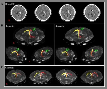
One-month DTT for the fornix showed a discontinuation of the left fornical crus, which was not observed in four male ageand sex-matched right handed normal control subjects (mean age 33.25 years (range 29-39 years )) and the discontinued left fornical crus was degenerated toward the fornical body as shown on 4-month DTT (Figure 1B). By contrast, the left fornical column was extended to the left medial temporal lobe on both 1- and 4-month DTTs.
In the current study, we reported on a patient who showed selective verbal memory impairment due to left fornical crus injury on DTT. The patient showed left fornical crus injury on both 1- and 4-month DTTs. We evaluated the patient’s cognitive function using two neuropsychological tests: the MMSE and the MAS. The MMSE is a screening test for general cognitive function by evaluating the subject’s orientation, memory, attention, calculation, visuospatial, and language abilities (Folstein et al., 1975). The MAS is a comprehensive standardized memory assessment test that yields scores for four factors: total memory, short-term memory, verbal memory, and visual memory (Williams, 1991). The patient’s general cognitive function was within normal range with a full MMSE score of 30 at both 1 and 4months after onset. However, the patient showed impairment in both visual and verbal memory at 1 month after onset (more severe impairment in verbal memory); in contrast, at 4 months after onset, the patient showed selective verbal memory impairment with marked improvement of visual memory. Because the left medial temporal lobe is known to be specialized for verbal memory, so the patient’s selective verbal memory impairment was ascribed to the left fornical crus injury (Tucker et al., 1988; Aggleton et al., 1999). The extension of the left fornical column to the left medial temporal lobe appears to be attributed to neural reorganization following injury of the left fornical crus (Yeo and Jang, 2013).

Figure 1 Brain CT and diffusion tensor tractography (DTT) images of a 33-year-old male patient with injury of the left fornical crus following intraventricular hemorrhage.
The hematoma in the present case was mainly located on the lateral side of the left fornix. Several studies have reported that periventricular neural tissue could be injured by intraventricular hemorrhage through mechanical or chemical mechanisms: (1) mechanical: following intraventricular hemorrhage, the increased intracranial pressure or direct mass effect can reduce cerebral perfusion pressure and cause secondary ischemic injury to periventricular white matter. (2) Chemical: a blood clot itself can cause extensive damage to the ependymal layer, subependymal layer, or periventricular tissues by release of potentially damaging substances, such as free iron, which may generate free radicals or in fl ammatory cytokines (Wasserman and Schlichter, 2008; Chua et al., 2009; Dai et al., 2009; Yeo et al., 2011). Therefore, we presumed that the left fornix crus was injured by the hematoma, although we could not determine whether the injury could be ascribed to a mechanical or chemical pathophysiological mechanism.
In conclusion, we report on a patient who showed selectiveverbal memory impairment due to injury of the left fornical crus following intraventricular hemorrhage. This study is limited to a single case report. Further studies involving a larger number of patients are required.
Han Do Lee, Sung Ho Jang Department of Physical Medicine and Rehabilitation, College of Medicine, Yeungnam University, Namku, Daegu, Republic of Korea
Aggleton JP, Brown MW (1999) Episodic memory, amnesia, and the hippocampal-anterior thalamic axis. Behav Brain Sci 22:425-444.
(2)对比二维模型与三维模型计算结果显示,二维模型与三维模型计算结果较为相近,各项指标误差率均小于15%。而三维模型建模复杂程度、计算时间要远大于二维模型,因此本文从钢管桁架—沉箱基础装配式新型码头结构能否满足安全使用要求的角度出发,:博上部钢管桁架结构与下部重力式沉箱基础分开计算,计算结果显示,上部钢管桁架受力、位移特征值均满足规范要求;下部基础的抗滑、抗倾稳定性均符合规范要求;基床顶面的最大应力也远小于工程区域地基实际承载能力,可见钢管桁架—沉箱基础装配式新型码头结构设计合理,能够很好的适应大水位差山区河流。
Aggleton JP, McMackin D, Carpenter K, Hornak J, Kapur N, Halpin S, Wiles CM, Kamel H, Brennan P, Carton S, Gaffan D (2000) Differential cognitive effects of colloid cysts in the third ventricle that spare or compromise the fornix. Brain 123:800-815.
Chang MC, Kim SH, Kim OL, Bai DS, Jang SH (2010) The relation between fornix injury and memory impairment in patients with diffuse axonal injury: a diffusion tensor imaging study. NeuroRehabilitation 26:347-353.
Chua CO, Chahboune H, Braun A, Dummula K, Chua CE, Yu J, Ungvari Z, Sherbany AA, Hyder F, Ballabh P (2009) Consequences of intraventricular hemorrhage in a rabbit pup model. Stroke 40:3369-3377.
Wechsler D (1991) Manual for the wechsler adult intelligence scale-revised. New York: The Psychological Corporation.
Dai J, Li S, Li X, Xiong W, Qiu Y (2009) The mechanism of pathological changes of intraventricular hemorrhage in dogs. Neurol India 57:567-577.
Folstein MF, Folstein SE, McHugh PR (1975) “Mini-mental state”. A practical method for grading the cognitive state of patients for the clinician. J Psychiatric Res 12:189-198.
Hong JH, Jang SH (2010) Left fornical crus injury and verbal memory impairment in a patient with head trauma. Eur Neurol 63:252.
Jang SH, Kim SH, Kim OL (2009) Fornix injury in a patient with diffuse axonal injury. Arch Neurol 66:1424-1425.
Nakayama N, Okumura A, Shinoda J, Yasokawa YT, Miwa K, Yoshimura SI, Iwama T (2006) Evidence for white matter disruption in traumatic brain injury without macroscopic lesions. J Neurol Neurosurg Psychiatry 77:850-855.
Sugiyama K, Kondo T, Higano S, Endo M, Watanabe H, Shindo K, Izumi S (2007) Diffusion tensor imaging fi ber tractography for evaluating diffuse axonal injury. Brain Inj 21:413-419.
Tsivilis D, Vann SD, Denby C, Roberts N, Mayes AR, Montaldi D, Aggleton JP (2008) A disproportionate role for the fornix and mammillary bodies in recall versus recognition memory. Nat Neurosci 11:834-842.
Tucker DM, Roeltgen DP, Tully R, Hartmann J, Boxell C (1988) Memory dysfunction following unilateral transection of the fornix: a hippocampal disconnection syndrome. Cortex 24:465-472.
Wang JY, Bakhadirov K, Devous MD, Sr., Abdi H, McColl R, Moore C, Marquez de la Plata CD, Ding K, Whittemore A, Babcock E, Rickbeil T, Dobervich J, Kroll D, Dao B, Mohindra N, Madden CJ, Diaz-Arrastia R (2008) Diffusion tensor tractography of traumatic diffuse axonal injury. Arch Neurol 65:619-626.
Wasserman JK, Schlichter LC (2008) White matter injury in young and aged rats after intracerebral hemorrhage. Exp Neurol 214:266-275.
Williams JM (1991) MAS:Memory Assessment Scales: professional manual. Odessa, Fla.: Psychological Assessment Resources.
Yeo SS, Choi BY, Chang CH, Jung YJ, Ahn SH, Son SM, Byun WM, Jang SH (2011) Periventricular white matter injury by primary intraventricular hemorrhage: a diffusion tensor imaging study. Eur Neurol 66:235-241.
Yeo SS, Jang SH (2013) Neural reorganization following bilateral injury of the fornix crus in a patient with traumatic brain injury. J Rehabil Med 45:595-598.
Copyedited by Li CH, Song LP, Zhao M
Sung Ho Jang, M.D., Department of Physical Medicine and Rehabilitation, College of Medicine, Yeungnam University 317-1, Daemyungdong, Namku, Daegu, 705-717, Republic of Korea, strokerehab@hanmail.net.
10.4103/1673-5374.137579 http://www.nrronline.org/
Funding: This study was supported by Basic Science Research Program through the National Research Foundation of Korea (NRF) funded by the Ministry of Education, Science and Technology, No. 2012R1A1A4A01001873.
Author contributions: Lee HD was responsible for data acquisition and analysis, and manuscript writing, and provided technical assistance. Jang SH was responsible for conception and design of this study, fundraising, data acquisition, and manuscript development and authorization. Both of these two authors approved the final version of this manuscript.
Con fl icts of interest: None declared.
Accepted: 2014-06-06
Lee HD, Jang SH. Selective verbal memory impairment due to left fornical crus injury in a patient with intraventricular hemorrhage. Neural Regen Res. 2014;9(13):1313-1315.
猜你喜欢
杂志排行
中国神经再生研究(英文版)的其它文章
- Stem cell therapy for central nerve system injuries: glial cells hold the key
- Beyond taxol: microtubule-based strategies for promoting nerve regeneration after injury
- Neuroprotective effect of the traditional Chinese herbal formula Tongxinluo: a PET imaging study in rats
- Neuroprotective effects of Asiaticoside
- Treating Alzheimer’s disease with Yizhijiannao granules by regulating expression of multiple proteins in temporal lobe
- Autophagy activation aggravates neuronal injury in the hippocampus of vascular dementia rats
