头颈癌生物标志物研究进展
2014-03-26付立伟张小涛田永峰胡清源综述侯宏卫审校
付立伟,张小涛,田永峰,胡清源(综述),陈 欢,侯宏卫(审校)
(国家烟草质量监督检验中心,郑州 450001)
头颈部肿瘤包括除眼、脑、耳、甲状腺以及食管外的头颈部组织或器官的肿瘤,超过90%的头颈部肿瘤为鳞状细胞癌[1]。由于缺乏有效的早期检测和风险评估手段,超过50%的头颈癌患者确诊时已属晚期,尽管化疗、放疗等手段可在一定程度上缓解病情,但5年生存期仍低于50%[2]。全球每年约有60万例新发头颈部肿瘤病例,其中30多万例死亡[3]。
头颈部肿瘤的主要诱因是烟草、酒精和人类乳头瘤病毒(human papillomavirus,HPV)[4],烟草是头颈癌最主要的诱因。流行病学研究表明,至少75%的头颈癌患者有吸烟和(或)饮酒史;头颈癌吸烟患者病死率较戒烟或非吸烟患者高4倍[5]。饮酒是头颈癌的第二大诱因,每日饮酒量>50 g,患癌风险率提高5.5%,但适度饮酒(每日10~19 g)不会增加患头颈癌的风险[6]。HPV是除吸烟、饮酒外的头颈癌的另一大诱因,因HPV感染而形成的口咽鳞状细胞癌正逐年增多,超过50%的口咽癌与HPV感染有关[7]。HPV呈阳性的头颈癌患者与其他头颈癌患者相比呈现年轻化,其烟酒摄入量少,有相对更多的性伴侣[8]。
在头颈部肿瘤形成初期,淋巴结转移较少,对肿瘤的治疗也最为有效,然而大约2/3的头颈部肿瘤患者确诊时已属晚期[9],因此早期确诊对于改善患者的存活率有重要意义。目前由于缺乏有效的早期诊断的生物标志物,头颈癌的早期筛查和发现仍然是难题,头颈癌生物标志物的深入研究将为今后改善头颈癌的早期诊断、分级、复发及预后评估提供可能性。
1 DNA甲基化
DNA甲基化过程可使甲基添加到DNA分子上,其能够在不改变DNA序列的前提下,改变遗传表现。DNA甲基化早于细胞的恶性增生,DNA甲基化的诊断对于肿瘤早期预测有重要意义。目前发现的与头颈部肿瘤相关的甲基化基因包括:p16、p15、p14、死亡相关蛋白激酶、6-氧甲基鸟嘌呤-DNA甲基转移酶、钙黏蛋白等(表1)。Carvalho等[10]采用甲基化特异聚合酶链反应(methylation specific polymerase chain reaction,MSP)检测大量头颈癌患者的血清和唾液样本发现其特异度高达90%;Sanchez-Cespedes等[11]通过检测头颈癌患者肿瘤组织和唾液中p16、6-氧甲基鸟嘌呤-DNA甲基转移酶、死亡相关蛋白激酶的甲基化发现,在肿瘤组织不同病理分化阶段均存在DNA甲基化,对于口腔部位的肿瘤,唾液中DNA甲基化率相对较高,而在肿瘤组织中未发现DNA甲基化的患者其相应的唾液样本中也未出现甲基化现象[11]。对于做过肿瘤切除手术的头颈部肿瘤患者,采用MSP检测其唾液样本中DNA甲基化率对其病情监测有一定的临床应用前景。
Chang等[12]研究发现,头颈癌患者体液和肿瘤组织中p15、p16甲基化程度明显高于健康对照组,有长期吸烟或饮酒习惯的头颈癌患者肿瘤组织中p15的甲基化程度高于头颈癌非吸烟患者。有研究认为,烟草暴露可能会造成CpG岛的特异性损害或降低DNA甲基转移酶的活性[13]。

表1 头颈部肿瘤相关基因甲基化
CYP1A1:细胞色素P450酶;CYP2AI3:细胞色素P450酶;GSTMI:谷胱甘肽转移酶M1;EDNRB:内皮素B受体;KIF1A:微管驱动蛋白家族成员1A;CDKN:周期蛋白依赖性蛋白激酶抑制剂;RUNX3:RUNT相关转录因子3;SFN:人分层蛋白基因;MSP:甲基化特异聚合酶链反应
2 基质金属蛋白酶
基质金属蛋白酶(matrix metalloproteinases,MMPs)几乎能降解细胞外基质中所有的蛋白成分,能降解破坏肿瘤细胞侵袭、转移所需要越过的组织学屏障,在肿瘤侵袭转移中起关键作用[20]。研究发现,头颈癌者体内易出现MMPs过表达现象。Ruokolainen等[21]通过比较头颈癌患者与健康人群血清中MMP-9的水平发现,MMP-9水平偏高的头颈癌患者相比其他患者死亡风险更高,并认为MMP-9有希望成为头颈癌诊断的生物标志物。Kuropkat等[22]研究发现,头颈癌患者血清中MMP-3、MMP-8、MMP-9的水平明显高于健康人群,其中血清中MMP-8的水平与肿瘤所处的阶段相关。Kawata等[23]发现,正常黏膜与肿瘤组织中MMP-2的水平无明显差异,但出现淋巴结转移的肿瘤组织其MMP-2水平明显高于未发生转移者。其他研究也发现,MMP-2和MMP-14的过表达与头颈癌淋巴结转移有关,其对于头颈癌的预后有重要价值[24]。与头颈部肿瘤相关的标志物包括MMPs、Ki-67、人类表皮生长因子受体2等(表2)。
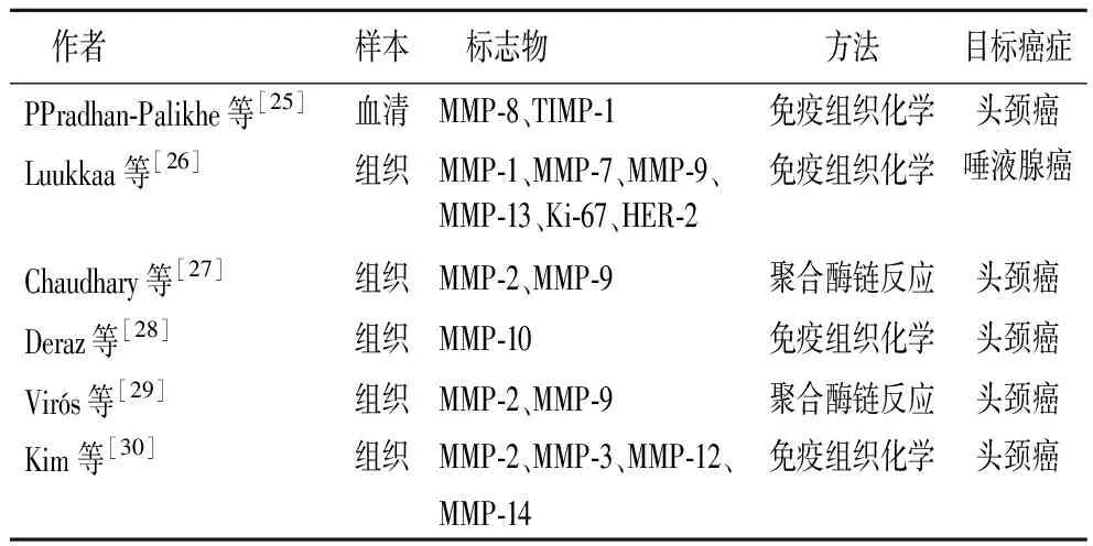
表2 与头颈部肿瘤相关的MMPs
MMP:基质金属蛋白酶;TIMP-1:基质金属蛋白酶组织抑制因子;HER-2:人类表皮生长因子受体2;
3 细胞因子
细胞因子是调节炎症与血管生成的重要蛋白质,目前已被广泛认可的与头颈癌检测、预后以及监控有关的细胞因子包括血管内皮生长因子(vascular endothelial growth factor,VEGF)[31]、白细胞介素6(interleukin-6,IL-6)等(表3)。
Chen等[32]检测出头颈癌患者唾液中IL-6、IL-8和VEGF的水平高于健康人群。Mineta等[33]将12项关于VEGF过表达与头颈癌存活率的数据进行统计学分析表明,VEGF阳性患者2年内死亡的风险率提高1.88倍,VEGF过表达对头颈癌淋巴结转移有预测价值。Akmansu等[34]对放射治疗前后头颈癌患者唾液中IL-6的水平进行检测发现,放射治疗后患者唾液中IL-6的水平明显升高。Duffy等[35]认为,IL-6是预测癌症复发及存活率的独立的生物标志物,其对于头颈癌的诊断和治疗有重要意义。Gokhale等[36]发现,唾液中IL-8的水平在头颈癌复发和转移阶段明显高于头颈癌形成初期,其对头颈癌的预后有重要意义。Korostoff等[37]采用定量酶联免疫吸附法检测喉鳞癌患者和对照组唾液中多种细胞因子的水平发现,外生性喉鳞癌患者唾液中IL-8的水平明显高于健康对照组。唾液及血清中IL-6和IL-8的测定通常采用酶联免疫吸附法,其敏感性和特异性较好,且操作简便、快速。IL-6、IL-8和VEGF等细胞因子对于头颈癌的分级和预后有重要研究价值。

表3 头颈部肿瘤相关细胞因子
VEGF:血管内皮生长因子;IL:白细胞介素;HGF:肝细胞生长因子;MMP:基质金属蛋白酶;TNF:肿瘤坏死因子
4 小 结
p16基因甲基化、MMP家族成员、IL-6、IL-8等生物标志物已被证明在头颈癌患者体液和组织中存在异常表达。这些生物标志物的发现为今后改善头颈癌早期诊断、分级以及预后等方面有重要意义。但头颈癌生物标志物真正的应用于临床还有较长的路要走,且临床诊断要求标志物分析过程准确、简单及标准化,多数生物标志物仍然需要进一步的研究以提供准确而规范标准化的分析流程。
[1] Schmitz S,Machiels JP.Molecular biology of squamous cell carcinoma of the head and neck relevance and therapeutic implications[J].Expert Rev Anticancer Ther,2010,10(9):1471-1484.
[2] Schaaij-Visser TB,Brakenhoff RH,Leemans CR,etal.Protein biomarker discovery for head and neck cancer[J].J Proteomics,2010,73(10):1790-1803.
[3] Lee KD,Lee HS,Jeon CH.Body fluid biomarkers for early detection of head andneck squamous cell carcinomas[J].Anticancer Res,2011,31(4):1161-1167.
[4] Rezende TM,de Souza Freire M,Franco OL.Head and neck cancer:proteomic advances and biomarker achievements[J].Cancer,2010,116(21):4914-4925.
[5] Thompson TL,Pageder NA,Karnell LH,etal.Factors associated with mortality in 2-year survivors of head and neck cancer[J].Arch Otolaryngol Head Neck Surg,2011,137(11):1100-1105.
[6] Lewin F,Norell SE,Johansson H,etal.Smoking Tobacco,oral snuff,and alcohol in the etiology of squamous cell carcinoma of the head and neck[J].Cancer,1998,82(7):1367-1375.
[7] Fakhry C,Gillisson ML.Clinical implications of human papillomavirus in head and neck cancers[J].J Clin Oncol,2006,24(17):2606-2611.
[8] Gillison ML,D′Souza G,Westra W,etal.Distinct risk factor profiles for human papillomavirus type 16-positive and human papillomavirus type 16-negative head and neck cancers[J].J Natl Cancer Inst,2008,100(6):407-420.
[9] Mydlarz WK,Hennessey PT,Califano JA.Advances and perspectives in the molecular diagnosis of head and neck cancer[J].Expert Opin Med Diagn,2010,4(1):53-65.
[10] Carvalho AL,Jeronimo C,Kim MM,etal.Evaluation of promoter hypermethylation detection in body fluids as a screening/diagnosis tool for head and neck squamous cell carcinoma[J].Clin Cancer Res,2008,14(1):97-107.
[11] Sanchez-Cespedes M,Esteller M,Wu L,etal.Gene promoter hypermet-hylation in tumors and serum of head and neck cancer patients[J].Cancer Res,2000,60(4):892-895.
[12] Chang HW,Ling GS,Wei WI,etal.Smoking and drinking can induce p15 methylation in the upper aerodigestive tract of healthy individuals and patients with head and neck squamous cell carcinoma[J].Cancer,2004,101(1):125-131.
[13] Kim DH,Nelson HH,Wiencke JK,etal.p16(INK4a) and histology-specific methylation of CpG islands by exposure to tobacco smoke in non-small cell lung cancer[J].Cancer Res,2001,61(8):3419-3424.
[14] Takeshima M,Saitoh M,Kusano K,etal.High frequency of hypermethylation of p14,p15 and p oral pre-cancerous lesions associated with betel-quid chewing in Sri Lanka[J].J Oral Pathol Med,2008,37(8):475-479.
[15] Sharma R,Panda NK,Khullar M.Hypermethylation of carcinogen metabolism genes,CYP1,CYP13 and GSTM1 genes in head and neckCancer[J].Oral Dis,2010,16(7):668-673.
[16] Kaur J,Demokan S,Tripathi SC.Promoter hypermethylation in Indian primary oral squamous cell carcinoma[J].Int J Cancer,2010,127(10):2367-2373.
[17] Langevin SM,Stone RA,Bunker CH,etal.MicroRNA-137 promoter methylation is associated with poorer overall survival in patients with squamous cell carcinoma of the head and neck[J].Cancer,2011,117(7):1454-1462.
[18] de Freitas Cordeiro-Silva M,Stur E,Agostini LP,etal.Promoter hypermethylation in primary squamous cell carcinoma of the oral cavity and oropharynx:a study of a Brazilian cohort[J].Mol Biol Rep,2012,39(12):10111-10119.

[20] Rodrigo JP,Ferlito A,Suárez C,etal.New molecular diagnostic methods in head and neck cancer[J].Head Neck,2005,27(11):995-1003.
[21] Ruokolainen H,Pääkkö P,Turpeenniemi-Hujanen T.Serum matrix metalloproteinase-9 in head and neck squamous cell carcinoma is a prognostic marker[J].Int J Cancer,2005,116(3):422-427.
[22] Kuropkat C,Duenne AA,Herz U,etal.Significant correlation of matrix metalloproteinase and macrophage colony-stimulating factor serum concentrations in patients with head and neck cancer[J].Neoplasma,2004,51(5):375-378.
[23] Kawata R,Shimada T,Maruyama S,etal.Enhanced production of matrix metalloproteinase human head and neck carcinomas is correlated with lymph node metastasis[J].Acta Otolaryngol,2002,122(1):101-106.
[24] Hong SD,Hong SP,Lee JI,etal.Expression of matrix metalloproteinase-2 and oral squamous cell carcinomas with regard to the metastatic potential[J].Oral Oncol,2000,36(2):207-213.
[25] Pradhan-Palikhe P,Vesterinen T,Tarkkanen J,etal.Plasma level of tissue inhibitor of matrix metalloproteinase-1 but not that of matrix metalloproteinase-8 predicts survival in head and neck squamous cell cancer[J].Oral Oncol,2010,46(7):514-518.
[26] Luukkaa H,Klemi P,Leivo I,etal.Expression of matrix metalloproteinase-1,-7,-9,-13,Ki-67,and HER-2 in epithelial-myoepithelial salivary gland cancer[J].Head Neck,2010,32(8):1019-1027.
[27] Chaudhary AK,Pandya S,Mehrotra R,etal.Role of functional polymorphism of matrix metalloproteinase-2 (-1306 C/T and -168 G/T) and MMP-9 (-1562 C/T) promoter in oral submucous fibrosis and head and neck squamous cell carcinoma in an Indian population[J].Biomarkers,2011,16(7):577-586.
[28] Deraz EM,Kudo Y,Yoshida M,etal.MMP-10/stromelysin-2 promotes invasion of head and neck cancer[J].PLoS One,2011,6(10):e25438.
[29] Virós D,Camacho M,Zarraonandia I,etal.Prognostic role of MMP-9 expression in head and neck carcinoma patients treated with radiotherapy or chemoradiotherapy[J].Oral Oncol,2013,49(4):322-325.
[30] Kim JM,Kim HJ,Koo BS,etal.Expression of matrix metalloproteinase-12 is correlated with extracapsular spread of tumor from nodes with metastasis in head and neck squamous cell carcinoma[J].Eur Arch Otorhinolaryngol,2013,270(3):1137-1142.
[31] Kyzas PA,Cunha IW,Ioannidis JP,etal.Prognostic significance of vascular endothelial growth factor immunohistochemical expression in head and neck squamous cell carcinoma:a meta-analysis[J].Clin Cancer Res,2005,11(4):1434-1440.
[32] Chen Z,Malhotra PS,Thomas GR,etal.Expression of proinflammatory and proangiogenic cytokines in patients with head and neck cancer[J].Clin Cancer Res,1999,5(6):1369-1379.
[33] Mineta H,Miura K,Ogino T,etal.Prognostic value of vascular endothelial growth factor(VEGF) in head and neck squamous cell carcinomas[J].Br J Cancer,2000,83(6):775-781.
[34] Akmansu M,Unsal D,Bora H,etal.Influence of locoregional radiation treatment on tumor necrosis factor-α and interleukin-6 in the serum of patients with head and neck cancer[J].Cytokine,2005,31(1):41-45.
[35] Duffy SA,Taylor JMG,Terrell JE,etal.Interleukin-6 predicts recurrence and survival among head and neck cancer patients[J].Cancer,2008,113(4):750-757.
[36] Gokhale AS,Haddad RI,Cavacini LA,etal.Serum concentrations of interleukin-8,vascularendothelial growth factor,and epidermal growthfactor receptor in patients with squamouscell cancer of the head and neck[J].Oral Oncol,2005,41(1):70-76.
[37] Korostoff A,Reder L,Masood R,etal.The role of salivary cytokine biomarkers in tongue cancer invasion and mortality[J].Oral Oncol,2011,47(4):282-287.
[38] Strauss L,Volland D,Kunkel M,etal.Dual role of VEGF family members in the pathogenesis of head and neck cancer (HNSCC):possible link between angiogenesis and immune tolerance[J].Med Sci Monit,2005,11(8):280-292.
[39] Druzgal CH,Chen Z,Yeh NT,etal.A pilot study of longitudinal serum cytokine and angiogenesis factor levels as markers of therapeutic response and survival in patients with head and neck squamous cell carcinoma[J].Head Neck,2005,27(9):771-784.
[40] Martin SG,Orridge C,Mukherjee A,etal.Vascular endothelial growth factor expression predicts outcome after primary radiotherapy for head and neck squamous cell cancer[J].Clin Oncol(R Coll Radiol),2007,19(1):71-76.
[41] Hong DY,Lee BJ,Lee JC,etal.Expression of VEGF,HGF,IL-6,IL-8,MMP-9,telomerase in peripheral blood of patients with head and neck squamous cell carcinoma[J].Clin Exp Otorhinolaryngol,2009,2(4):186-192.
[42] Alhamarneh O,Agada F,Madden L,etal.Serum IL10 and circulating CD4+CD25high regulatory T cell numbers as predictors of clinical outcome and survival in patients with head and neck squamous cell carcinoma[J].Head Neck,2011,33(3):415-423.