Automatic Vessel Segmentation on Retinal Images
2014-03-24ChunYuanYuChiaJenChangYenJuYaoandShyrShenYu
Chun-Yuan Yu, Chia-Jen Chang, Yen-Ju Yao, and Shyr-Shen Yu
Automatic Vessel Segmentation on Retinal Images
Chun-Yuan Yu, Chia-Jen Chang, Yen-Ju Yao, and Shyr-Shen Yu
——Several features of retinal vessels can be used to monitor the progression of diseases. Changes in vascular structures, for example, vessel caliber, branching angle, and tortuosity, are portents of many diseases such as diabetic retinopathy and arterial hypertension. This paper proposes an automatic retinal vessel segmentation method based on morphological closing and multi-scale line detection. First, an illumination correction is performed on the green band retinal image. Next, the morphological closing and subtraction processing are applied to obtain the crude retinal vessel image. Then, the multi-scale line detection is used to fine the vessel image. Finally, the binary vasculature is extracted by the Otsu algorithm. In this paper, for improving the drawbacks of multi-scale line detection, only the line detectors at 4 scales are used. The experimental results show that the accuracy is 0.939 for DRIVE (digital retinal images for vessel extraction) retinal database, which is much better than other methods.
Index Terms——Line detector, morphological closing, retinal vessel, segmentation.
1. Introduction
Diabetic retinopathy is a chronic disease which is the primary cause of blindness to the working population of the developed world[1]. Therefore, the early detection of diabetic retinopathy is an important task from retinal images. Considering the cost and benefit, the development of computer assistant detection system should received more attention and concern.
The procedure of a computer assistant system of retinal disease detection is to automatically locate the anatomic structures (i.e. optic disc (OD), vasculature, fovea, macula etc.) correctly. Several features of retinal vessels can be used to monitor the progression of diseases[2]. Changes in vascular structures, for example, vessel caliber, branching angle, and tortuosity, are portents of many diseases such as diabetic retinopathy and arterial hypertension[3],[4].
Most of the work on retinal vessel segmentation methods can be divided into two sets: rule-based methods and supervised methods[5]. The rule-based methods are based on vessel tracking[6], mathematical morphology[7],[8]or matched filter[9],[10]. However, supervised methods are based on pixel classification, which classifies each pixel as vessel or background[5],[11].
Ricciet al. used a line detector on the negative green channel images[12]. The line detector is based on the measurement of the average grey level along lines of fixed length passing through the target pixel at different orientation. Nguyenet al. proposed a multi-scale line detector for retinal vessel segmentation which is based on the linear combination of line detectors at varying line length[13]. However, it tends to produce a false vessel detection around the optic disk and pathological regions. As shown in Fig. 1 (b), the border of the optic disc and the fovea at the center of the image present as vessel pixels. It decreases the accuracy of vessel segmentation.
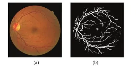
Fig. 1. Retinal image: (a) the color retinal image (DRIVE database #1) and (b) the binary vessel segmentation by Nguyenet al. method.
In this paper, for overcoming these drawbacks, a novel method is proposed. The morphological closing and subtraction processing are introduced before multi-scale line detecting. Furthermore, the weight of each line responses is tuned for getting the best performance at the linear combination process. The experimental results show that the accuracy is better than that by other methods.
The remainder of this paper is organized as follows. Section 2 describes the proposed vessel segmentation method. Section 3 presents the experimental results. Conclusions are given in Section 4.
2. Proposed Method
This paper proposes an automatic scheme to extract the retinal vessels. Fig. 2 shows a flow chart of the proposed scheme.

Fig. 2. Flow chart of the proposed scheme.
2.1 Illumination Correction
Non-uniform illumination caused by different photograph angle is always one of the major reasons that influence the feature selecting on retinal image. It makes optic disc not the brightest region and the vessels unclear. The intensities of vessel pixels would even be higher than optic disc. Green channel highlights the vessels best, so we first extract the green band from the original color image. And then, an illumination correction is defined by (1) to remove uneven background intensity[1].

whereI′ denotes the illumination corrected image andIis the green band image.μN(x,y) andσN(x,y) are the mean and the standard deviation of aN×Nsquare neighborhood centered at the pixel (x,y) , respectively.
2.2 Crude Vessel Image
Since the vessels are darker in retinal images, the morphological closing operation is applied on the illumination corrected image to directly remove vessels and leave the optic disc and background. The grayscale closing of imagefby structuring elementbis the dilation offbyb, followed by an erosion of the result withb[15]. The grayscale dilation at any location (x,y) offbybis given by (2), and the grayscale erosion is defined by (3),


The coverage of the structuring elementbof the closing operator cannot be too large; otherwise the fundus image will lose its original feature. On the other hand, if it is too small, vessels cannot be completely removed. Here,bis set as a circle with 11 pixels as the diameter. For choosing the structuring element, 20 images in DRIVE (digital retinal images for vessel extraction) database are used to estimate the parameterb. The image after closing is called the vessel-removed image.
Now, the illumination corrected image is subtracted by the vessel-removed image. The result is a crude vessel structure image, in which the background and optic disc are no longer obvious.
2.3 Multi-Scale Line Detection
Based on the fact that the vessels are line-like and smooth, a multi-scale line detector[12],[13]is applied to crude the vessel structure image. The line detector is a window of sizew×w, in which 12 segments of lengthlpixels orient at 12 different directions (interval 15°) passing through the center pixelp(x,y).
The segments are defined by (4) and restricted by (5):

where (i,j) denotes a neighbor ofp, andθ= 0°, 15°, 30°, …, 165°, is the slope angle of each segment. Fig. 3 illustrates a line detector withw=15 andl=13.
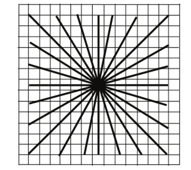
Fig. 3. Line detector withw=15 andl=13.
The average intensity of each segment is computed. The line strength of the pixelp, denoted as, is defined as follows:

wherelandware the length of segment and window size, respectively, and 1≤l≤w.is the maximum average among those of 12 segments, andis the mean intensity of thew×wsquare window. After subtraction operation mentioned in Subsection 2.2, thevessels are brighter as being compared with the background, so the strength of target pixelpcan be used for discriminating this pixel whether it is a vessel pixel or not.
Longer length line detectors are effective when handling the vessel central light reflex. However, it tends to merge close vessels and produces false vessel points near the thick vessel[13]. Shorter length line detectors overcome those drawbacks, but background noise is produced in the image. Nguyenet al.[13]linearly combined the line strengths at varying scales (lfrom 1 to 15 with a step of 2) with the same weight. However, it tends to produce false vessel detection around the optic disk and pathological regions.
This paper abandons the line strengths of middle length line detectors to eliminate the drawbacks, and tunes the weights of varying line strengths for getting better performance. The linear combination of line strengths is formulated as follow:

whereIneg_gis the negative green band retinal image,αlandβare the weights ofand, respectively, andN=∑αl+β.
2.4 Otsu Operation
After multi-scale line detection, the Otsu algorithm is used to get the binary vasculature image. An example of the vessel segmentation is shown in Fig. 4.
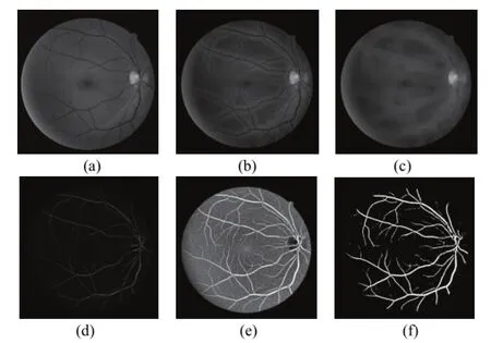
Fig. 4. Retinal vessel segmentation: (a) green band retinal image, (b) illumination corrected image, (c) closing processed image, (d) crude vessel structure image, (e) image after multi-scale line detector, and (f) binary vasculature image.
3. Experimental Results
All images used in this work come from the public retinal database DRIVE[14], which contains two sets: the test set and the training set. The test set provides manual vessel segmentation by two observes. The performance of the presented method is evaluated on the test set (20 images). The manual segmentations of the first observer are given as ground truth, and those of the second observer are used for comparison.
The performance is evaluated in terms of accuracy (Acc). The metric is defined as

where TP, TN, FP, and FN are true positive, true negative, false positive, and false negative, respectively.
In this paper, for improving the disadvantages of multi-scale line detection, the middle length line detectors are abandoned, and only the line detectors at 4 scales are used (w=15;l=1, 3, 13, and 15). By experiments, the weights of varying line strengths are set as follows:α1=α15= 2,α3=α13= 1, andβ=2.
Fig. 5 (a) and Fig. 5 (b) present some examples of vessel segmentation by the proposed method and Nguyenet al.method[13], respectively. Fig. 5 (c) and Fig. 5 (d) are the hand-drawn retinal vasculatures by observer 1 and 2. It is obvious that many false positive pixels appear at the center of the image and around the border of the optic disc in the Fig. 5 (b). Conversely, Fig. 5 (a) shows better segmentation.
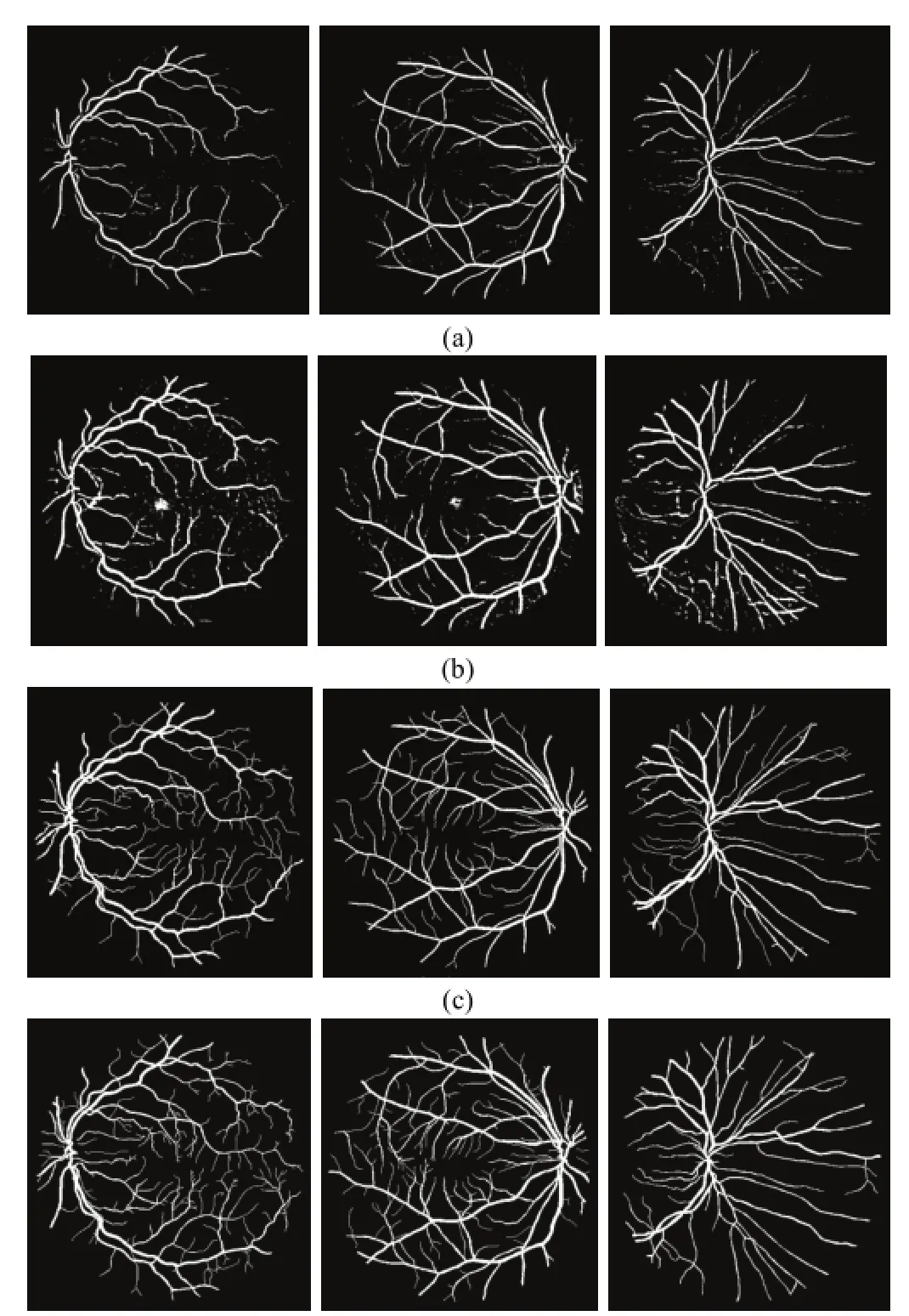
Fig. 5. Vessel segmentation by: (a) the proposed method, (b) Nguyen et al. method, (c) observer 1, and (d) observer 2.
Table 1 compares the proposed method with other methods in terms of accuracy. As mentioned above, the manual segmentation of second observer also participates in comparison. The accuracy of Ricciet al.’s method and observer 2 is taken from [13]. Concerning Nguyenet al.’s method, it was implemented by the authors of this paper according to the processes described in [13]. The results show that the performance of the proposed method is better than that of other methods.

Table 1: Comparison in terms ofAccon DRIVE database
4. Conclusions
In this paper, we present an automatic vessel segmentation method to extract vasculature from retinal image. The illumination correction processing generates significant contrast between the vessels and background. Then, the morphological closing and subtraction processing are applied to obtain the crude vessel structure before multi-scale line detecting. In order to improve the disadvantages of multi-scale line detection, only the line detectors at 4 scales (l= 1, 3, 13, and 15) are used. We also tune the weights of line strengths obtained from these 4 line detectors at the linear combination step. The experimental results show that the accuracy is 0.939 for the 20 test images in the DRIVE database. It is better than that of other methods.
[1] H.-K. Hsiao, C.-C. Liu, C.-Y. Yu, S.-W. Kuo, and S.-S. Yu,“A novel optic disc detection scheme on retinal images,”Expert Systems with Applications, vol. 39, pp.10600-10606, Sep. 2012.
[2] M. E. Martinez-Perez, A. D. Hughes, A. V. Stanton, S. A. Thom, N. Chapman, A. B. Bharath, and K. H. Parker,“Retinal vascular tree morphology: A semi-automatic quantification,”IEEE Trans. Biomed. Eng., vol. 49, pp. 912-917, Aug. 2002.
[3] B. Wasan, A. Cerutti, S. Ford, and R. Marsh, “Vascular network changes in the retina with age and hypertension,”Journal of Hypertension, vol. 13, pp. 1724-1728, Dec. 1995.
[4] T. Y. Wong and R. McIntosh, “Hypertensive retinopathy signs as risk indicators of cardiovascular morbidity and mortality,”British Medical Bulletin, vol. 73, pp. 57-70, Sep. 2005.
[5] D. Marín, A. Aquino, M. E. Gegúndez-Arias, and J. M. Bravo, “A new supervised method for blood vessel segmentation in retinal images by using gray-level and moment invariants-based features,”IEEE Trans. on Med. Imaging, vol. 30, pp. 146-158, Jan. 2011.
[6] Y. Yin, M. Adel, and S. Bourennane, “Retinal vessel segmentation using a probabilistic tracking method,”Pattern Recognition, vol. 45, pp. 1235-1244, Apr. 2012.
[7] A. M. Mendonça and A. Campilho, “Segmentation of retinal blood vessels by combining the detection of centerlines and morphological reconstruction,”IEEE Trans. Med. Imaging, vol. 25, no. 9, pp.1200-1213, 2006.
[8] F. Zana and J. C. Klein, “Segmentation of vessel-like patterns using mathematical morphology and curvature evaluation,”IEEE Trans. Image Process., vol. 10, no. 7, pp. 1010-1019, 2001.
[9] M. Al-Rawi, M. Qutaishat, and M. Arrar, “An improved matched filter for blood vessel detection of digital retinal images,”Comput. Biol. Med., vol. 37, pp. 262-267, Feb. 2007.
[10] S. Chaudhuri, S. Chatterjee, N. Katz, M. Nelson, and M. Goldbaum, “Detection of blood vessels in retinal images using two-dimensional matched filters,”IEEE Trans. on Medical Imaging, vol. 8, no. 3, pp. 263-269, 1989.
[11] J. V. B. Soares, J. J. G. Leandro, R. M. Cesar, H. F. Jelinek, and M. J. Cree, “Retinal vessel segmentation using the 2-d gabor wavelet and supervised classification,”IEEE Trans. on Medical Imaging, vol. 25, no. 9, pp. 1214-1222, 2006.
[12] E. Ricci and R. Perfetti, “Retinal blood vessel segmentation using line operators and support vector classification,”IEEE Trans. on Medical Imaging, vol. 26, pp.1357-1365, Oct. 2007.
[13] U. T. V. Nguyen, A. Bhuiyan, L. A. F. Park, and K. Ramamohanarao, “An effective retinal blood vessel segmentation method using multi-scale line detection,”Pattern Recognition, vol. 46, pp. 703-715, Mar. 2013.
[14]Digital Retinal Images for Vessel Extraction: DRIVE Database, Image Science Institute. [Online]. Available: http://www.isi.uu.nl/Research/Databases
[15] R. C. Gonzalez and R. E. Woods,Digital Image Processing, 3rd ed., New Jersey: Pearson, 2010, ch. 9.

Chun-Yuan Yu was born in Lukang in 1957. He received his M.S. degree in mining, metallurgical & material engineering from National Chen-Kung University, Tainan in 1983. He has been an instructor with the Department of Computer and Communication engineering, Nan-Kai University of Technology, Tsaotun since 1983. Currently, he is working toward the Ph.D. degree with the Department of Computer Science and Engineering, National Chung-Hsing University, Taichung. His research interests focus on medical image processing, programming, and pattern recognition.

Chia-Jen Chang was born in Taichung in 1977. He received the M.D. degree in medicine from the National Cheng Kung University, Tainan in 2002 and the Ph.D. degree in applied chemistry from the Chaoyang University of Technology, Taichung in 2012. He is currently an ophthalmologist with Taichung Veterans General Hospital. His research interests include medical images, ophthalmology, and air pollution.

Yen-Ju Yao was born in Chiayi in 1990. She is currently pursuing the M.S. degree with the Department of Computer Science and Engineering, National Chung-Hsing University, Taichung. Her research interest is retinal images processing.

Shyr-Shen Yu received the Ph.D. degree in computer science from Western Ontario University, Ontario in 1990. Currently, he is a professor with the Department of Computer Science and Engineering, National Chung-Hsing University, Taichung. His research work focuses on image processing, bioinformatics, pattern recognition, and data mining.
Manuscript received December 13, 2013; revised March 14, 2014. This work was supported by the NSC under Grant NSC 102-2221-E-005-082.
C.-Y. Yu is with the Department of Digital Living Innovation, Nan Kai University of Technology, Tsaotun, and also with the Department of Computer Science and Engineering, National Chung-Hsing University, Taichung (e-mail: t112@nkut.edu.tw).
C.-J. Chang is with the Department of Ophthalmology, Taichung Veterans General Hospital, Taichung (e-mail: capmchang@seed.net.tw)
Y.-J. Yao is with the Department of Computer Science and Engineering, National Chung-Hsing University, Taichung (e-mail: lisa79517@ hotmail.com).
S.-S. Yu is with the Department of Computer Science and Engineering, National Chung-Hsing University, Taichung (Corresponding author e-mail: pyu@nchu.edu.tw).
Digital Object Identifier: 10.3969/j.issn.1674-862X.2014.04.011
杂志排行
Journal of Electronic Science and Technology的其它文章
- Study on Temperature Distribution of Specimens Tested on the Gleeble 3800 at Hot Forming Conditions
- Non-Linearity of Visual Sensitivity and Pursuit Velocity during Smooth Pursuit Eye Movements
- Family Competition Pheromone Genetic Algorithm for Comparative Genome Assembly
- Quantification of Cranial Asymmetry in Infants by Facial Feature Extraction
- Intrinsic Limits of Electron Mobility inModulation-Doped AlGaN/GaN 2D Electron Gas by Phonon Scattering
- Real-Time Hand Motion Parameter Estimation with Feature Point Detection Using Kinect
