用于海洋原位浮游生物探测的同轴数字全息显微技术研究
2014-03-18聂亚茹刘惠萍王金城
于 佳,聂亚茹,王 添,刘惠萍,王金城
(中国海洋大学 光学光电子实验室,山东 青岛266100)
1 Introduction
Plankton,including phytoplankton,zooplankton and bacterio-plankton,is a huge and diverse group of marine organisms.As a main source of food to many aquatic species,it plays an irreplaceable role in the balance and adjustment process of the marine eco-system and even that of the entire global eco-system.Apart from being the bottom of the marine food chain,plankton also plays an important role in marine bio-chemical cycle,e.g.the ocean’s carbon cycle.Nowadays,the study of plankton has become one major part of marine research and is substantially involved in many other research fields such as environmental protection,global climate changing,marine gene science,etc.
The critical need to understand the dynamics of plankton ecosystems has led to the development of equipments to help scientists collect high-resolution data on plankton species-specific population structures.There are two primary methods for plankton investigation,sampling and in-situ detection.Compared with traditional sampling method,in-situ detection is a real-time,nonintrusive,non-destructive and highly efficient technology that can acquire the original status of the plankton without causing damage.Various optical imaging systems,such as Video Plankton Recorder[1],the Underwater Video Profiler System,the ZooVis System,the 3D Zooplankton Observatory,the Optical Plankton Counter and the Laser Optical Plankton Counter[2]had been usually used for direct observation of plankton,especially for zooplankton.These systems can acquire the images of plankton with resolution as high as the size at micrometers and information regarding identification and abundance can be retrieved,however they tend to sample with very small volumes because of the shallow field depth of the microscopic imaging optical system.One possible solution to this problem is holography.Holography has been used in observing zooplankton[3],air bubbles[4]and sediments[5].Holographic Camera systems for the study of plankton has been put into practice in bay and Island[6]using in-line holography.Then the holographic camera seeks to simultaneously record in-line and off-axis holograms of the same scene[7].Electronic holographic camera has been developed for researches of the distribution of plankton[8].Digital inline holographic microscope was designed for the imaging of particles in size from 50 μm to several millimeters.[9]It is possible for digital holographic imaging system to sense marine plankton.[10]These studies have proved that holography is an effective method for the in-situ detection of plankton and other species’without intrusion and destruction and can achieve high accurate spatial distribution,image fidelity,and large depth of field.Recently,a underwater system has been developed for the detection of plankton in deep sea.[11]
In this paper,a Digital in-line holographic microscopy(DIHM)developed in our laboratory was introduced and used to record the holographic images of the plankton fields.The magnification,resolution and the depth of field of this developed system were discussed in some detail.
2 Experimental setup and sampling
The Digital in-line holographic microscopy(DIHM)system described in this paper is based on the in-line holographic optical set up shown in Fig.1.In this system,the objects in sample cell are illuminated with an expanded and collimated laser beam.And the diffracted waves from these objects are collected by a microscopic objective hence to form an enlarged diffractive wave field which,then,is recorded by a computer controlled CCD detector.
The specifications of main components used in this DIHM system are shown in Table 1.As shown in Table 1,a semiconductor CW laser,with the output power 50 mw at the wavelength of 532 nm,was used.For an inline holography setup,all the optical components,such as lens,beam expander and objectives,and the CCD should set to be co-axial.The laser beam is expanded and collimated into a parallel light beam with a diameter about 40 mm.This beam goes into the sample cell perpendicularly,enlightens the planktons and other particles in water and is then diffracted.The diffracted wave is enlarged with a microscopy objective(10X,NA=0.25)and forms a fringe pattern which hits the CCD detector.The holographic image of the field is then recorded by this computer controlled CCD(Amazon IMx-7050G)which has the pixel number of 2448×2048 with the pixel size of 3.45×3.45μm2.To minimize the instability of the system,the exposure time is limited to several micro-seconds.With one single exposure,a magnified diffractive pattern of the plankton in water is recorded as a digital hologram,and the serial of holograms of the planktons at motion are recorded with the highest frame rate of 15 fps.

Fig.1 Schematic diagram of DIHM using microscopic objective
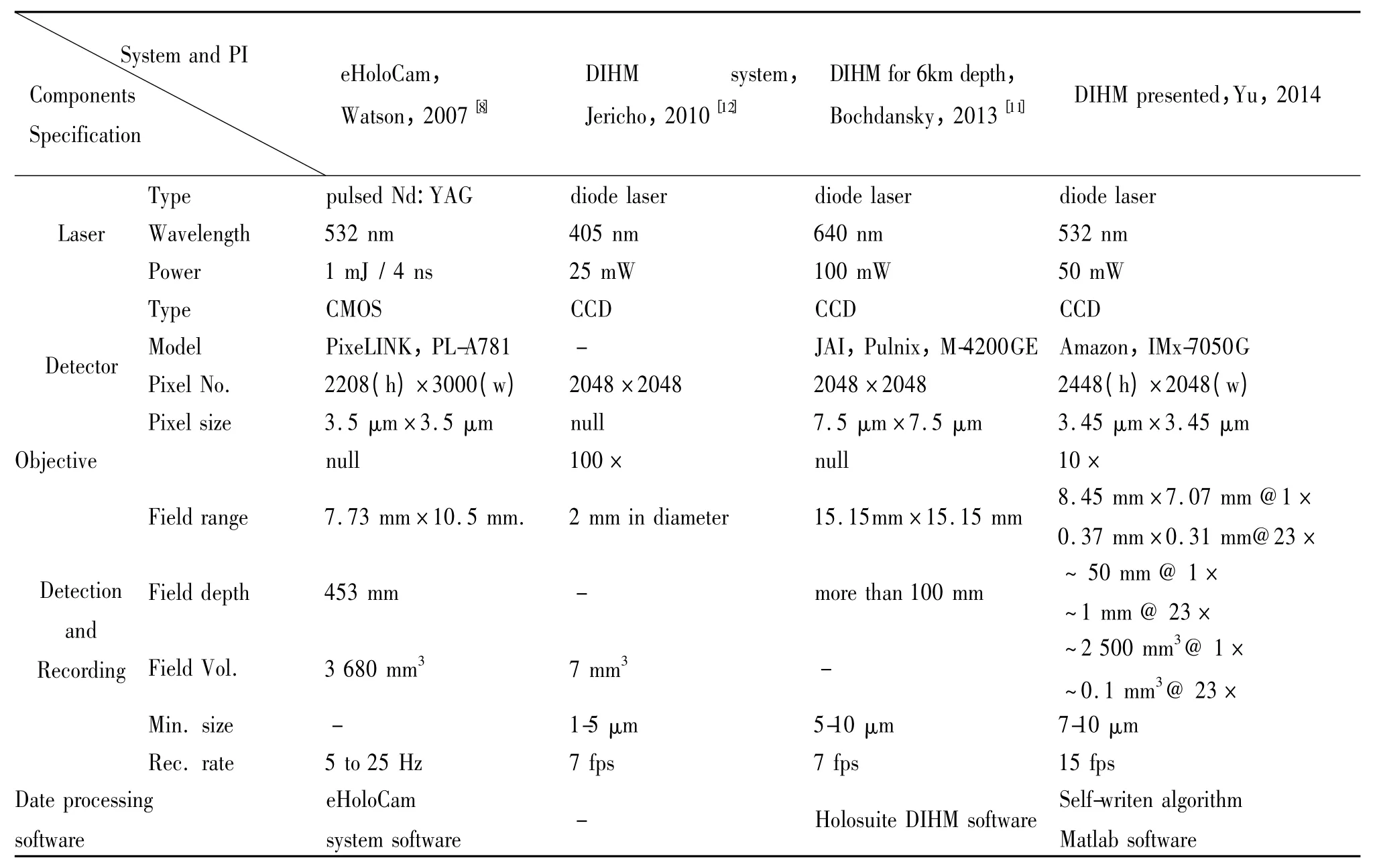
Tab.1 The Specification and performance of the developed DIHM system with the comparison of those given in Ref.
The plankton samples were drawn from the upper layer of the near-shore sea water in Qingdao,using the plankton net.The volume of the sea water could be estimated by measuring the dredging distance(normally 3-5 meters in our investigation)and the size of the plankton net.In laboratory,the sampling water was put into a sample cell and the holography recording experiment is carried out in time as to ensure the activeness of the plankton.
3 Image Reproducing
In the image reproducing stage,the recorded diffractive pattern is analyzed and calculated according to the procedure shown in Fig.2.The original optical field of the planktons in water is finally reproduced.According to the follow-chart,the light field recorded by CCD is input as the hologram and the parameters of reproducing,such as the wavelength,the size of the hologram and the pixels size of the CCD are also input based on the experimental setup.Then,after the distance ziof reproducing is given,three algorithms are tested and compared,in order to get the optimal result of the reproduced image.Since the distance z changes within a certain range,the cycle needs to be used and then a sequence of reproduced images will be achieved with each values of ziand the each image in the sequence corresponds to a slice in water in certain field depth along the z direction which is also the axial of light.Although not included in this paper,we have plans in our future work in image processing and object recognition based on the reproduced images,as to achieve the information of the plankton and reconstruct the three-dimensional distribution and shape of plankton.

Fig.2 The procedure of digital hologram reproducing
4 Results and discussion
4.1 The reproduced image of plankton
Fig.3 is a comparison of the images taken by Olympus microscope(on the left)and reproduced by DIHM system(on the right).As shown in Fig.3,the plankton with the size of 180μm×120μm can be recorded and the detail information of plankton can be seen clearly.
Fig.4 shows the reconstruction images of several typical planktons,noctiluca,algae and copepods.In those reproduced images,the shape and internal structure of plankton can be seen clearly,with the detailed tentacles and flagella for copepods.The understanding of the morphology of plankton helps to reflect the changes of marine and climate.
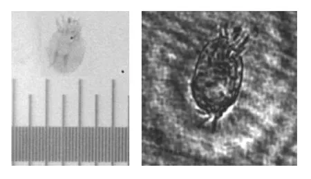
Fig.3 A comparison of images taken by Olympus(left)and reproduced by DIHM system(right)
4.2 The resolution of the DIHM system
In order to verify the resolution of our DHIM system,a resolution board USAF 1951 has been used as the object.The hologram and the reproduced image of the group 6 and group 7 are shown in Fig.5.The result dem onstrates that minimum size of the object to be resolved by this developed DIHM system is 7.8125μm,which corresponding to the element 1 of group 7 in the resolution board.

Fig.4 Reconstruction images of typical planktons
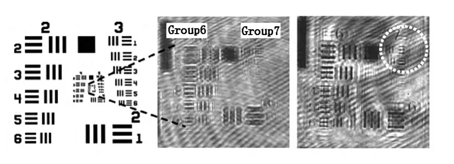
Fig.5 The resolution board(left)and its hologram(middle)as well as reproduced image(right)
4.3 The field depth of the DIHM system
Fig.6 shows the relationship between magnification and depth of field of DIHM system.As is well-known that the field depth of a microscopic system decreases with the magnification,the DIHM system faces the same challenge as shown as in Fig.6.However combining with holographic technology,the field depth of DIHM has significantly improved.Even at the magnification as high as 12.7×,the depth of field is 4mm,much larger than that an ordinary system could achieve.On dropping the magnification to 6×,the depth of field goes up to 10mm.The advantage of this DIHM method is obvious.Such a method could ensure sufficient magnification and obtain a large depth of field exploration simultaneously.
The detection field of the system is varied with magnification with 8.45 mm×7.07 mm at 1×,and drops to 0.37 mm×0.31 mm at magnification of 23×,as shown as in Table 1.The resulted detection volume of a single record has been determined to be 2 500 mm3with about 50 mm field depth at the magnification of 1×,and drops to0.1 mm3with about 1mm field depth at the magnification of 23×.
Fig.7 shows a hologram reconstruction by a number of different depth images.As can be seen from Fig.7,at different reconstruction distance,different plankton can be seen clearly in the field,as such,the spatial distribution of plankton could be obtained.The results demonstrate that the system has a potential to be used for the detection of three-dimensional distribution.
5 Conclusion
In this paper,a newly developed digital In-line Holographic Microscopy(DIHM)system was presented and the experiments of marine plankton imaging based on DIHM had been carried out.It is demonstrated that DIHM is an appropriate and highly effective method for aquatic plankton study.With this system,a sufficient magnification could be ensured with a relatively large depth of field exploration.The resulted resolution was determined to be 7-10 m which meets the demands of detection for zooplankton and phytoplankton.An in-situ underwater DIHM instrument is hoped to be developed for field study of marine plankton in near future.
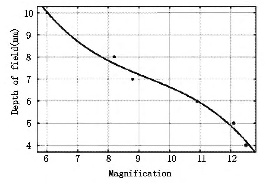
Fig.6 The depth of field as a function of magnification
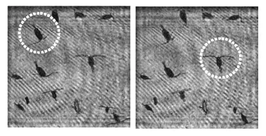
Fig.7 In-focus plankton at the reconstruction distances of 196 mm(left)and 204 mm(right)
[1] DAVISC S,THWAITESF T,GALLAGER SM,et al.A threeaxis fast-tow digital Video Plankton Recorder for rapid surveys of plankton taxa and hydrography[J].Limnology and Oceanography:Methods,2005,3:59-74.
[2] HERMAN A W,BEANLANDS B,PHILLIPS E F.The next generation of Optical Plankton Counter:the Laser-OPC[J].Journal of Plankton Research,2004,26(10):1135-1145.
[3] HEFLINGER L O,STEWART G L,BOOTH C R.Holographic motion pictures of microscopic plankton[J].Applied Optics,1978,17(6):951-954.
[4] O’HERN T J,D’AGOSTINO L,ACOSTA A J.Comparison of Holographic and Coulter Counter measurements of cavitation nuclei in the ocean[J].Trans ASME J Fluids Eng,1988,110(2):200-207.
[5] BLACK K S,SUN H,CRAIG G,et al.Incipient erosion of biostabilized sediments examined using particle-field optical holography[J].Environ Sci Technol,2001,35(11):2275-2281.
[6] KATZ J,DONAGHAY PL,ZHANG J,et al.Submersible holocamera for detection of particle characteristics and motions in the ocean[J].Deep-Sea Research,1999,46(8):1455-1481.
[7] HOBSON PR,WATSON J.The principles and practice of holographic recording of plankton[J].J Opt A:Pure Appl Opt,2002,4(4):S34-S49.
[8] SUN H,HENDRY D C,PLAYER M A,et al.In situ underwater electronic holographic camera for studies of plankton[J].IEEE Journal of Oceanic Engineering,2007,32(2):373-382.
[9] WATSON J,ALEXANDER S,CRAIG G,et al.Simultaneous in-line and off-axis subsea holographic recording of plankton and other marine particles[J].Meas Sci Technol,2001,12(8):L9-L15.
[10] TAN S,WANGS.An approach for sensing marine plankton using digital holographic imaging[J].Optik,2013,124(24):6611-6614.
[11] BOCHDANSKY A B,JERICHO M H,HERNDL G J.Development and deployment of a point-source digital inline holographic microscope for the study of plankton and particles to a depth of 6 000 m[J].Limnology and Oceanographt:Methods,2013,11(1):28-40.
[12] JERICHO SK,KLAGESP,NADEAU J,et al.In-line digital holographic microscopy for terrestrial land exobiological research[J].Planetary and Space Science,2010,58(4):701-705.
