右膈下动脉64层螺旋CT三维重建在肝癌介入治疗中的应用
2014-02-07刘长春董景辉安维民宿贝贝高原智李勇武
刘长春 董景辉 安维民 宿贝贝 高原智 李勇武 马 威*
右膈下动脉64层螺旋CT三维重建在肝癌介入治疗中的应用
刘长春①董景辉①安维民①宿贝贝①高原智①李勇武①马 威①*
目的:分析右侧膈下动脉(RIPA)在多层螺旋CT血管三维重建中的表现,探查其解剖与变异在肝癌介入治疗中的作用。方法:回顾性分析935例患者行腹部螺旋CT三维重建的临床资料,采用容积再现(VR)、曲面重组(MPR)和最大密度投影(MIP)3种方法进行血管重建,总结RIPA的起源和分布对经导管动脉栓塞化疗(TACE)的指导意义。结果:935例进行右膈下动脉螺旋CT三维重建的病例均可成功显示膈下动脉起源(占100%),其中发自于双侧膈下动脉共干者为163例(占17.4%),单独发自于RIPA者为772例,其中发自于腹主动脉、腹腔动脉干、右侧肾及副肾动脉、胃左动脉、肠系膜上动脉和肝动脉分别为367(占39.3%)、320例(占34.2%)、196例(占21.0%)、27例(占2.9%)、11例(占1.1%)和14例(占1.5%)。RIPA参与原发性肝癌(HCC)供血者达12.5%(32/255)。结论:螺旋CT三维重建可在介入术前了解右膈下动脉的起源与分布,并判断有无肝癌侧枝供血,对于临床TACE具有指导意义。
右膈下动脉;肝细胞癌;多排螺旋CT;化疗栓塞
刘长春,男,(1984- ),本科学历,主治医师。解放军第302医院医学影像中心,从事腹部影像诊断工作。
膈下动脉尤其是右膈下动脉是肝癌最常见的肝外侧枝供血动脉,目前越来越多的介入医生逐渐认识到膈下动脉的栓塞对肝癌经导管动脉栓塞化疗(transcatheter arterial chemo embolization,TACE)的疗效有重要意义,甚至把膈下动脉造影作为与腹腔动脉与肠系膜上动脉造影并列的肝癌介入治疗的常规造影,熟悉膈下动脉的起源及变异、术前掌握膈下动脉的走行对介入医生减少术中曝光剂量大有裨益。本研究回顾性分析了在解放军第302医院行腹部螺旋CT三维重建的935例病例资料,其中32例患者进行了右膈下动脉栓塞。
1 资料与方法
1.1 病例资料
检索解放军第302医院从2011年1月至2012年10月对935例进行腹部螺旋CT三维重建的病例资料,其中男性653例,女性282例;年龄24~77岁,平均年龄为51.8岁。其中肝硬化患者628例,肝癌伴肝硬化患者255例,正常者52人。32例肝癌患者进行了肝癌的右膈下动脉栓塞。
1.2 仪器设备
采用GE LightSpeedVCT 64 MSCT行肝脏CT平扫及增强扫描,采集层厚0.625 mm,矩阵512×512,重建矩阵1024×1024,螺距1∶1。
1.3 扫描方法
使用高压注射器经右肘正中静脉注射对比剂(碘普罗胺370 mg I/ml),对比剂用量为1.5 ml/kg,注射速率为3.5~5 ml/s,从A筒注入对比剂后以相同速率从B筒注入生理盐水50 ml。动脉期扫描方向为从头侧至足侧,扫描范围自膈上至髂前上棘;采用smart prep智能追踪技术,在膈上胸主动脉层面设定感兴趣区(region of interest,ROI),将ROI中心放置于主动脉中心偏内上方,触发阈值设定为140 Hu,Smart Prep监测延迟时间10 s,达到阈值后延迟3 s(语音时间)自动进入扫描程序。动脉期三维后处理图像重建层厚0.625 mm,间隔0.625 mm。采用GE ADW4.5工作站进行动脉血管的多平面重组(multiplanar reformation,MPR)、最大密度投影(maximum intensity projection,MIP)和容积再现(volume rendering,VR)图像重建。由2名有经验的放射科医生进行动脉期血管三维重建,并共同确认右膈下动脉开口及走行。
2 结果
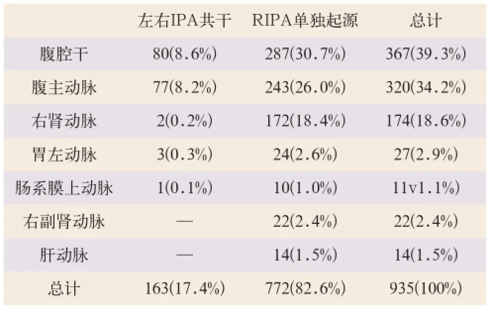
935例患者右侧膈下动脉(right inferior phrenic artery,RIPA)的起源见表1,RIPA参与HCC供血达12.5%(32/255),其供血的肝癌所在肝段为SⅦ者24例、SⅧ者5例、SⅣ者2例、SⅡ者1例。32例RIPA均成功进行了TACE治疗,手术成功率为100%,寻找RIPA所用曝光时间为(1.1±0.6)min。

表1 935例RIPA起源情况[例(%)]
VR图像重建对RIPA的显示率为64.9%(607/935),MPR及MIP图像重建的显示率分别为97.1%(908/935)、96.8%(905/935),MPR及MIP较VR能更好的显示RIPA的起源及走行。
MIP图像可显示出肝顶部占位介入术后仍可见强化,并可见增粗的RIPA供血原发性肝癌(hepato cellular carcinoma,HCC),VR图像背面观可清晰显示RIPA的走行,与DSA结果一致(如图1、图2所示)。

图2 双侧IPA分别发自肾动脉的MIP及VR图像
3 讨论
TACE目前已成为不能手术切除的肝癌的首选治疗方法,完全栓塞所有肝癌滋养动脉是决定疗效的关键因素[1-2]。肝癌肝外供血血管主要包括膈下动脉、胃左动脉、网膜动脉、胆囊动脉、内乳动脉、肋间动脉、右肾上腺动脉及肾动脉等12种侧枝,其中膈下动脉是肝癌肝外供血最主要的侧枝血管,约占所有肝癌侧枝供血血管的52.5%[3-4]。在介入治疗操作过程中,发现完全栓塞肿瘤供血动脉是介入医生面临的首要问题。在此过程中介入医生受X射线照射剂量明显增大,且常难以发现全部侧枝血管。而多排螺旋CT薄层及三维重建,可在介入治疗前对患者的所有动脉血管情况有较为全面的了解,术中可有的放矢,减少曝光时间,从而降低介入术中的辐射剂量。
膈下动脉的起源通常位于第12胸椎至第2腰椎间,多数起源于腹腔动脉干及腹主动脉,左、右膈下动脉可共干或单独发出;少数发自肾动脉,极少数发自胃左动脉、肠系膜上动脉及肝动脉[5]。本研究发现,发自肾动脉及副肾动脉的RIPA并不少见,占到RIPA起源的1/5,且多位于右肾动脉开口处。RIPA主要供应包括与肝脏裸区相邻的大部分膈肌,分支包括升支(前支)、降支(后支)、肾上腺上动脉及肾上腺中动脉,还发出下腔静脉支及膈肌支,其分支有时可与肝动脉、内乳动脉、肋间动脉、膈肌动脉及心包膈动脉分支相吻合[6]。
膈下动脉主要供给肝SⅡ、SⅦ及SⅧ这些更靠进肝裸区部位的肿瘤,但同时文献报道膈下动脉还可供应肝SⅠ、SⅢ、SⅣ及SⅥ部位的肿瘤,甚至有SⅥ的肝肿瘤可完全由RIPA单独供血的报道[7-8]。因此,在任何靠近肝表面并且肝动脉造影显影不完全的病灶,均应考虑到肝癌膈下动脉供血。有文献表明,膈下动脉的变异很多,这极大的增加了介入手术时寻找膈下动脉的难度,在术前进行膈下动脉螺旋CT三维重建十分必要[9-12]。本研究发现,如果膈下动脉参与肝癌的供血,可100%在横断面薄层重组图像中找到膈下动脉,对其起源可大致了解,通过VR图像,可清晰直观的显示其分布与走行,但VR图像的显示成功率较低,本研究只有64.9%的病例可显示其分布与走行,可能与病例资料多为肝硬化、腹水患者有关,而大量的腹水对VR图像的显示有一定的影响。
在介入手术术前进行RIPA的螺旋CT三维重建可明确RIPA参与肿瘤供血的情况,并了解其解剖及变异,有利于介入医生手术操作,可作为介入手术前的常规检查。
[1]Lau WY,Lai EC.Hepatocellular carcinoma:current management and recent advances[J].Hepatobiliary Pancreat Dis Int,2008,7(3):237-257.
[2]Llovet JM,Bruix J.Systematic review of randomized trials for unresectable hepatocellular carcinoma:chemoembolization improves survival[J].Hepatology,2003,37(2):429-442.
[3]Miyayama S,Matsui O,Taki K,et al.Extrahepatic blood supply to hepatocellular carcinoma:Angiographic demonstration and transcatheter arterial chemoembolization[J]. Cardiovasc Intervent Radiol,2006,29(1):39-48.
[4]Kim HC,Chung JW,Lee W,et al.Recognizing extrahepatic collateralVessels that supply hepatocellular carcinoma to avoid complications of transcatheter arterial chemoembolization[J]. Radiographics,2005,25(Suppl 1):S25-S39.
[5]Ozbulbul NI,Yurdakul M,Tola M,et al.Can multidetector row CTVisualize the right and left inferior phrenic artery in a population without disease of theliver?[J].Surg Radiol Anat,2009,31(9):681-685.
[6]Cheng LF,Ma KF,Fan WC,et al.Hepatocellular carcinoma with extrahepatic collateral arterial supply[J].J Med Imaging Radiat Oncol,2010,54(1):26-34.
[7]Miyayama S,Yamashiro M,Okuda M,et al.The march of extrahepatic collaterals:analysis of blood supply to hepatocellular carcinoma located in the bare area of the liver after chemoembolization[J].Cardiovasc Intervent Radiol,2010,33(3):513-522.
[8]Gwon DI,Ko GY,Yoon HK,et al.Inferior phrenic artery:anatomy,variations,pathologic conditions,and interventional management[J].Ra diographics,2007,27(3):687-705.
[9]Loukas M,Hullett J,Wagner T.Clinical anatomy of the inferior phrenic artery[J].Clin Anat,2005,18(5):357-365.
[10]Miyayama S,Yamashiro M,Yoshie Y,et al.Inferior phrenic arteries:angiographic anatomy,variations and catheterization techniques for transcatheter arterial chemoembolization[J].Jpn J Radiol,2010,28(7):502-511.
[11]Matusz P,Loukas M,Lacob N,et al.Common stem origin of left gastric,right and left inferior phrenic arteries,in association with a hepatosplenomesenteric trunk,independently arising from the abdominal aorta:Case report using MDCT angiography[J].Clin Anat,2013,26(8):980-983.
[12]Matusz P,Loukas M,Lacob N,et al.Rare case of the trunk of the inferior phrenic arteries originating from a common stem with a superior additional left renal artery from the abdominal aorta[J].Clinical Anatomy,2012,25(8):979-982.
The application of right inferior phrenic artery reconstruction by using 64-slice 3D-MDCT in interventional treatment of hepatocellular carcinoma
LIU Chang-chun,DONG Jing-hui, AN Wei-min, et al// China Medical Equipment,2014,11(5):87-89.
Objective:To analysis the anatomy andVariations of the right inferior phrenic artery (RIPA) in 3D-MDCT reconstruction and discuss the application in interventional treatment of HCC.Methods:The origin and distribution of RIPAs in 935 cases(628 cases of hepatic cirrhosis, 255 cases of HCC, 52 cases of normal people) were analyzed retrospectively by usingVolume rendering (VR), multiplanar reconstruction (MPR) and maximum intensity projection (MIP) to summarize the applications before and after transcatheter arterial chemoembolization (TACE).Results:The RIPA origin was detected in all cases (sensitivity 100%). The RIPA and LPIA have the common trunk in 163 cases, and arise separately in other 772 cases. RIPAs originated from the aorta (39.3%), celiac trunk (34.2%), right renal artery or accessory renal artery (21.0%), left gastric artery (2.9%), superior mesenteric artery (1.1%) and hepatic artery (1.5%). Thirty-two of 255 cases demonstrate the supplement of HCC.Conclusion:3D-MDCT reconstruction can be used to recognize the origin and distribution of RIPA and judge the extrahepatic collateralVessels by interventional radiologists before TACE.
Right inferior phrenic artery; Hepatocellular carcinoma; Multidetector computed tomography; Chemoembolization
1672-8270(2014)05-0087-03
R735.7
A
10.3969/J.ISSN.1672-8270.2014.05.033

2014-01-29
①解放军第302医院医学影像中心 北京 100039
*通讯作者:mawei302@sohu.com
[First-author’s address]Medical Imaging Center, The 302 Hospital of PLA, Beijing 100039, China.
