国际科技信息
2013-09-21
“生物支架”可修复先天性心脏缺损
近日,美国莱斯大学和德州儿童医院的研究人员成功制成了一种名为“生物支架”的生物相容性补丁,能够修复婴儿的先天性心脏缺损。其能与活性心肌细胞相容,并可支撑新的健康组织的生长。随时间推移,它也会逐渐降解。相关研究报告发表在近期出版的《生物材料学报》上。
一个好的支架需能够完美地执行多种功能:它需要强大到足以承受心脏跳动的压力,还能良好地扩展和收缩。另外还需要具有孔洞支持新的心脏细胞的迁移,使它们自发建立连接,形成自然支架以取代植入的补丁。同时其还要足够坚韧以支持缝合过程,并能在自然组织发挥作用后适时进行生物降解。
目前用于治疗先天性心脏缺损的修复补丁多由合成纤维制成,或取材自乳牛或病患本体。但这种补丁并不具有活性,其就如塑料一般,无法与病患的心脏组织融为一体,并具有引发心力衰竭、心律失常和纤维性颤动等症状的风险。
此次制成的三明治结构支架的中间一层为聚己内酯(PCL)聚合物,其能逐渐硬化形成拉伸带。而混合两种具有不同分子质量的PCL,可在粗糙表面形成微小的孔隙。外面的两层则是由明胶和脱乙酰壳多糖一比一混合而成的水性凝胶,两种原材料都很常见。
心肌细胞能在水性凝胶表面增殖,逐渐形成网络,并能契合心脏组织的舒张和收缩。虽然细胞不能穿过PCL的孔洞,但这些孔隙却能使营养物质从一侧传输至另一侧,它们也能使凝胶紧紧附着在PCL的核心。
研究人员在水中测试了PCL的生物降解特质。50天后,约有15%的P C L分散开来,形成了不规则的薄片。他们表示如果PCL被植入人体内,这一降解速度将会变慢,需要数月才能完成。但这个过程也很稳定,可支持肌肉组织的逐步构建。科学家称,他们希望未来“生物支架”能够与干细胞衍生的心脏细胞相结合,在植入婴儿体内后,快速缝合心脏缺陷处,缓解心室压力,并在之后观测时表现得与正常组织无异。研究人员还透露,干细胞的可能来源包括羊水和新生儿母体等,这将确保心脏补丁的基因一致性,他们目前亦在进行相关的研究。下一步,科研人员还将进行数年测试,直至这种支架顺利进入人体试验阶段。
New biocompatible patch can repair congenital heart defects
A painstaking effort to create a biocompatible patch to heal infant hearts is paying off at Rice University and Texas Children's Hospital.
The proof is in a petri dish in Jeffrey Jacot's lab, where a small slab of gelatinous material beats with the rhythm of a living heart.
Jacot, lead author Seokwon Pok, a postdoctoral researcher at Rice, and their tissue-engineering colleagues have published the results ofyears of effort to produce a material called a bioscaffold that could be sutured into the hearts of infants suffering from birth defects. The scaffold, seeded with living cardiac cells, is designed to support the growth of healthy new tissue. Over time, it would degrade and leave a repaired heart.
The research was detailed in the Elsevier journal Acta Biomaterialia.
Patches used now to repair congenital heart defects are made of synthetic fabrics or are taken from cows or from the patient's own body. About one in 125 babies born in the United States suffers such a defect; three to six of every 10,000 have what's known as a defect called Tetralogy of Fallot, a cause of "blue baby syndrome" that requires the surgical placement of a patch across the heart's right ventricular outflow tract.
Current strategies work well until the patches, which do not grow with the patient, need to be replaced, said Jacot, an assistant professor of bioengineering at Rice University, director of the Pediatric Cardiac Bioengineering Laboratory at the Congenital Heart Surgery Service at Texas Children's Hospital and an adjunct professor at Baylor College of Medicine.
"None of those patches are alive," Jacot said, including the biologically derived patches that are "more like a plastic" and are not incorporated into the heart tissue.
"They're in a muscular area in the heart that's important for contraction and, more so, for electrical conduction," he said. "Electrical signals have to go around this area of dead tissue. And having dead tissue means the heart produces less force, so it's not surprising that children with these types of repairs are more at risk for developing heart failure, arrhythmias and fibrillation.
"What we're making can replace current patches in an operation that surgeons are already familiar with and that has a very high short- and medium-term success rate, but with long-term complications," he said.
A better scaffold would have to perform many functions perfectly. It must be strong enough to withstand the pressures delivered by a beating heart yet flexible enough to expand and contract; porous enough to allow new heart cells to migrate, make connections and excrete their own natural scaffold to replace the patch; and tough enough to handle sutures but still be able to biodegrade over just the right amount of time for natural tissue to take over.
The sandwich the researchers created seems to fill the bill on all counts. In the middle is a selfassembled polycaprolactone (PCL) polymer that hardens into a tough but stretchable ribbon. Mixing two types of PCL with different molecular weights allows tiny pores to form along the rough surface. The "bread" is a hydrogel made from a 50/50 mixture of gelatin and chitosan, a widely used material made from the shells of crustaceans like shrimp.
Heart cells cultured on the hydrogel surface were able to thrive and formed networks and ultimately beat. Though cells could not attach to the surface or pass through the pores of the PCL, the pores do allow nutrients to migrate from one side to the other, Jacot said. They also allow the hydrogel to hold on to the PCL core.
新荧光成像技术可清晰呈现血管脉动
美国斯坦福大学的科学家开发出一种荧光成像技术,能够使活体动物血管脉动以前所未有的清晰度呈现。与传统的影像技术相比,其增加的清晰度类似于擦拭掉眼镜前的迷雾一般。该研究结果发表在最新一期的《自然医学》杂志在线版上。
该技术被称为近红外-Ⅱ成像,或NIR-Ⅱ。研究人员首先将水溶性碳纳米管注射到活体的血液中,然后用激光照射要观察的对象,如小白鼠。
激光的波长在近红外范围内,约为0.8微米,可导致专门设计的碳纳米管发出1微米至1.4微米的波长更长的荧光,用于检测确定血管的结构。
碳纳米管发出的荧光波长要比传统成像技术更长,这是实现令人惊叹的微小血管清晰图像的关键。由于更长波长光散射较少,因此形成了更清晰的血管图像。
此外,这种技术使图像呈现更精致的细节,允许研究人员能够获得一个快速的图像采集速度,近乎实时地测量血流量。
同时获得血流信息和看到清晰血管对于动脉疾病动物模型的研究将特别有用,如血流是如何受到动脉阻塞和收缩诱发的影响,还有其他事项如中风和心脏病发作的影响。
研究人员说:“对于医学研究而言,这是一个非常好的观察小动物特征的工具。其将有助于我们更好地理解一些血管疾病,以及其对于治疗的反应和如何可以设计出更好的治疗。”
由于NIR-Ⅱ至多只能穿透身体1厘米,所以它不会取代其他成像技术,而是X射线、CT、MRI和激光多普勒技术的补充。不过,它却是一个用于研究动物模型的强大方法。
研究人员说,下一步将使这项技术在人体内更容易接受应用,并探索可替代的荧光分子。
他们希望找到小于碳纳米管又能够发出同样波长光的物质,以便使其可以很容易地从体内排出,消除任何毒性的担忧。
New technique for visualizing blood flow involves carbon nanotubes and lasers
Stanford scientists have developed a fluorescence imaging technique that allows them to view the pulsing blood vessels of living animals with unprecedented clarity. Compared with conventional imaging techniques, the increase in sharpness is akin to wiping fog off your glasses.
The technique, called near infrared-II imaging, or NIR-II, involves first injecting water-soluble carbon nanotubes into the living subject's bloodstream.
The researchers then shine a laser (its light is in the near-infrared range, a wavelength of about 0.8 micron) over the subject; in this case, a mouse.The light causes the specially designed nanotubes to fluoresce at a longer wavelength of 1-1.4 microns, which is then detected to determine the blood vessels' structure.
That the nanotubes fluoresce at substantially longer wavelengths than conventional imaging techniques is critical in achieving the stunningly clear images of the tiny blood vessels: longer wavelength light scatters less, and thus creates sharper images of the vessels. Another benefit of detecting such long wavelength light is that the detector registers less background noise since the body does not does not produce autofluorescence in this wavelength range.
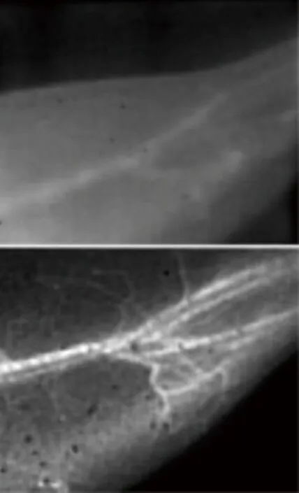
In addition to providing fine details, the technique - developed by Stanford scientists Hongjie Dai, professor of chemistry; John Cooke, professor of cardiovascular medicine; and Ngan Huang, acting assistant professor of cardiothoracic surgery - has a fast image acquisition rate, allowing researchers to measure blood flow in near real time.
The ability to obtain both blood flow information and blood vessel clarity was not previously possible, and will be particularly useful in studying animal models of arterial disease, such as how blood flow is affected by the arterial blockages and constrictions that cause, among other things, strokes and heart attacks.
"For medical research, it's a very nice tool for looking at features in small animals," Dai said. "It will help us better understand some vasculature diseases and how they respond to therapy, and how we might devise better treatments."
Because NIR-II can only penetrate a centimeter, at most, into the body, it won't replace other imaging techniques for humans, but it will be a powerful method for studying animal models by replacing or complementing X-ray, CT, MRI and laser Doppler techniques.
The next step for the research, and one that will make the technology more easily accepted for use in humans, is to explore alternative fluorescent molecules, Dai said. "We'd like to find something smaller than the carbon nanotubes but that emit light at the same long wavelength, so that they can be easily excreted from the body and we can eliminate any toxicity concerns."
The lead authors of the study are graduate student Guosong Hong of the Department of Chemistry and research assistant Jerry Lee of the School of Medicine. Other co-authors include graduate student Joshua Robinson and postdoctoral scholars Uwe Raaz and Liming Xie. The work was supported by the National Cancer Institute, the National Heart, Lung and Blood Institute and a Stanford Graduate Fellowship.
“三明治”新结构创建出“有来无回”光陷阱
美国普林斯顿大学的研究人员发现了一种可将有机太阳能电池效率提高近两倍的新方法。科学家们认为,这种廉价的柔性塑料装置或将成为太阳能发电的未来。该研究成果将刊登在近期《光学快报》网络版上。
由普林斯顿大学机电工程系纳米结构实验室主任周郁(音译)教授领导的研究团队,通过使用纳米结构的金属和塑料“三明治”来收集和诱捕光线,将有机太阳能电池效率提升了175%。
周教授表示,这项技术也应能提高传统的无机太阳能集热器,如标准的硅太阳能电池板的效率。
导致太阳能电池损失能量的两个主要问题是:电池的光反射及无法充分捕捉进入电池的光线。
研究团队的金属“三明治”新结构——次波长等离子腔可同时解决这两个问题,其具有抑制反射和捕获光线的非凡能力。
新电池的顶层(即窗口层)使用极精细金属网,金属厚度为30纳米,网孔直径为175纳米,间隔为25纳米。该金属网取代了以往由铟—锡—氧化物(ITO)材料制成的窗口层。
该网格状窗口层与“三明治”结构的底层非常接近,底层使用的是与传统太阳能电池中相同的金属膜。两个金属板之间夹杂着太阳能电池板中使用的半导体材料薄带。其可以是硅、塑料或砷化镓中的任一种。周郁研究团队使用的是85纳米厚的塑料。
太阳能电池的网孔间隔,“三明治”结构厚度乃至网孔直径,都要小于所收集的光的波长。研究团队发现,使用这些次波长结构,使他们能够创建出一个几乎“有来无回”的光陷阱。
此项新技术使研究团队最终创造出一个仅反射4%光线,即光吸收率高达96%的太阳能电池。在阳光直射情况下,其光电转换效率要比常规太阳能电池高出52%;在阴天或电池不直接面向太阳,光线以更大角度入射到太阳能板时,该结构可获得更高的效率。通过捕捉斜射光线,新结构可额外提升81%的效率,从而使最终的效率增长达到了175%。
Tiny structure gives big boost to solar power
Princeton researchers have found a simple and economical way to nearly triple the efficiency of organic solar cells, the cheap and flexible plastic devices that many scientists believe could be the future of solar power.
The researchers, led by electrical engineer Stephen Chou, were able to increase the efficiency of the solar cells 175 percent by using a nanostructured "sandwich" of metal and plastic that collects and traps light. Chou said the technology also should increase the efficiency of conventional inorganic solar collectors, such as standard silicon solar panels, although he cautioned that his team has not yet completed research with inorganic devices.
Chou, the Joseph C. Elgin Professor of Engineering, said the research team used nanotechnology to overcome two primary challenges that cause solar cells to lose energy: light reflecting from the cell, and the inability to fully capture light that enters the cell.
With their new metallic sandwich, the researchers were able to address both problems. The sandwich — called a subwavelength plasmonic cavity — has an extraordinary ability to dampen reflection and trap light. The new technique allowed Chou's team to create a solar cell that only reflects about 4 percent of light and absorbs as much as 96 percent. It demonstrates 52 percent higher efficiency in converting light to electrical energy than a conventional solar cell.
That is for direct sunlight. The structure achieves even more efficiency for light that strikes the solar cell at large angles, which occurs on cloudy days or when the cell is not directly facing the sun. By capturing these angled rays, the new structure boosts efficiency by an additional 81 percent, leading to the 175 percent total increase. Chou said the system is ready for commercial use although, as with any new product, there will be a transition period in moving from the lab to mass production.
The physics behind the innovation is formidably complex. But the device structure, in concept, is fairly simple.
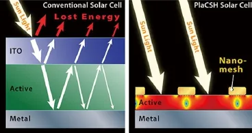
The top layer, known as the window layer, of the new solar cell uses an incredibly fine metal mesh: the metal is 30 nanometers thick, and each hole is 175 nanometers in diameter and 25 nanometers apart. (A nanometer is a billionth of a meter and about one hundredthousandth the width of human hair). This mesh replaces the conventional window layer typically made of a material called indiumtin-oxide (ITO).
The mesh window layer is placed very close to the bottom layer of the sandwich, the same metal film used in conventional solar cells. In between the two metal sheets is a thin strip of semiconducting material used in solar panels. It can be any type — silicon, plastic or gallium arsenide — although Chou's team used an 85-nanometer-thick plastic.
The solar cell's features — the spacing of the mesh, the thickness of the sandwich, the diameter of the holes — are all smaller than the wavelength of the light being collected. This is critical because light behaves in very unusual ways in subwavelength structures. Chou's team discovered that using these subwavelength structures allowed them to create a trap in which light enters, with almost no reflection, and does not leave.
"It is like a black hole for light," Chou said. "It traps it."
The team calls the system a "plasmonic cavity with subwavelength hole array" or PlaCSH. Photos of the surface of the PlaCSH solar cells demonstrate this light-absorbing effect: under sunlight, a standard solar power cell looks tinted in color due to light reflecting from its surface, but the PlaCSH looks deep black because of the extremely low light reflection.
借助基因修改的HIV成功治疗一名白血病患者
近日,美国费城儿童医院的医生宣称,他们从病魔手中成功夺回了一个罹患白血病的7岁女孩的生命,赢得这场胜利多亏了一个可能会让所有人感到意外的“帮手”——经过基因修改的HIV(人类免疫缺陷病毒)。不过,主治医生强调,治疗过程不会出现感染艾滋病的风险。
这个名叫艾米莉的小女孩接受了近两年的化疗,其间病情两次复发,医生们认为她治愈的“希望渺茫”。
于是在今年2月,他们开始对她实施这一“以毒攻毒”的实验性治疗方案(官方名称为CTL019疗法)。
医生们将HIV中引发艾滋病的因素剔除,借助这些经过基因修改的HIV,艾米莉自己的免疫细胞占据了优势,一举击溃了入侵的白血病细胞。艾米莉是参与CTL019疗法的为数不多的志愿者中唯一的儿童。费城儿童医院强调,该疗法还不能被称为“神奇的子弹”,但至少在艾米莉身上显示出了巨大的成功。
在治疗过程中,医生先将艾米莉自己的数百万免疫系统细胞移除,接着用经过基因修改的HIV将一种能够增强免疫细胞的新基因送入她体内,这种基因如同导弹一样,可以帮助免疫细胞“锁定”潜藏起来的白血病细胞并发动攻击。
主治医生、儿科肿瘤学家斯蒂芬·格拉普说,负责运送新基因的HIV中能够导致艾滋病的部分全都被剔除了,因此治疗过程中不会出现任何感染艾滋病的风险。
目前艾米莉已经回到学校继续学习,还能进行遛狗、踢足球等活动。
医生表示,艾米莉的治疗结果是“完满的”,最重要的是,增强了的免疫防护系统仍然“存留在她体内,以防止癌症复发”。
“任何我们能够做的检测,即使是最敏感的测试都表明,她身体里已经没有白血病症状了。”格拉普说,“我们还需要观察几年看看病情缓解情况,才能考虑她是否已被治愈。目前下断言还为时尚早。”
格拉普表示,这是他第一次看到治疗白血病的标准手段骨髓移植可能有了替代方案,细胞疗法有望最终取代昂贵而痛苦的骨髓移植治疗。
Doctors ‘cure’ leukemia by using HIV to rewire immune system
Doctors have successfully used a disabled version of HIV to modify a 7-year-old leukemia patient’s white blood cells to attack her cancer. The breakthrough procedure could potentially replace bone marrow transplant as a leukemia treatment.
Emma Whitehead was selected as a patient for the experimental technique after two years of battling with acute lymphoblastic leukemia, the New York Times reported. Chemotherapy failed to either cure the disease or result in a period of remission long enough for a bone marrow transplant.
The process was previously tested only on adult patients. In April, Whitehead became the first child to undergo the treatment, her medical team revealed during an annual meeting of the American Society of Hematology in Atlanta last weekend. She was also the first patient to be treated for her kind of leukemia.
Doctors from the University of Pennsylvania and the Children's Hospital of Philadelphia manipulated Whitehead’s immune system to make it target cancer cells. They took a batch of her own T cells - a kind of white blood cell - and genetically engineered them to kill the B cells - another kind of white blood cell - responsible for her disease.
To do this, the doctors used a modified and disabled form of HIV, the virus responsible for AIDS, to alter the T cells’ genes, making them produce a protein called a chimeric antigen receptor on their surface. This artificial protein matches another protein encountered only on the surface of B cells. The alteration allows T cells to attach to B cells, and destroy them. The genetically engineered T cells were then injected back into Whitehead’s blood, where they could reproduce on their own.
Two months after the procedure, testing revealed there was no sign of cancer in the girl’s body. The altered T cells were still present in her blood, but in smaller quantities than during treatment. Six months later, Whitehead is still in remission and is now back in school.
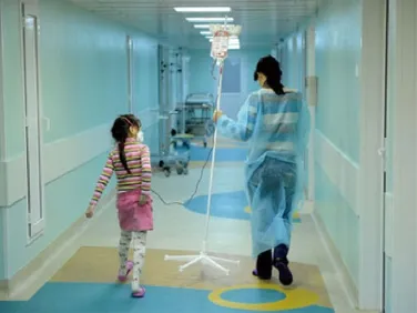
The experimental treatment has not yet been fully tested: Whitehead nearly died when the procedure caused a spontaneous high fever, and other near-fatal symptoms.
Not all of the 12 patients in the clinical trial responded to the treatment as well as Whitehead: Three adults with chronic leukemia had complete remissions; four improved their condition, but did not beat the disease completely; one is still in too early a stage to evaluate; two patients saw no effect from the treatment; another child initially responded, but eventually relapsed.
The treatment also kills healthy B cells along with malignant ones, making patients vulnerable to certain types of infections; patients require regular treatments of immune globulins to prevent illness.
T cell therapy appears to be a promising medical breakthrough that may replace older bone marrow treatments, the researchers said. They plan to conduct additional trials with at least a half-dozen patients over the next year.
基因发出“死神预言”:何时死去可以预知

当一个人临近死亡的时候,如果有人告诉他,你将会在某天的上午或下午死去,这将是一件多么恐怖的事情。
然而,这样的事情可能就会发生了,而发出这样预言的其实是一种控制人体生理节律的基因,因为临近死亡时,人的身体会还原到一种更加自然的生理节律,这也许就是死神的预言吧。
基因“开关”决定了我们身体的很多特征,包括头发颜色、血型等等,而且对于某些疾病非常敏感。现在研究人员认为他们已经发现了一种能够决定更加怪异事情的基因:一个人可能离世的时间。
在发表于2012年11月《神经学年鉴》杂志的一项研究中,研究人体生物钟(也称作生理节律)的科学家们声称发现了基因变体,不仅能够确定你能否成为一个早起的人,而且也能够以令人不安的精确度预测出你可能去世的时间。
根据哈佛医学院公布的一份声明,这种基因可能存在三种核苷酸组合(四种核苷酸构建了DNA模块):腺嘌呤与腺嘌呤组合(A-A)、腺嘌呤与鸟嘌呤组合(A-G)、鸟嘌呤与鸟嘌呤组合(G-G)。
撒珀尔博士在声明中写道:“这种特别的基因类型几乎影响每个人的睡觉和觉醒模式。而且它拥有一种相当深远的效果,拥有A-A基因类型的人们比那些拥有G-G基因类型的人们要早起大约1个小时,而A-G类型的人醒来的时间几乎正好就在中间。”
此外,研究人员已经发现1200名参与实验的老年人中有一些人的去世时间与这些核苷酸序列所准确预测的时间相差只有几个小时。具有A-A和A-G基因类型的病人在上午11点之前去世,而拥有G-G组合的人趋向于下午6点左右去世。
撒珀尔博士说道:“因此真的有一种基因预测你去世的时间。不是日期,而是一天中的时刻。”据《大西洋月刊》报告,研究人员相信他们的结果或许是当死亡接近的时候人体会还原到一种更加自然的生理节律感应阶段,而不是生活习惯所产生的循环。
Gene Predicts Time Of Death Down To Hour, Study Suggests
Genetic "switches" determine much about our bodies, including hair color, blood type, and susceptibility to certain diseases. Now, researchers believe they have found a gene that regulates something far more eerie: the time of day a person is likely to die.
In an study published in the November 2012 issue of the Annals of Neurology scientists studying the body's biological clock (a.k.a. the circadian rhythm) report the discovery of gene variant that not only determines the likelihood of your being a morning person, but also predicts, with unsettling accuracy, your likely time of death.
The gene typically allows for three possible combinations of nucleotides (the four molecular building blocks of DNA): adenineadenine (A-A), adenine-guanine (A-G), and guanine-guanine (G-G), according to a written statement released by Harvard Medical School.
"This particular genotype affects the sleep-wake pattern of virtually everyone walking around," Dr. Clifford Saper, chief of neurology at Beth Israel Deaconess Medical Center in Boston, wrote in the statement. "And it is a fairly profound effect so that the people who have the A-A genotype wake up about an hour earlier than the people who have the G-G genotype, and the A-Gs wake up almost exactly in the middle."
Moreover, investigators realized as some of the 1,200 older subjects in the project died that these nucleotide sequences were accurate predictors of their time of death, within a range of only a few hours.
Patients with the A-A and A-G genotypes typically died just before 11 a.m., while subjects with the G-G combination tended to die near 6 p.m.
"So there is really a gene that predicts the time of day that you’ll die. Not the date, fortunately, but the time of day," said Saper.
The Atlantic reports researchers believe their results may be due to the human body reverting to its more natural, circadian rhythm-induced state as death approaches, instead of the cycle created by social commitments.
零重力下生长氦晶体获得成功
最近,日本物理学家在研究零重力条件下晶体生长的过程中,突破了实验室条件限制,在飞机上用氦-4超流液生成了长达10毫米的氦晶体,检验了氦晶体在更宽广条件下生长的特殊动力学,这是普通材料无法实现的。相关论文发表在当日的《新物理学杂志》上。
该氦晶体是利用高压,在0.6K(约零下272℃)的极低温度下,以超流液泼溅的方式生长出来。超流液是一种量子态物质,其性质像液体但黏度为零,所以在高压下也能畅通无阻。超流液能流过极微小的缝隙而没有任何摩擦阻力。
晶体生长过程也叫“奥斯特瓦尔德熟化”(Ostwald ripening),即在溶液中,较小的结晶会溶解并再次沉积到较大的结晶上。“奥斯特瓦尔德熟化通常是一种很慢的过程,短时间内在较大晶体中几乎无法看到。”论文领导作者、东京工业大学教授野村龙司(音译)说,“对于普通的传统晶体,要形成最后的固定形状可能要花几千年。而低温氦晶体1秒钟就能达到它们的最终形状。一般氦晶体生长超过1毫米时,就会由于重力的作用而变形,这也是我们为何要在飞机上做该实验的原因。”
当飞机以特殊轨迹,也就是沿抛物线飞行时,飞机上的重力为零。研究人员与日本航空研究开发机构(JAXA)合作,在一架小型喷气式飞机上进行了实验。该飞机能提供20秒的零重力条件,在一次两小时的飞行中,大约能做8个实验。
他们在飞机上设计了一个专用的小冰箱,上面装有窗口以观察晶体形成。他们把一块较大的氦晶体放在高压室中,用声波将其击碎成微小颗粒,然后泼入氦-4超流液,较小颗粒融解其中,较大的则迅速生长,很快形成一个10毫米长的独立晶体。
“氦晶体能极快地从超流液中生长出来,因为氦原子是由非常灵敏迅捷的超流液携带,结晶过程不受任何阻碍。这是一种研究晶体形成基本问题的理想材料,因为结晶速度极快。”龙司说。这项研究有助于人们理解晶体生长背后的基本物理机制,也揭示了隐藏在重力下面的一些现象。
点评
通常情况下,生成氦晶体并不容易,不仅需要把液态氦-4转换为固体的冷冻温度,而且还必须将液态氦-4加压到至少25个标准大气压。此外氦晶体生长超过1毫米时,还会受到重力的作用而变形。而日本物理学家独辟蹊径,在飞机上零重力的条件下,生成长达10毫米的氦晶体,不能不说是一大突破。前些年,像幽灵一样互相穿过对方的晶体——“超固态氦”的发现,引起巨大反响,而这次零重力下生长氦晶体的成功,无疑又是一记重磅炸弹。
Japanese physicists grow helium crystals in zero gravity
Helium crystals are grown under zero gravity conditions by a team of Japanese physicists with the help of a phenomenon called parabolic flight.
A team of Japanese physicists have grown helium crystals in zero gravity conditions. The experiments took place in a small jet under parabolic flight conditions. During parabolic flight, zero gravity was achieved for a duration of 20 seconds. During the two-hour flight, approximately eight experiments were conducted.
Using low temperatures and high pressures, the crystals were grown and splashed with a superfluid. Superfluids are a form of quantum matter which contain zero viscosity and maintain the behavior of a fluid, flowing frictionfree through miniscule gaps.
"Helium crystals can grow from a superfluid extremely fast because the helium atoms are carried by a swift superflow, so it cannot hinder the crystallization process. It has been an ideal material to study the fundamental issues of crystal shape because the crystals form so quickly,” says lead author Professor Ryuji Nomura from The Tokyo Institute of Technology. Nomura states that helium crystals grow from superfluids rapidly because the atoms are carried by a “swift superflow” that does not interfere with crystallization. He adds that because the crystals form so fast, superfluids are an “ideal material” to study crystal shape.
"It can take thousands of years for ordinary classic crystals to reach their final shape; however, at very low temperatures helium crystals can reach their final shape within a second. When helium crystals grow larger than 1 mm they can be easily deformed by gravity, which is why we did our experiments on a plane,” says Nomura.
A small refrigerator fitted with observation windows was taken on the plane. A high-pressure chamber was used to hold large crystals which were then crushed into small pieces with an acoustic wave. A helium-4 superfluid was then applied to the crystal pieces. After being crushed, the tiny crystals were melted, causing larger crystals to grow quickly, with one surviving 10 mm crystal. The other crystals melted in seconds.
The process under which the crystal was grown is called Ostwald ripening. This phenomenon can be seen on ice cream when it’s been in the freezer too long. Large ice crystals form over time, a process commonly known as “freezer burn.”
"Ostwald ripening is usually a very slow process and has never been seen in such huge crystals in a very short period," says Nomura.
The results of the experiments were presented December 13 in the German Physical Society’s New Journal of Physics and the Institute of Physics.
Researchers believe the results of the helium crystal experiments may uncover the fundamentals behind crystal development without the hindrance of gravity.
科学家首次从GRACE信号中成功分离出北美北欧近十年的水量变化趋势
2002年欧美发射的重力恢复和气候实验(GRACE)卫星在探测陆地水储量变化、冰雪消融取得极大的成功,但是探测北美、北欧和南极地区的现今质量变化趋势却遭遇严重的挑战。在末次冰期这些地区发育了巨厚的冰盖,2万年以来由于古冰盖的消融,导致现今地壳回弹、地幔物质回流。这种冰川均衡调整引起的质量增加在卫星重力观测中产生很强的干扰,甚至完全掩盖了所探测的现今物质平衡信号,虽然GRACE发射超过十年,但在这些地区的相关研究一直没有取得进展。
中科院测量与地球物理研究所汪汉胜研究员及其负荷研究团队,在国家杰出青年科学基金、国家基金创新研究群体科学基金等资助下,与加拿大卡尔加里大学胡百卓教授、瑞典国土测量局Holger Steffen博士合作,对冰川均衡调整理论进行深入研究,首次提出了GRACE联合GPS观测网络分离现今物质平衡信号的有效途径,在北美中部的加拿大大草原(艾伯塔、萨斯喀彻温和马尼托巴省)、五大湖地区,发现过去十年陆地水量剧增,每年增加(43.0±5.0)x吨,在北欧斯堪的纳维亚半岛南部也发现陆地水量增加,每年增加(2.3±0.8)x 吨。最大的水量增加出现在萨斯喀彻温省,每年达20mm,揭示了加拿大草原1999年~2005年发生极端干旱后的水量恢复过程。GRACE 所揭示的水储量变化均为验潮站和井中水位观测所证实,而且倾向于支持欧洲的WGHM水文模型。更为重要的是,这里发现显著陆地水量上升,意味着冰融水和降水流进海洋的量减少,因此,如果用海平面上升评估全球变化,则会低估全球变暖的响应。
该研究成果于2012年12月2日发表在《自然-地球科学》上,论文题目为《GRACE卫星重力数据揭示北美和斯堪的纳维亚半岛南部水储量增加》,该成果被选为“研究亮点”,同时入选与该期刊同期Nature Climate Change的共同网络焦点(水资源利用)。该研究所提出的途径能够从GRACE卫星重力信号中排除冰川均衡调整的巨大干扰,从而有效分离出相关研究地区水储量变化及其趋势,所给出的结果有利于了解北美北欧当前知之甚少的区域水储量变化趋势,进一步显示了卫星重力探测地球系统质量变化与迁移的巨大能力。该研究对了解地球系统质量变化和迁移,特别是对于全球水循环及其与大气圈、水圈和海洋的交换过程,具有重要创新性贡献,也对水资源利用和海平面上升等研究具有重要意义。
Scientists found water storage Increased in North America and Scandinavia from GRACE gravity data
Natural and anthropogenic stresses such as climate change, drought and deluge, increasing water use, land use and agricultural practices affect surface and groundwater resources globally. A small change in the hydrological cycle may have a significant socio-economic impact, which, for example, has recently been observed in the form of groundwater depletion in northwest India probably leading to reduction of agricultural output and shortages of potable water. It is thus of importance to determine the spatial and temporal variability in continental water storage.
The Gravity Recovery and Climate Experiment (GRACE) satellite mission has proved to be an invaluable tool in monitoring such hydrological changes with global coverage and sufficient spatial and temporal resolution. Gravity data from GRACE have revealed trends in present-day continental water storage in many parts of the world. In North America and northern Europe, it has been difficult to provide reliable estimates because of the strong background signals of glacial isostatic adjustment. Attempts to separate the hydrologic signal from the background with numerical models are affected by uncertainties in our understanding of the precise glacial history and mantle viscosity.
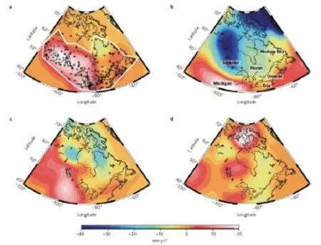
Wang Hansheng, a leading scientist in the research project, used a combination of GRACE data and measurements from the global positioning system to separate the hydrological signals without any model assumptions. According to their estimates, water storage in central North America increased by 43.0±5.0 Gt yr-1 over the past decade (Fig1). They attribute this increase to a recovery in terrestrial water storage after the extreme Canadian Prairies drought between 1999 and 2005. They find a smaller rise in water storage in southern Scandinavia, by 2.3±0.8 Gt yr-1. In both North America and Scandinavia, their computed increases in water storage are consistent with longterm observations of terrestrial water level. They suggest that the detected mass gains in terrestrial water storage need to be taken into account in studies on global sea-level rise.Figure1. Hydrology trend rates in North America in equivalent water thickness. a, Using CSR GRACE data and GPS data from ref. 29 in the separation approach. b, Using CSR GRACE data but with ICE-5G(VM2) (ref. 16) model predictions for correction. c,WGHM (ref. 13). d, GLDAS/NOAH (ref. 12) hydrology model. The label A marks the trend peak in the Canadian Prairies and B marks that in the Great Lakes for investigation in Fig. 3. Crosses denote GPS sites. A white line surrounds the investigation area, which depends on a dense GPS network. The area west of the dashed blue line is used for mass change calculation. See Supplementary Fig. S12 for uncertainties.
