Effects of 4-Hydroxyproline Stereochemistry on α-Conotoxin Solution Conformation
2013-07-25ZHANGBingBingZHAOCongWANGXueSongHELeiDUWeiHong
ZHANG Bing-Bing ZHAO Cong WANG Xue-Song HE Lei DU Wei-Hong
(Department of Chemistry,Renmin University of China,Beijing 100872,P.R.China)
1 lntroduction
Conotoxins,commonly extracted from the venom of conus snails,are a rich resource of novel peptides that can specifically target distinct membrane receptors,ion channels,and nervous system transporter.1-5Conotoxins that target neuronal or muscle-type nicotinic acetylcholine receptors(nAChRs)are classified into the α-conotoxin family.6More than half of the known α-conotoxins belong to the α4/7 subfamily,which refers to the number of residues in the two intercysteine loops.Almost all α4/7 conotoxins share a well-defined structural motif in the form of a helical region centered around the third cysteine residue.7
In α4/7 conotoxins,a vast array of post-translational modifications(PTM),such as C-terminal amidation,4-hydroxyproline(Hyp),gamma carboxylic glutamic acid(Gla),and D-type amino acids,have been discovered,8as in other conotoxin families.Chemical modifications of conotoxins create structural and functional diversity,serving as valuable tools in improving their stability and pharmaceutical properties;such properties make conotoxins attractive leads for biological research and drug discovery and development.9-12Particularly,hydroxylation have been studied in many fields,demonstrating interesting effects on the structural characteristics and biological functions of conotoxins.13,14
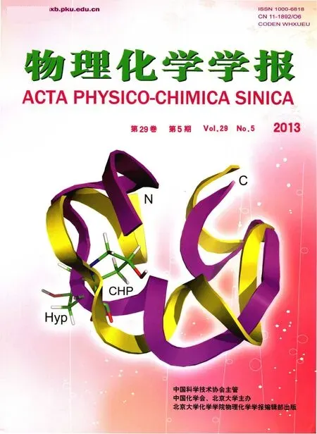
The hydroxyl group appears pervasively throughout organic chemical and biochemical structures.It plays a vital role in maintaining structural stability,protein-ligand interaction,and biological function.15-17An examination of the hydroxylation of proline in thein vitrooxidative folding and biological activity of conotoxins has led to the discovery that the modification of the conserved residue Pro6 to Hyp6 greatly improves the stability of native conotoxins.13α-Conotoxin Vc1.1(i.e.,the socalled[O6P/γ14E]Vc1A,Table 118-24),which contains 16 amino acids with typical α4/7 intercysteine loops,has been discovered from the venom dusts ofConus victoriae.It is almost identical with the native peptide Vc1A and the intermediate analog[P6O]Vc1.1 in three-dimensional(3D)structure due to the similarities in their NMR CαH and CβH shifts.25Scanning mutagenesis,a very powerful technique in identifying notable residues that play a crucial role in the structural and biological characteristics of peptides,has revealed that residues at positions 4 and 9 are crucial in the bioactivity of Vc1.1 at α9α10 nAChR.19,26SrIA and SrIB,Hyp-contained α-conotoxins with 4/7-type intercysteine loops,were first isolated fromConus spuriusand showed comparable biological functions with EI.18Although the[γ15E]Sr1B mutant has demonstrated the slight little difference between the bioactivities of Sr1A and Sr1B,the role of Hyp7 in conotoxin structure and bioactivity is still unknown.In addition,our previous research14revealed that thecis/transisomerization of 4-hydroxyproline has remarkable effects on the conformation of some conopeptides.
In the current work,we selected three conopeptides containingcis/trans-4-hydroxyproline(Fig.1)and determined their 3D solution structures to investigate the effects of stereochemistry of 4-hydroxyproline on the folding and structure of α-conotoxin.These α-conopeptides were[γ15E]Sr1B,[O7O′/γ15E]Sr1B,and[O6O′/γ14E]Vc1A,in which a Glu residue was used instead of Gla,because studies on the impacts of gamma carboxylic glutamic acid on some highlighted conotoxins,such as conantokin-G,RVIIIA,VxXXB,Vc1A,SrIA,and SrIB,indicated that the mutation of Gla to Glu did not affect the structural property and pharmacological activity of those conotoxins.18-21,27-30The results of such chemical modifications may lead to exciting discoveries in research on conformational change and potential bioactivity regulation of α-conotoxins.

Table 1 Sequences and their receptors of some α4/7-conotoxins showing highly conserved proline residue(P,O or O′)in the first intercysteine loop

Fig.1 Structures of cis-4-hydroxyproline(A)and trans-4-hydroxyproline(B)
2 Materials and methods
2.1 Reagents
The used acetonitrile-d3(deuterated ratio(D)>99.5%)was from Sigma-Aldrich,USA.D2O(D>99.9%,purity>99.99%)was from Cambridge Isotope Lab,MA,USA.Trifluoroacetic acid-d(TFA,D>99.5%,purity>99.99%)was from Deuterium Laboratory,Peking University,China.All other reagents were of analytical grade.
2.2 Peptide synthesis
Two peptides(China-Peptides Co.,Ltd.,Shanghai,China),namely,[γ15E]Sr1B and[O7O′/γ15E]Sr1B,were chemically synthesized and identified in order to obtain enough NMR samples.Another mutant(SBS Co.,Ltd.,Beijing,China),namely,[O6O′/γ14E]Vc1A,was also chemically synthesized.Furthermore,all the peptides were identified by high-performance liquid chromatography(HPLC)and mass spectrometry(MS)with more than 95%certainty.
2.3 NMR experiments
The[γ15E]Sr1B and[O7O′/γ15E]Sr1B NMR samples were prepared by dissolving peptides in 400 μL of either 99.99%D2O(Cambridge Isotope Lab,MA,USA)or 9:1(V/V)H2O/D2O with 0.01%trifluoroacetic acid(TFA,St.Louis,USA)at pH 3.0.[O6O′/γ14E]Vc1A were dissolved in 400 μL 3:2(V/V)acetonitrile-d3/H2O or acetonitrile-d3/D2O due to its low solubility in aqueous solution,and the solution was adjusted to pH 3.0.The final peptide concentration was approximately 4.0 mmol·L-1.
NMR measurements were performed using standard pulse sequences and phase cycling on Bruker Avance 400 and 600 MHz NMR spectrometers at 293 K.Due to the existence of disulfide bonds in conopeptides,the weak effect of temperature and solvent was ignored.The homonuclear double quantum filtered correlation spectroscopy(DQF-COSY),rotating frame nuclear overhauser effect spectroscopy(ROESY),nuclear overhauser effect spectroscopy(NOESY),and total correlation spectroscopy(TOCSY)were obtained in a phase-sensitive mode using time-proportional phase incrementation for quadrature detection in the t1(evolution time)dimension.Presaturation during the relaxation delay period was used to suppress the solvent resonance,unless specified otherwise.All 2D spectra were obtained using a spectral width of 6000.00 Hz(Δδ=10).ROESY and NOESY spectra were obtained with a mixing time of 300 ms.TOCSY spectra were obtained using the MLEV-17 pulse scheme for a spin lock of 120 ms.31NOESY/ROESY experiments were obtained using gradients for water saturation.32Each sample lyophilized from the hydrogen-containing solution was redissolved in a deuterium-containing solution in order to identify the slow exchange of backbone amide protons.1D1H NMR spectra were measured after 3 min and every 10 min thereafter for 3 h.All chemical shifts were referenced to the methyl resonance of 4,4-dimethyl-4-silapentane-1-sulfonic acid used as internal standard.The spectra were processed using Bruker Topspin 2.1 and analyzed by Sparky 3.1.33Final matrix sizes were usually 4096×2048 real points.
2.4 Distance restraints and structural calculations
Three sets of distance constraints were derived from the ROESY spectra of[γ15E]Sr1B,[O7O′/γ15E]Sr1B,and[O6O′/γ14E]Vc1A,respectively.Distance constraints,representing unambiguously assigned dipolar couplings,were used for structural calculations using Cyana 2.1 software.34Dihedral angle restraints were determined based on the3JHN-Hαcoupling constants derived from the DQF-COSY spectral analysis and 1D H/D exchange experiments if possible.Theφangle constraints for some residues were set to-120°±40°for3JNHα>8.0 Hz and-65°±25°for3JNHα<5.5 Hz.Backbone dihedral constraints were not applied for3JNHαvalues ranging from 5.5 to 8.0 Hz.Based on the slow exchange of amide protons in hydrogen-deuterium exchange experiments,distance constraints of the hydrogen bond were added as target values of 0.22 and 0.32 nm for the NH(i)-O(j)and N(i)-O(j)bonds,respectively.Due to the significant influence of C-terminal amidation on the folding tendency and bioactivity of conotoxin,we reproduced it as a new residue in the Cyana library in order to calculate the structures.According to the primary sequence,100 random structures were generated to fit covalent and spatial requirements.The 20 lowest energy conformers from 100 calculated structures were submitted to a molecular dynamics refinement procedure using the Sander module of the Amber 9 program.The final outcomes were used for structural quality analysis using MolMol software.35The data,including chemical shifts,were submitted to the BMRB database with access codes 18382,18383,and 18384 for[O7O′/γ15E]Sr1B,[γ15E]Sr1B,and[O6O′/063514E]Vc1A,respectively.Further,all the constraints data and coordinates of three structures could be supplied if necessary.
3 Results and discussion
3.1 NMR assignments
In the present work,α-conopeptides containing 4-hydroxyproline were chosen to explore the role of PTM in conotoxin conformational change and potential bioactivity regulation.The solution structures of the three chemically synthesized conopeptides[γ15E]Sr1B,[O7O′/γ15E]Sr1B,and[O6O′/γ14E]Vc1A were identified experimentally by 2D NMR method.The respective sequences of[γ15E]Sr1B,[O7O′/γ15E]Sr1B,[O6Oʹ/γ14E]Vc1A,Vc1.1,and some other selected α-conotoxins are shown in Table 1.

Fig.2 Assignments of residues with unique spin systems in part of TOCSY spectrum for[γ15E]Sr1B
The sequence-specific resonance assignments were achieved using the traditional visual analysis method.36The spin systems of most amino acids were resolved by TOCSY and DQF-COSY spectra.Fig.2 shows the representative amino acid spin systems of the[γ15E]Sr1B TOCSY spectrum in H2O.A total of 15 expected cross peaks between the amide proton and CαH were observed.Other spin systems were found in the fingerprint region of the TOCSY spectrum,and their assignments were verified in the fingerprint region of the DQF-COSY spectrum.The sequential assignments of amino acids in the primary sequence began with the unique residues Ser5,Tyr13,and Leu16.The NOE sequential walk identified residues from Thr2 to Glu15 and from Leu16 to Gly18 toward the N-terminus and the C-terminus,respectively.Owing to the rapid exchange in water and a missing amide proton,the first N-terminal residue Arg1 was finally assigned based on its spin system.The final chemical shifts of[γ15E]Sr1B were deposited in BMRB(access code 18383).
Similar to[γ15E]Sr1B,15 of the 18 spin systems were found in the fingerprint region of the 120 ms TOCSY spectrum for[O7O′/γ15E]Sr1B(Fig.S1,see Supporting Information).The sequential assignments of amino acids in the primary sequence started with the unique residues Ser5,Tyr13,and Leu16.Hence,nuclear overhauser effect(NOE)walks toward the N-terminus and the C-terminus were identified.The final chemical shifts of[O7O′/γ15E]Sr1B were deposited in BMRB(access code 18382).
As for[O6O′/γ14E]Vc1A,multiple components were found in the solution.cis-4-Hydroxy-proline induced the conformational equilibrium to[O6O′/γ14E]Vc1A(Fig.3,in which clearly showing three conformations in solution).Three sets of resonance signals were observed for some residues.The TOCSY spectrum showed the spin systems for the major isomer of[O6O′/γ14E]Vc1A in H2O(Fig.S2,see Supporting Information).A total of 12 expected cross peaks between the amide proton and CαH were observed for the major component.Other spin systems were also found in the fingerprint region of the TOCSY spectrum,and their assignments were verified in the fingerprint region of the DQF-COSY spectrum.The sequential assignments started with the unique residues Ser4,Arg7,and Ile15.NOE walks from residues Cys2 to Asn9 and from Tyr10 to Cys16,except residues CHP6(CHP:cis-4-hydroxyproline)and Pro13,were identified.Given the rapid exchange in water and a missing amide proton,the first N-terminal residue Gly1 was finally assigned based on its spin system.The final chemical shifts of[O6O′/γ14E]Vc1A were deposited in BMRB(access code 18384).
3.2 Structural calculation and evaluation

Fig.3 Portion of the TOCSY spectrum of[O6O′/γ14E]Vc1Awith respect to Ile15
The constraints for determining the three conopeptide-solution structures were obtained from a survey of NMR data.A total of 191 distance constraints were used,and the NOE root mean square violation was no more than 0.02 nm for[γ15E]Sr1B.Moreover,7φangle constraints(i.e.,Ser5,Cys9,Glu12,Tyr13,Leu16,Cys17,and Gly18),5 hydrogen bond restraints(i.e.,carbonyl O atoms of Arg6,Hyp7,Thr8,Tyr13,and Pro14 corresponding to amide protons of Cys9,Arg10,Met11,Leu16 and Cys17 respectively)and 2 pairs of disulfide bonds(i.e.,Cys3-Cys9 and Cys4-Cys17)defining the globular skeleton were inputted for the molecular modeling protocol of the Cyana algorithm.These constraints were sufficient for the structural calculation of such a small-sized peptide.As for[O7Oʹ/γ15E]Sr1B,192 distance constraints,5 dihedral restraints(i.e.,Cys9,Glu12,Tyr13,Cys17,and Gly18),4 hydrogen bond restraints,and 2 pairs of disulfide bonds were used to build up the structures.The H-bond restraints were from the carbonyl O atoms of Arg10,Tyr13 and Pro14 corresponding to amide protons of Tyr13,Leu16 and Cys17 respectively,and from the Oδatomof CHP7 corresponding to the amide proton of Thr8.Furthermore,102 distance constraints,5 dihedral restraints(i.e.,Cys2,Ser4,Cys8,Tyr10,and C16),4 hydrogen bond restraints(i.e.,carbonyl O atoms of Ser4,Arg7,Cys8,and Asn9 corresponding to amide protons of Arg7,Tyr10,Asp11,and His12 respectively),and 2 pairs of disulfide bonds(i.e.,Cys2-Cys8 and Cys3-Cys16)were used to build up the structure for the major conformer of[O6O′/γ14E]Vc1A.
First,we computed 100 solution structures to evaluate the folding of the peptidic chain using medium-distance constraints with more than 2 bond intervals|i-j|>2.Subsequently,all distance constraints and dihedral restraints were used.H-bond constraints were then introduced into the calculations.The simulated annealing calculations began with 100 random structures.Finally,the ensemble of 20 best resulting models with the lowest residual target function and the minimum root mean square deviation were selected.The resulting conformers contained no significant violation of any constraint.The Ramachandran plots were chosen to represent the 3D folding of[γ15E]Sr1B,[O7O′/γ15E]Sr1B,and[O6O′/γ14E]Vc1A in solution(Fig.S3,see Supporting Information).A summary of statistics for the converged structures evaluated in terms of structural parameters for the three conopeptides are listed in Table 2.Compared with[γ15E]Sr1B and[O7O′/γ15E]Sr1B,the mean global RMSD of backbone atoms of[O6O′/γ14E]Vc1A was a bit high.However,the constraints for[O6O′/γ14E]Vc1A was enough to get reasonable structure and the Procheck data was satisfied.
3.3 Characterization of NMR-derived structures of[γ15E]Sr1B,[O7O′/γ15E]Sr1B,and[O6O′/γ14E]Vc1A
Three ensembles of the 20 best overlay structures calculated using NMR-derived constraints are shown in Fig.4.[γ15E]Sr1B(Fig.4(a))and[O7O′/γ15E]Sr1B(Fig.4(b))belonged to the α4/7 intercysteine spacing subfamily and shared the same disulfide framework of Cys1-Cys3 and Cys2-Cys4.Despite the γ15E mutation,a 310helix between Hyp7 and Arg10 was formed in[γ15E]Sr1B(Fig.5(a)),comprising a conserved“ω”structure as other native α4/7 conotoxin.The helix structure could be evidenced by the H-bond formation from the residue pairs Arg6-Cys9,Hyp7-Arg10,and Thr8-Met11.The NH protons of Cys9,Arg10,and Met11 showed slow exchange rate,which was observed during H/D exchange experiment.In addition,a type I β turn was formed between Pro14 and Cys17.The H-bond was observed between carbonyl O atom of Pro14and amide H atom of Cys17.

Table 2 Structural statistics for the 20 best structures of[γ15E]Sr1B,[O7O′/γ15E]Sr1B,and[O6O′/γ14E]Vc1A

Fig.4 Overlays of the backbone atoms for the 20 converged structures of conopeptides[γ15E]Sr1B(a),[O7O′/γ15E]Sr1B(b),and[O6O′/γ14E]Vc1A(c)

Fig.5 Comparison of the backbone structures between[γ15E]Sr1B(a)and[O7O′/γ15E]Sr1B(b)with side chain orientation of residues Hyp7/CHP7 andArg10 shown in stick;surface representations of[γ15E]Sr1B(c)and[O7O′/γ15E]Sr1B(d)shown in front views
In[O7O′/γ15E]Sr1B,no helix was observed in the center of the peptide.Interestingly,a new 310helix was formed between Pro14 and Leu16 around the C-terminus(Fig.5(b)).The NOE connectivity for the sequence 13-18 was illustrated in Fig.6.Furthermore,a γ turn between Thr2 and Cys4,and a type I β turn between Arg10 and Tyr13 were also observed.Surface representations of[γ15E]Sr1B and[O7O′/γ15E]Sr1B also indicated their distinct structural characteristics in terms of hydrophilicity and hydrophobicity(Fig.5(c,d)).
In[O6O′/γ14E]Vc1A,the residue 6 was converted fromtrans-4-hydroxyproline tocis-4-hydroxyproline,this disturbed the H-bond network,making the α-helix shrink from residue Pro6-Asp11 in Vc1.1 to a 310helix between residues Cys8 and Asp10 in[O6O′/γ14E]Vc1A.The helix structure was evidenced by the observation of 1D H/D exchange experiment.The 3D structure of[O6O′/γ14E]Vc1A(Fig.7(a)),showed a distorted“ω”structure compared with other typical α-conotoxins.In addition,CHP6 took part in the formation of a γ turn between residues Ser4 and CHP6.In contrast to Vc1.1,the side chain orientations of CHP6 and His12 in[O6O′/γ14E]Vc1A changed from the outside to inside of the backbone plane(Figs.7(a)and 7(b),respectively).Moreover,the surface representations of the two conopeptides were used to indicate the various interesting hydrophilic and hydrophobic characteristics(Fig.7(c,d)).

Fig.6 Portion of the NOE connectivity for the conopeptide[O7O′/γ15E]Sr1B

Fig.7 Comparison of the backbone structures between[O6O′/γ14E]Vc1A(a)and Vc1.1(b,PDB 2H8S)with side chain orientation of residues Hyp6/CHP6 and His12 shown in stick;surface representations of[O6O′/γ14E]Vc1A(c)and Vc1.1(d)shown in front views
3.4 Roles of Hyp/CHP residue in α-conotoxins
PTMs including C-terminal amidation,D-type amino acids,and hydroxylation are widely found in various conotoxin families.13,37-39Proline residue is hydroxylated in many α-conotoxins.However,the conserved proline behind 2 consecutive cysteines,which represents residue 6 in Vc1A,PnIA,and MII and residue 7 in Sr1B,is thought to play a notable role in structural stability and key hydrophobic binding interaction with the β-subunit of nAChR by docking simulations.22,40Position 7 after two consecutive cysteines also influenced the selectivity for α3β2 and α3β4 over the α7 nAChR subtype as well as the affinity between the conotoxin-receptor complex.25,40The NMR-derived structures of[γ15E]Sr1B and[O7O′/γ15E]Sr1B display remarkable distinctions in secondary structure element,side chain orientations of some key residues,and surface hydrophobic properties(Fig.5).Comparing the residue characteristics of[γ15E]Sr1B and[O7O′/γ15E]Sr1B,the side chain orientations of Hyp7/CHP7 and Arg10 changed remarkably from outside to inside the big inner loop,the helical secondary structure between residues 7 and 10 vanished due to thecis-transfer of Hyp7 hydroxyl group.The original H-bond between carbonyl O of Hyp7 and amide H of Arg10 in[γ15E]Sr1B disappeared,and the distance between carbonyl O of Hyp7 and amide H of Arg10 changed from 0.23 nm in[γ15E]Sr1B to 0.42 nm in[O7O′/γ15E]Sr1B(Fig.8).These results elucidated the vital role of Hyp7 in the secondary structure and peptide folding as well as in potential bioactivity influence.It is reported that[γ15E]Sr1B is not significantly different from Sr1B on the biological function.18However,the further chemically modification of[O7O′/γ15E]Sr1B and its role in nAChR activity need to be further explored,and the work is still under investigation.

Fig.8 H-bond distance between Hyp7 andArg10 in[γ15E]Sr1B(A)and the change in[O7O′/γ15E]Sr1B(B)
Vc1.1 presented conserved“ω”structural characteristics of α-conotoxin.The other two post-translationally modified derivatives of Vc1.1,Vc1A,and[P6O]Vc1.1 were structurally analogous to Vc1.1.25Hence,we examined the[P6O′]Vc1.1 structure(same as that of[O6O′/γ14E]Vc1A)and compared it with the former analogue(Vc1.1)to investigate the structural impacts of thetrans-andcis-4-hydroxyproline conformations.The mutation fromtrans-tocis-4-hydroxyproline disrupted the inner hydrogen bond network,thus inducing the conformational change of[O6O′/γ14E]Vc1A and[O6O′/r14E]Vc1A]lost the turn structure around the N-/C-termini;in comparison,the mutation from proline totrans-4-hydroxyproline did not result in any distinct structural change in Vc1.1.The significant changes in the CαH secondary chemical shifts of mutant[P6K]Vc1.1 indicated that the mutation induced the formation of 2 isomers that were different from other mutants or the original conopeptide Vc1.1.19In[O6O′/γ14E]Vc1A,3 isomers were also observed from the separated cross-peaks between the NH and CαH of residue Ile15 in the 2D TOCSY spectrum of[O6O′/γ14E]Vc1A(Fig.3).The differences in the NH chemical shifts of some residues implied various conformations of[O6O′/γ 14E]Vc1Ain the solution(data not shown).
In addition,the[O6O′]mutation of Vc1A had an indirect influence on the secondary structure around key residue Arg7.An investigation of crystal structures and docking simulations of IMI-AChBP indicated that Arg7 played a critical role in the binding of conotoxins to nAChR.41Four hydrogen bonds were formed on the principal binding side(loop C)of AChBP,3 of which involved the contacts of IMI Arg7 and Tyr91(loop A),Trp145(loop B),and Ile194(loop C),respectively.Furthermore,extensive van der Waals interactions of Ser144,Val146,Tyr147,and Tyr193 with IMI Asp5 were observed by forming an intramolecular salt bridge.Arg7 was also involved in van der Waals interactions with α7-Tyr195.The mutation of IMI Arg7 to Gln broke the charge interactions between α7-Tyr197 and IMI-Asp5,which was consistent with the decrease of experimental affinity.42,43Considering the importance of residue Arg7 in conotoxin interaction with the α7 subunit,the helix shrinking due to O6O′mutation around Arg7(Fig.7)may have prompted the biofunctional differences between Vc1.1 and[O6O′/γ14E]Vc1A.Moreover,the bioactivity of[O6O′/γ14E]Vc1Ais under study.
4 Conclusions
In this study,we determined the solution structures of[γ15E]Sr1B,[O7O′/γ15E]Sr1B,and[O6O′/γ14E]Vc1A.The impacts of the chemical modification ofcis/trans-4-hydroxyproline on conopeptide structure were remarkable.The modulation fromtrans-tocis-4-hydroxyproline led to notable conformational changes,including the secondary structure elements,the side chain orientation of key residues,and H-bond formation,which might have affected their pharmacological properties.Furthermore,this work could help deepen our understanding of the chemical modification of conopeptides,which can be useful in elucidating the structure-bioactivity relationships of α-conotoxins and in regulating potential biological activity for better peptide drug design.
(1) Terlau,H.;Olivera,B.M.Physiol.Rev.2004,84,41.doi:10.1152/physrev.00020.2003
(2) Halai,R.;Craik,D.J.Nat.Prod.Rep.2009,26,526.doi:10.1039/b819311h
(3)Azam,L.;McIntosh,J.M.Acta Pharmacol.Sin.2009,30,771.doi:10.1038/aps.2009.47
(4) Kaas,Q.;Yu,R.;Jin,A.H.;Dutertre,S.;Craik,D.J.Nucleic Acids Res.2012,40,D325.
(5) Livett,B.G.;Sandall,D.W.;Keays,D.;Down,J.;Gayler,K.R.;Satkunanathan,N.;Khalil,Z.Toxicon2006,48,810.doi:10.1016/j.toxicon.2006.07.023
(6) Myers,R.A.;Cruz,L.J.;Rivier,J.E.;Olivera,B.M.Chem.Rev.1993,93,1923.doi:10.1021/cr00021a013
(7) Jin,A.H.;Daly,N.L.;Nevin,S.T.;Wang,C.A.;Dutertre,S.;Lewis,R.J.;Adams,D.J.;Craik,D.J.;Alewood,P.F.J.Med.Chem.2008,51,5575.doi:10.1021/jm800278k
(8) Buczek,O.;Bulaj,G.;Olivera,B.M.Cell Mol.Life Sci.2005,62,3067.doi:10.1007/s00018-005-5283-0
(9) Craik,D.J.;Adams,D.J.ACS Chem.Biol.2007,2,457.doi:10.1021/cb700091j
(10)Armishaw,C.J.Toxins2010,2,1471.doi:10.3390/toxins2061471
(11) Clark,R.J.;Jensen,J.;Nevin,S.T.;Callaghan,B.P.;Adams,D.J.;Craik,D.J.Angew.Chem.Int.Edit.2010,49,6545.doi:10.1002/anie.201000620
(12) Muttenthaler,M.;Nevin,S.T.;Grishin,A.A.;Ngo,S.T.;Choy,P.T.;Daly,N.L.;Hu,S.H.;Armishaw,C.J.;Wang,C.I.A.;Lewis,R.J.;Martin,J.L.;Noakes,P.G.;Craik,D.J.;Adams,D.J.;Alewood,P.F.J.Am.Chem.Soc.2010,132,3514.doi:10.1021/ja910602h
(13) Lopez-Vera,E.;Walewska,A.;Skalicky,J.J.;Olivera,B.M.;Bulaj,G.Biochemistry2008,47,1741.doi:10.1021/bi701934m
(14)Xu,J.;Wang,Y.L.;Zhang,B.B.;Wang,B.H.;Du,W.H.Chem.Commun.2010,46,5467.doi:10.1039/c0cc00075b
(15) Calzolari,A.;Cicero,G.;Cavazzoni,C.;Di Felice,R.;Atellani,C.A.;Corni,S.J.Am.Chem.Soc.2010,132,4790.doi:10.1021/ja909823n
(16)Takeda,M.;Jee,J.;Ono,A.M.;Terauchi,T.;Kainosho,M.J.Am.Chem.Soc.2011,133,17420.doi:10.1021/ja206799v
(17)Denning,E.J.;MacKerell,A.D.,Jr.J.Am.Chem.Soc.2012,134,2800.doi:10.1021/ja211328g
(18) López-Vera,E.;Aguilar,M.B.;Schiavon,E.;Marinzi,C.;Ortiz,E.;Cassulini,R.R.;Batista,C.V.F.;Possani,L.D.;de la Cotera,E.P.H.;Peri,F.;Becerril,B.;Wanke,E.FEBS J.2007,274,3972.doi:10.1111/j.1742-4658.2007.05931.x
(19) Halai,R.;Clark,R.J.;Nevin,S.T.;Jensen,J.E.;Adams,D.J.;Craik,D.J.J.Biol.Chem.2009,284,20275.doi:10.1074/jbc.M109.015339
(20) Sandall,D.W.;Satkunanathan,N.;Keays,D.A.;Polidano,M.A.;Liping,X.;Pham,V.;Down,J.G.;Khalil,Z.;Livett,B.G.;Gayler,K.R.Biochemistry2003,42,6904.doi:10.1021/bi034043e
(21)Townsend,A.;Livett,B.G.;Bingham,J.P.;Truong,H.T.;Karas,J.A.;O'Donnell,P.;Williamson,N.A.;Purcell,A.W.;Scanlon,D.Inter.J.Pep.Res.Ther.2009,15,195.doi:10.1007/s10989-009-9173-4
(22)Armishaw,C.;Jensen,A.A.;Balle,T.;Clark,R.J.;Harpsøe,K.;Skonberg,C.;Liljefors,T.;Strømgaard,K.J.Biol.Chem.2009,284,9498.doi:10.1074/jbc.M806136200
(23) Luo,S.;Nguyen,T.A.;Cartier,G.E.;Olivera,B.M.;Yoshikami,D.;McIntosh,J.M.Biochemistry1999,38,14542.doi:10.1021/bi991252j
(24) Park,K.H.;Suk,J.E.;Jacobsen,R.;Gray,W.R.;McIntosh,J.M.;Han,K.H.J.Biol.Chem.2001,276,49028.doi:10.1074/jbc.M107798200
(25) Clark,R.J.;Fischer,H.;Nevin,S.T.;Adams,D.J.;Craik,D.J.J.Biol.Chem.2006,281,23254.doi:10.1074/jbc.M604550200
(26) Nevin,S.T.;Clark,R.J.;Klimis,H.;Christie,M.J.;Craik,D.J.;Adams,D.J.Mol.Pharmacol.2007,72,1406.doi:10.1124/mol.107.040568
(27) Skjærbæk,N.;Nielsen,K.J.;Lewis,R.J.;Alewood,P.F.;Craik,D.J.J.Biol.Chem.1997,272,2291.doi:10.1074/jbc.272.4.2291
(28)Teichert,R.W.;Jimenez,E.C.;Olivera,B.M.Biochemistry2005,44,7897.doi:10.1021/bi047274+
(29) Loughnan,M.;Nicke,A.;Jones,A.;Schroeder,C.I.;Nevin,S.T.;Adams,D.J.;Alewood,P.F.;Lewis,R.J.J.Biol.Chem.2006,281,24745.doi:10.1074/jbc.M603703200
(30) Jakubowski,J.A.;Keays,D.A.;Kelley,W.P.;Sandall,D.W.;Bingham,J.P.;Livett,B.G.;Gayler,K.R.;Sweedler,J.V.J.Mass Spectrom.2004,39,548.
(31) Bax,A.;Davis,D.G.J.Magn.Reson.1985,65,355.
(32) Jeener,J.;Meier,B.H.;Bachmann,P.;Ernst,R.R.J.Chem.Phys.1979,71,4546.doi:10.1063/1.438208
(33) Goddard,T.D.;Kneller,D.G.Sparky 3;University of California,San Francisco,CA,2007.
(34) Güntert,P.;Mumenthaler,C.;Wüthrich,K.J.Mol.Biol.1997,273,283.doi:10.1006/jmbi.1997.1284
(35) Koradi,R.;Billeter,M.;Wüthrich,K.J.Mol.Graph.1996,14,51.doi:10.1016/0263-7855(96)00009-4
(36)Wüthrich,K.NMR of Proteins and Nucleic Acids;Wiley:New York,1986.
(37)Huang,F.J.;Du,W.H.;Wang,B.B.Acta Phys.-Chim.Sin.2008,24,1558.[黄飞娟,杜为红,王保怀.物理化学学报,2008,24,1558.]doi:10.1016/S1872-1508(08)60064-9
(38) Zhang,B.B.;Huang,F.J.;Du,W.H.Amino Acids2012,43,389.doi:10.1007/s00726-011-1093-x
(39) Chi,C.W.Chin.Sci.Bull.2009,54,2734.[戚正武.科学通报,2009,54,2734.]doi:10.1360/972009-1582
(40) Dutertre,S.;Nicke,A.;Lewis,R.J.J.Biol.Chem.2005,280,30460.doi:10.1074/jbc.M504229200
(41) Ulens,C.;Hogg,R.C.;Celie,P.H.;Bertrand,D.;Tsetlin,V.;Smit,A.B.;Sixma,T.K.Proc.Natl.Acad.Sci.U.S.A.2006,103,3615.doi:10.1073/pnas.0507889103
(42) Quiram,P.A.;Sine,S.M.J.Biol.Chem.1998,273,11007.doi:10.1074/jbc.273.18.11007
(43)Yu,R.;Craik,D.J.;Kaas,Q.PLoS Comput.Biol.2011,7,e1002011.
