AFP level and histologic differentiation predict the survival of patients with liver transplantation for hepatocellular carcinoma
2012-07-07
Istanbul, Turkey
AFP level and histologic differentiation predict the survival of patients with liver transplantation for hepatocellular carcinoma
Onur Yaprak, Murat Akyildiz, Murat Dayangac, Baha Tolga Demirbas, Necdet Guler, Gulen Bulbul Dogusoy, Yildiray Yuzer and Yaman Tokat
Istanbul, Turkey
BACKGROUND: In liver transplantation or resection for hepatocellular carcinoma (HCC), patient selection depends on morphological features. In patients with HCC, we performed a clinicopathological analysis of risk factors that affected survival after liver transplantation.
METHODS: In 389 liver transplantations performed from 2004 to 2010, 102 were for HCC patients. Data were collected retrospectively from the Organ Transplantation Center Database. Variables were as follows: age, gender, preoperative alpha-fetoprotein (AFP) levels, Child-Pugh and MELD scores, prognostic staging criteria (Milan and UCSF), etiology, number of tumors, the largest tumor size, total tumor size, multifocality, intrahepatic portal vein tumor thrombosis, bilobarity, and histological differentiation.
RESULTS: One hundred and two patients were evaluated. The 5-year overall survival rate was 56.5%. According to the UCSF criteria, 63% of the patients were within and 37% were beyond UCSF (P=0.03). Ten patients were excluded (one with fibrolamellary HCC and 9 because of early postoperative death without HCC recurrence), and 92 patients were assessed. The mean age of the patients was 56.5±6.9 years. Sixty-two patients underwent living donor liver transplantations. The mean follow-up time was 29.4±22.6 months. Fifteen patients (16.3%) died in the follow-up period due to HCC recurrence. Univariate analysis showed that AFP level, intrahepatic portal vein tumor thrombosis, histologic differentiation and UCSF criteria were significant factors related to survival and tumor recurrence. The 5-year estimated overall survival rate was 62.2% in all patients. According to the UCSF criteria, and the 5-year overall survival rate was 66.7% within and 52.7% beyond the criteria (P=0.04). Multivariate analysis showed that AFP level and poor differentiation were independent factors.
CONCLUSIONS: For proper patient selection in liver transplantation for HCC, prognostic criteria related to tumor biology (especially AFP level and histological differentiation) should be considered. Poor differentiation and higher AFP levels are indicators of poor prognosis after liver transplantation.
(Hepatobiliary Pancreat Dis Int 2012;11:256-261)
liver transplantation; hepatocellular carcinoma; alpha-fetoprotein
Introduction
Hepatocellular carcinoma (HCC) is the fifth most common cancer in the world resulting in more than 500 000 deaths per year.[1-6]Liver transplantation (LT) and surgical resection are curative treatment options in selected patients with HCC.[5-13]The decision for resection depends on the tumor size and the severity of liver disease dictated by the Child-Pugh score and the presence of portal hypertension for the risk of liver failure after operation. On the other hand, LT treats both cirrhosis and tumor at the same time. The initially reported results of LT for HCC were not satisfactory and patients had a 5-year survival rate of <40% because of large and bulky tumors.[5,6]Mazzaferro et al[8]reported a 5-year survival rate in 70% of the patients who had a single tumor of <5 cm or three tumors <3 cm in diameter and these data were defined as the Milan criteria. Subsequently the Milan criteria became the rule in selecting patients with HCC for LT. However, some patients with HCC who were beyond the Milan criteria were reported to have good results after LT.[9]In addition, organ shortage and increasednumber of patients on waiting lists for LT have resulted in the development of new criteria that extend the limits. Experience with LT for HCC demonstrated that except tumor size predicts the prognosis, histological findings and preoperative alpha-fetoprotein (AFP) levels are important prognostic markers of HCC recurrence.[14-19]
In this report, we present a clinicopathological analysis of the risk factors affecting patient survival and tumor recurrence after LT for HCC in a single center.
Methods
Between 2004 and 2010, 389 consecutive LTs (267 were living donor LT, 122 were deceased donor LT) were performed and 102 patients with HCC underwent LT at our center. The indication for LT was HCC with neither extrahepatic metastasis nor macroscopic portal vascular invasion shown by conventional imaging studies. Pretransplant tumor size and number were not regarded as limitations for LT.
The explanted livers were fixed in formalin and cut into 1-cm slices to note the number and largest size of tumor nodules. Hematoxylin and eosin-stained tumor sections were analyzed routinely for differentiation grade, growth pattern, microscopic and larger vessel invasions as well as lymph node metastases. Tumor differentiation was graded as well, moderate, or poor according to Edmonson and Steiner.[20]The patients were monitored regularly by measurement of serum AFP, Doppler ultrasonography was performed every 3 months in the first year then twice a year, and a thoracoabdominal CT scan was performed every year. MRI confirmation was performed when necessary. Immunosuppressive treatment after the transplant was prescribed with calcineurin inhibitors and rapid steroid tapering. Prednisolone was given at 500 mg intravenously (i.v.) in the operating room, followed by tapering from 100 to 20 mg within 8 days and from 20 to 10 mg after 2 months. The steroids were stopped in 6 months. Mycophenolate mofetil was used when lower doses of calcineurin inhibitors were required for renal dysfunction.
Data consisted of patient age, gender, preoperative AFP level, Child-Pugh and MELD scores, prognostic staging criteria (Milan and UCSF), etiology, number of tumors, largest tumor size, total tumor size, multifocality, intrahepatic portal vein tumor thrombosis, bilobarity, and histological differentiation.
Informed consent from the patients was waived since this was a retrospective study. But the study was approved by the institutional ethical committee. Deceased donors provided from the national organ donation system which is arranged by the Turkish Ministry of Health.
Statistical analysis
Continuous variables are expressed as mean±SD and comparisons between subgroups were done by the independent samplesttest. Categorical variables were compared using the Chi-square test or Fisher's exact test. Survival analysis was performed using the Kaplan-Meier method and the log-rank test. Univariate Cox-regression analysis was used to detect variables that had significant impact on the survival. Important risk factors were included in a multivariate analysis model. In this multivariate regression, aPvalue <0.05 was considered statistically significant.
Results
One hundred and two patients were evaluated. The 5-year overall survival of the patients was 56.5%. According to the UCSF criteria, 63% of the patients were within and 37% beyond the UCSF criteria (P=0.03). Similarly, 53.3% of the patients were within and 46.7% beyond the Milan criteria (P=0.19).
Nine patients who died from diseases other than HCC in the early postoperative period were excluded from the study (3 patients from sepsis, 2 from disseminated intravascular coagulation associated with massive blood transfusion, 2 from prolonged biliary sepsis and multiorgan failure, 1 from vena cava thrombosis, and 1 from recurrent HCV cirrhosis). One patient who had fibrolamellary HCC was excluded.
The data of the remaining 92 patients (76 males and 16 females, mean age 56.5±6.9 years) were analyzed (Table 1). Sixty-two patients received partial liver grafts from living donors, and 30 received full-size grafts from deceased donors.
The mean follow-up time was 29.4±22.6 months (range 1-76). Fifteen patients (16.3%) died during the follow-up. All mortalities were associated with recurrence of HCC. The effect of variables on mortality and their statistical analyses are shown in Tables 2-4.
Age, type of transplantation (living versus deceased donor LT), etiology of liver disease, preoperative Child-Pugh and MELD scores, the number of nodules, total tumor size, biggest tumor size, and the bilobarity of tumor involvement were not related to survival and/or tumor recurrence.
Univariate analysis revealed that AFP level, intrahepatic portal vein tumor thrombosis, histological differentiation and grade, and the UCSF criteria were significant factors affecting survival and recurrence.
The estimated 5-year overall survival rate for all patients was 62.2%, for those within the Milan criteriawas 64.2%, and for those beyond the Milan critteria was 52.2% (P=0.20).

Table 1. Demographic and clinicopathologic features of patients (n=92)
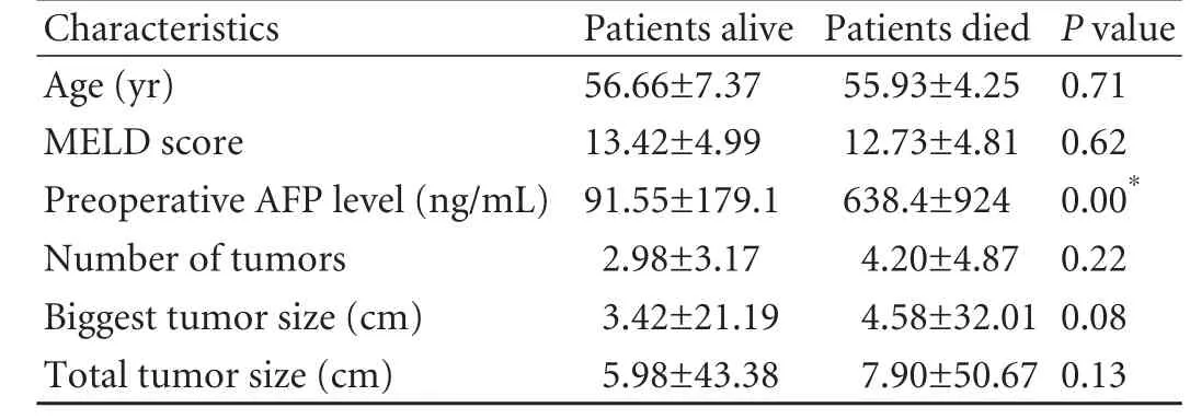
Table 2. Effect of quantitative variables on mortality
According to the UCSF criteria, the 5-year overall survival was 66.7% for patients within the criteria versus 52.7% for those beyond the criteria (P=0.04).
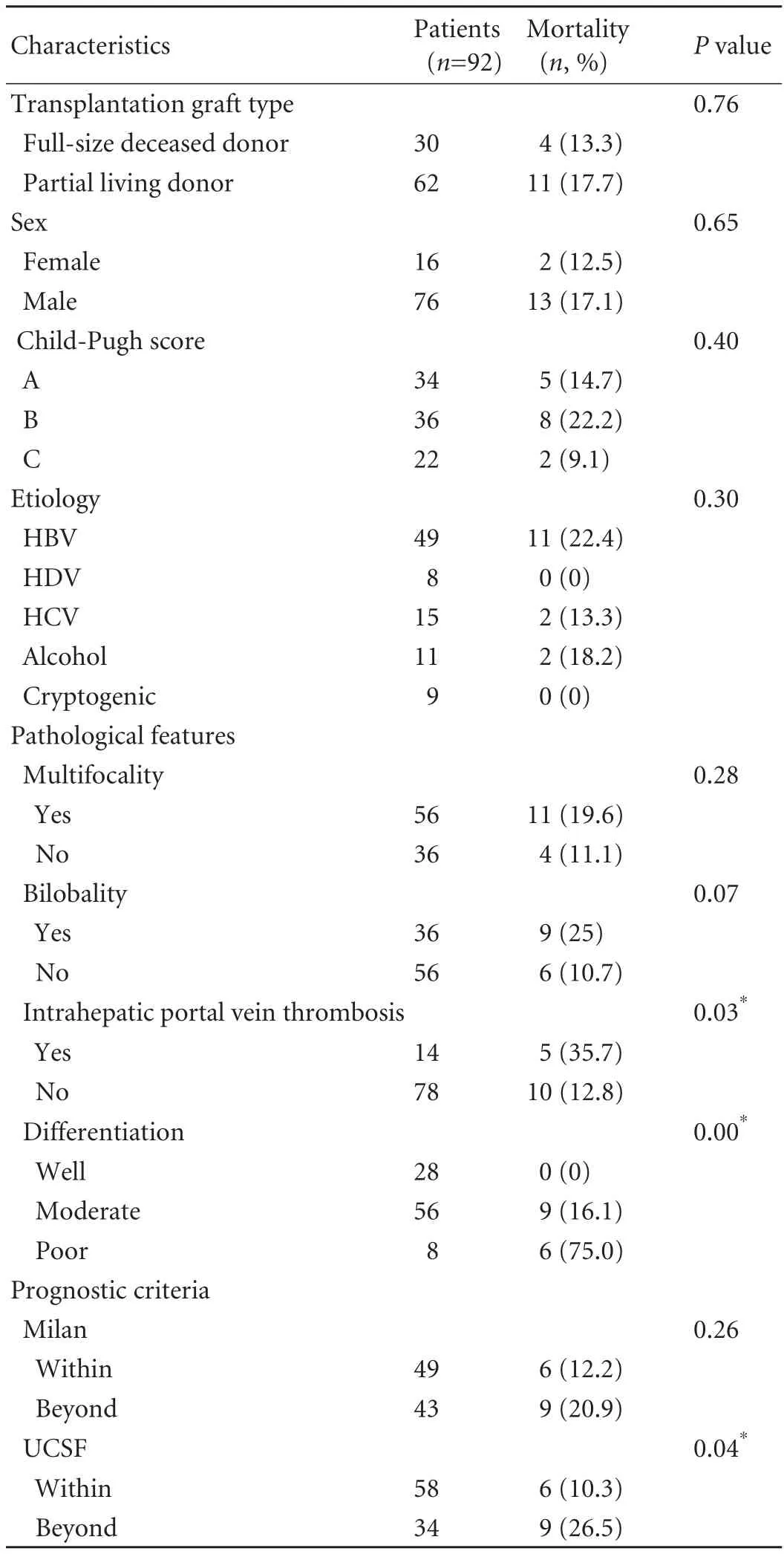
Table 3. Effect of the qualitative variables on mortality
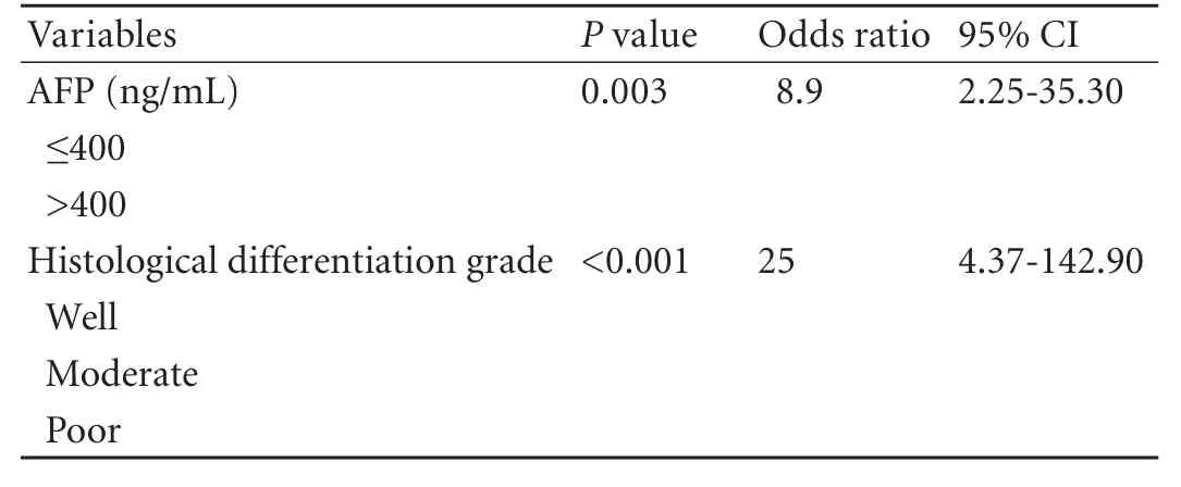
Table 4. Significant factors in multivariate analysis

Fig. 1. Five-year survival based on AFP level (ng/mL) (P=0.000).
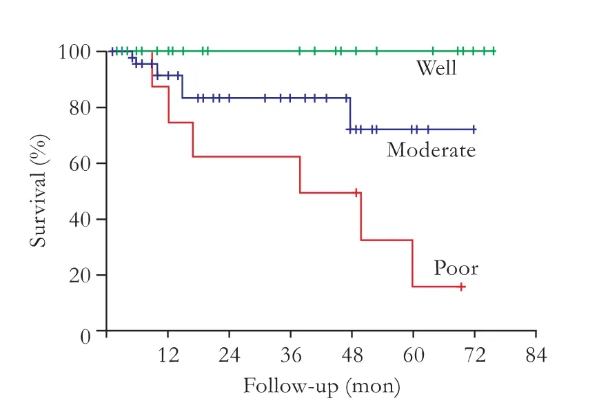
Fig. 2. Histological differentiation and survival (P=0.002).

Fig. 3. Survival plot according to AFP level (ng/mL) in patients beyond the Milan criteria (P=0.000).
The estimated 5-year disease-free survival rate for all patients was 60.4%. For patients within the Milan criteria the rate was 64.0% and for those beyond the criteria was 54.3% (P=0.07). According to the UCSF criteria, the 5-year disease-free survival rate was 66.7% for those within the criteria and 47.0% for those beyond the criteria (P=0.01).
The survival plots of the patients based on AFP level and histological differentiation are shown in Figs. 1 and 2. In the patients who were beyond the Milan criteria, the survival was significantly affected by AFP level, and the lower level may result in a higher survival rate (Fig. 3). Similarly, patients with well-differentiated tumors had a good prognosis although they were beyond the Milan criteria (Fig. 4).

Fig. 4. Survival plot according to histological differentiation in patients beyond the Milan criteria (P=0.001).
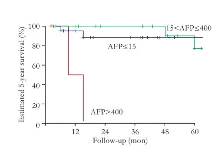
Fig. 5. Survival plot according to AFP level (ng/mL) in patients satisfying the Milan criteria.
The 5-year survival was 22.6% in patients with AFP levels >400 ng/mL. Moreover, patients with AFP levels>400 ng/mL had a significantly lower 5-year survival rate although they were within the Milan criteria (Fig. 5).
Multivariate analysis showed that AFP levels >400 ng/mL (P=0.003) and poor differentiation (P<0.001) were independent factors.
Discussion
In this study, we performed a clinicopathological analysis of HCC patients who underwent LT, and univariate analysis showed that the UCSF criteria, AFP level, poor histological differentiation and the presence of intrahepatic portal vein invasion significantly affected patient survival. Moreover, histological differentiation and AFP level predicted survival rates of patients independently of the tumor size, number and other clinical prognostic criteria such as the Milan and UCSF criteria.
The early reports on LT for HCC were very discouraging, and HCC was considered to be acontraindication for LT until the appearance of the Milan criteria in 1996.[1-5]Eligibility guidelines for transplantation, such as the Milan, UCSF, and Barcelona criteria have been adopted to reduce the post-transplant recurrence of HCC and the wastage of donor organs.[1-5]Tumor numbers and sizes were well known predictive variables which were described in the Milan, UCS and Barcelona criteria. Lee et al[17]reported expanded criteria of living donor LT for HCC from a single-center experience and defined them as the Asan criteria that consisted of the largest tumor diameter ≤5 cm, number of tumors ≤6 and no gross vascular invasion. In that trial, the 5-year patient survival rate was 76% within the criteria, similar to those within the Milan and UCSF criteria.[17]Recently, the “up to seven” criterion was reported as the sum of the combination of size and number ≤7 in the absence of gross vascular invasion with excellent outcomes.[14]In the present study, the UCSF criteria significantly affected the survival; however, multivariate analysis revealed that total tumor size, the largest tumor size and the number of tumors did not have significant effects on the survival.
At present, there are many prognostic criteria for LT and most are based on morphological features such as tumor number, diameter, total size and total volume. On the other hand, tumor biology and tumor markers such as AFP level, protein induced by vitamin K antagonism or absence (PIVKA) level and histologic differentiation are as important as the morphologic features that predict the survival after LT.[1-4,13-16,18-22]Many studies revealed that higher PIVKA-II levels predict poor prognosis, higher risk of HCC recurrence and vascular invasion.[20-22]Morover, Fujiki et al[22]reported that PIVKA-II level is an independent factor for HCC recurrence and defined it as the Kyoto criteria. However, we did not study PIVKA-II levels since we did not use it in the HCC work-up protocol.
The main purpose of all those studies was to increase the number of patients for LT and to decrease the number of recurrences after LT. According to the Hangzhou experience, macrovascular invasion, tumor size, preoperative AFP level, and histopathological grading are independent prognostic factors associated with patient survival.[16]Similarly, our results showed that AFP level and tumor differentiation were independent prognostic factors, rather than tumor size and number.
Many different cut-off points for preoperative AFP have been reported, ranging from 10 to 1000 ng/mL. Todo et al[18]studied living donor LT for HCC in 698 Japanese patients and found notably that the patients who had AFP levels ≤200 ng/mL and PIVKA-II levels ≤100 ng/mL had a 5-year disease-free survival rate of 85% despite being beyond the Milan criteria. Furthermore, according to the Asan criteria, the cut-off point was <3000 ng/mL and the 3-year recurrence rate was 12.4% in patients who had AFP levels <3000 ng/mL within the Asan criteria and 80.4% in those beyond the criteria.[17]In addition to that, DuBay et al[19]showed that an AFP level >400 ng/mL is an independent risk factor for HCC recurrence and mortality after LT. Recently, Pomfret et al[23]reported that AFP levels >1000 ng/mL should have been downstaged before being listed for LT. In that report, decreased AFP levels by downstaging resulted in better survival rates even if the levels were >1000 ng/mL at the beginning. In the present study, we found that AFP levels >400 ng/mL were an independent risk factor which indicated an 8.9-fold increased risk.
Tumor differentiation is a risk factor for recurrence after resection or LT and may be accepted as a selection criterion for LT especially in patients beyond the morphological criteria.[24-29]Currently, DuBay et al[19]reported that patients beyond the morphological criteria should be biopsied before the operation since poor tumor differentiation as a marker of tumor biology and poor prognosis predicts a poor outcome after LT or resection. Similarly, a poorly-differentiated histological grade increased the mortality risk by 25 times in our study; moreover well-differentiated histology presented better survival even if the patients were beyond the Milan criteria.
In conclusion, LT offers satisfactory long-term survival in the treatment of HCC. This study showed that AFP levels and tumor differentiation are independent factors affecting patient survival. The prognostic criteria related to tumor biology (especially AFP level and histological differentiation) should be considered in selection of patients and therapeutic strategies. Hence poor differantiation and higher AFP levels are indicators of poor prognosis after LT.
Contributors: YO and AM proposed the study, performed research and wrote the first draft. DM, DBT and GN collected and analyzed the data. DGB evaluated pathologic findings. All authors contributed to the design and interpretation of the study and to further drafts. YY and TY are the guarantors.
Funding: None.
Ethical approval: The study was approved by the institutional ethical committee.
Competing interest: No benefits in any form have been received or will be received from a commercial party related directly or indirectly to the subject of this article.
1 Bruix J, Sherman M; American Association for the Study of Liver Diseases. Management of hepatocellular carcinoma: anupdate. Hepatology 2011;53:1020-1022.
2 El-Serag HB, Marrero JA, Rudolph L, Reddy KR. Diagnosis and treatment of hepatocellular carcinoma. Gastroenterology 2008;134:1752-1763.
3 Shariff MI, Cox IJ, Gomaa AI, Khan SA, Gedroyc W, Taylor-Robinson SD. Hepatocellular carcinoma: current trends in worldwide epidemiology, risk factors, diagnosis and therapeutics. Expert Rev Gastroenterol Hepatol 2009;3:353-367.
4 Wong R, Frenette C. Updates in the management of hepatocellular carcinoma. Gastroenterol Hepatol (N Y) 2011; 7:16-24.
5 Ringe B, Pichlmayr R, Wittekind C, Tusch G. Surgical treatment of hepatocellular carcinoma: experience with liver resection and transplantation in 198 patients. World J Surg 1991;15:270-285.
6 Iwatsuki S, Starzl TE, Sheahan DG, Yokoyama I, Demetris AJ, Todo S, et al. Hepatic resection versus transplantation for hepatocellular carcinoma. Ann Surg 1991;214:221-229.
7 Bismuth H, Chiche L, Adam R, Castaing D, Diamond T, Dennison A. Liver resection versus transplantation for hepatocellular carcinoma in cirrhotic patients. Ann Surg 1993;218:145-151.
8 Mazzaferro V, Regalia E, Doci R, Andreola S, Pulvirenti A, Bozzetti F, et al. Liver transplantation for the treatment of small hepatocellular carcinomas in patients with cirrhosis. N Engl J Med 1996;334:693-699.
9 Yao FY, Ferrell L, Bass NM, Watson JJ, Bacchetti P, Venook A, et al. Liver transplantation for hepatocellular carcinoma: expansion of the tumor size limits does not adversely impact survival. Hepatology 2001;33:1394-1403.
10 Figueras J, Ibaäez L, Ramos E, Jaurrieta E, Ortiz-de-Urbina J, Pardo F, et al. Selection criteria for liver transplantation in early-stage hepatocellular carcinoma with cirrhosis: results of a multicenter study. Liver Transpl 2001;7:877-883.
11 Leung JY, Zhu AX, Gordon FD, Pratt DS, Mithoefer A, Garrigan K, et al. Liver transplantation outcomes for earlystage hepatocellular carcinoma: results of a multicenter study. Liver Transpl 2004;10:1343-1354.
12 Shetty K, Timmins K, Brensinger C, Furth EE, Rattan S, Sun W, et al. Liver transplantation for hepatocellular carcinoma validation of present selection criteria in predicting outcome. Liver Transpl 2004;10:911-918.
13 Zavaglia C, De Carlis L, Alberti AB, Minola E, Belli LS, Slim AO, et al. Predictors of long-term survival after liver transplantation for hepatocellular carcinoma. Am J Gastroenterol 2005;100:2708-2716.
14 Mazzaferro V, Llovet JM, Miceli R, Bhoori S, Schiavo M, Mariani L, et al. Predicting survival after liver transplantation in patients with hepatocellular carcinoma beyond the Milan criteria: a retrospective, exploratory analysis. Lancet Oncol 2009;10:35-43.
15 Jonas S, Bechstein WO, Steinmüller T, Herrmann M, Radke C, Berg T, et al. Vascular invasion and histopathologic grading determine outcome after liver transplantation for hepatocellular carcinoma in cirrhosis. Hepatology 2001;33:1080-1086.
16 Zheng SS, Xu X, Wu J, Chen J, Wang WL, Zhang M, et al. Liver transplantation for hepatocellular carcinoma: Hangzhou experiences. Transplantation 2008;85:1726-1732.
17 Lee SG, Hwang S, Moon DB, Ahn CS, Kim KH, Sung KB, et al. Expanded indication criteria of living donor liver transplantation for hepatocellular carcinoma at one largevolume center. Liver Transpl 2008;14:935-945.
18 Todo S, Furukawa H, Tada M; Japanese Liver Transplantation Study Group. Extending indication: role of living donor liver transplantation for hepatocellular carcinoma. Liver Transpl 2007;13:S48-54.
19 DuBay D, Sandroussi C, Sandhu L, Cleary S, Guba M, Cattral MS, et al. Liver transplantation for advanced hepatocellular carcinoma using poor tumor differentiation on biopsy as an exclusion criterion. Ann Surg 2011;253:166-172.
20 Shimada M, Yonemura Y, Ijichi H, Harada N, Shiotani S, Ninomiya M, et al. Living donor liver transplantation for hepatocellular carcinoma: a special reference to a preoperative des-gamma-carboxy prothrombin value. Transplant Proc 2005;37:1177-1179.
21 Taketomi A, Sanefuji K, Soejima Y, Yoshizumi T, Uhciyama H, Ikegami T, et al. Impact of des-gamma-carboxy prothrombin and tumor size on the recurrence of hepatocellular carcinoma after living donor liver transplantation. Transplantation 2009;87:531-537.
22 Fujiki M, Takada Y, Ogura Y, Oike F, Kaido T, Teramukai S, et al. Significance of des-gamma-carboxy prothrombin in selection criteria for living donor liver transplantation for hepatocellular carcinoma. Am J Transplant 2009;9:2362-2371.
23 Pomfret EA, Washburn K, Wald C, Nalesnik MA, Douglas D, Russo M, et al. Report of a national conference on liver allocation in patients with hepatocellular carcinoma in the United States. Liver Transpl 2010;16:262-278.
24 Cillo U, Vitale A, Bassanello M, Boccagni P, Brolese A, Zanus G, et al. Liver transplantation for the treatment of moderately or well-differentiated hepatocellular carcinoma. Ann Surg 2004;239:150-159.
25 Esnaola NF, Lauwers GY, Mirza NQ, Nagorney DM, Doherty D, Ikai I, et al. Predictors of microvascular invasion in patients with hepatocellular carcinoma who are candidates for orthotopic liver transplantation. J Gastrointest Surg 2002; 6:224-232.
26 Tamura S, Kato T, Berho M, Misiakos EP, O'Brien C, Reddy KR, et al. Impact of histological grade of hepatocellular carcinoma on the outcome of liver transplantation. Arch Surg 2001;136:25-31.
27 Marsh JW, Dvorchik I, Subotin M, Balan V, Rakela J, Popechitelev EP, et al. The prediction of risk of recurrence and time to recurrence of hepatocellular carcinoma after orthotopic liver transplantation: a pilot study. Hepatology 1997;26:444-450.
28 Klintmalm GB. Liver transplantation for hepatocellular carcinoma: a registry report of the impact of tumor characteristics on outcome. Ann Surg 1998;228:479-490.
29 Schwartz ME, D'Amico F, Vitale A, Emre S, Cillo U. Liver transplantation for hepatocellular carcinoma: Are the Milan criteria still valid? Eur J Surg Oncol 2008;34:256-262.
July 19, 2011
Accepted after revision November 30, 2011
Author Affiliations: Organ Transplantation Center, Sisli Florence Nightingale Hospital, Istanbul, Turkey (Yaprak O, Akyildiz M, Dayangac M, Demirbas BT, Guler N, Dogusoy GB, Yuzer Y and Tokat Y)
Onur Yaprak, MD, Organ Transplantation Center, Sisli Florence Nightingale Hospital, Istanbul, Turkey (Tel: 90-212-2258398; Email: onuryaprak@hotmail.com)
© 2012, Hepatobiliary Pancreat Dis Int. All rights reserved.
10.1016/S1499-3872(12)60157-X
杂志排行
Hepatobiliary & Pancreatic Diseases International的其它文章
- Management of splenic artery aneurysm associated with extrahepatic portal vein obstruction
- Pancreas-preserving segmental duodenectomy for gastrointestinal stromal tumor of the duodenum and splenectomy for splenic angiosarcoma
- Induction, modulation and potential targets of miR-210 in pancreatic cancer cells
- Quantitative analysis of intestinal gas in patients with acute pancreatitis
- Can the biliary enhancement of Gd-EOB-DTPA predict the degree of liver function?
- Quality control measures for lowering the seroconversion rate of hemodialysis patients with hepatitis B or C virus
