The Fabrication and Detection Performance of High Sensitivity Au-Ag Alloy Nanostar/Paper Flexible Surface Enhanced Raman Spectroscopy Sensors
2024-04-11DENGZhiyingWANGTianyiCAOShiyiZHAOYuanHANXiaoyuZHANGJihongXIEJun
DENG Zhiying ,WANG Tianyi ,CAO Shiyi ,ZHAO Yuan ,HAN Xiaoyu ,ZHANG Jihong,,5* ,XIE Jun,,5*
(1.State Key Laboratory of Silicate Materials for Architectures;2.Department of Materials Science and Engineering,Wuhan University of Technology,Wuhan 430070,China;3.Hainan Institute of Wuhan University of Technology,Sanya 572000,China;4.International School of Materials Science and Engineering,Wuhan University of Technology,Wuhan 430070,China;5.Hepu Research Center for Silicate Materials Industry Technology,Beihai 536100,China)
Abstract: Au-Ag alloy nanostars based flexible paper surface enhanced Raman spectroscopy sensors were fabricated through simple nanostar coating on regular office paper,and the surface enhanced Raman spectroscopy detection performances were investigated using crystal violet dye analyte.Au-Ag nanostars with sharp tips were synthesized via metal ions reduction method.Transmission electron microscope images,X-Ray diffraction pattern and energy dispersive spectroscopy elemental mapping confirmed the nanostar geometry and Au/Ag components of the nanostructure.UV-Vis-NIR absorption spectrum shows wide local surface plasmon resonance induced optical extinction.In addition,finite-difference time-domain simulation shows much stronger electromagnetic field from nanostars than from sphere nanoparticle.The effect of coating layer on Raman signal intensities was discussed,and optimized 5-layer coating with best Raman signal was obtained.The Au-Ag nanostatrs homogeneously distribute on paper fiber surface.The detection limit is 10-10 M,and the relationship between analyte concentrations and Raman signal intensities shows well linear,for potential quantitative analysis.The calculated enhancement factor is 4.795×10⁶.The flexible paper surface enhanced Raman spectroscopy sensors could be applied for trace chemical and biology molecule detection.
Key words: surface-enhanced raman;gold-silver alloy nanostars;paper-based SERS sensor;flexibility
1 Introduction
Surface-enhanced Raman spectroscopy (SERS),as a non-destructive trace or single molecular detection and structure characterization method,has attracted great attentions due to its potential applications for chemicals,biology,environmental,agriculture and others in recent years[1-3].Weak inelastic Raman scattering signals can be orders magnitude amplification from the interaction excitation beam,noble metal nanostructures,and attached analyte molecules for low concentration substance qualitative detection from characteristic fingerprint scattering peaks and quantitative analysis from peak intensities[4].The detection limit and sensitivity of SERS devices are highly determined by the components,sizes,morphologies of active noble metal or semiconductor nanostructures[5],also by the supporting substrate materials,from the distribution density and structure of active noble nanostructure and the transmission of SERS signals.Moreover,the supporting substrate also determines the ways of sensor usage under various circumstances.Many kinds of materials have been used as SERS supporting materials,including hard materials,such as glass,silica wafer,and soft flexible materials,for instance glass fiber,metal foil,film[6],cellulose[7],regular tape and paper,for portable,real-time,in-situ detection[8,9].Furthermore,largescale SERS detections require low-cost and disposable substrate with low detection limit as well.As the most common writing and printing media,regular paper for its low cost,lightness,thinness,porousness,flexibility,and more importantly,porous and cellulose fiber structure for probably dense active noble metal structure distributions can be SERS supporting materials for trace molecular detection[10].In fact,the investigation of paper based flexible SERS began from as early as 1984,Vo-Dinh and others[11]spin-coated polystyrene spheres on sub-micron silver coated filter papers for trace polycyclic aromatic hydrocarbons detection.After that,flexible paper based sensors received growing interest for various applications,such as rapid detection of biochemical compounds[12-14],methyl parathion on fruit surface[15],and food adulterant detection[16,17].The combination of different kinds of paper and Au and Ag metal nanostructure have been studied for decreasing limit of detection,and sensitivity improvement.In fact,the performance of paper-based SERS sensors should be further improved for extremely low concentration molecular detection.
The basic mechanisms of surface enhanced Raman scattering include local surface plasmon resonance(LSPR) induced electromagnetic enhancement and chemical enhancement from electron transfer between nanostructure and analyte[18].In general,chemical enhancement effect (~102) is much weaker than electromagnetic enhancement effect (~107),which can be negligible.In fact,the enhancement of SERS sensors is highly dependent on the LSPR electrical field intensities and hot-spot areas,further on the components,geometries,and sizes of noble mental nanostructures[19].Ag and Au are promising candidate metals for their unique electron structure,resulting in excellent plasmonic effect.Ag has a better LSPR effect than that of Au,but it is easy to be oxided in an ambient atmosphere[20,21].On the other hand,Au is environmentally stable[22],while has a weaker plasmonic effect,which makes it difficult to fabricate SERS sensors with low detection limit.Ag/Au alloy nanostructures can provide both effective LSPR effect for sensitive SERS detection and ambient stability.More importantly,the geometries of nanostructure also play critical roles for SERS behavior,due to the dependency of morphologies,sizes,and nano-gaps on LSPR effect.In general,nanostructures with sharp tips generate more intense plasma induced electromagnetic fields from the tip edges under incident beam excitation,than regular sphere nanoparticles,further for larger enhancement factors and better SERS performance[23,24].Various nanostructures,such nanocubes[25],nanorods[26-29],nanostars[30,31]have been synthesized and their LSPR effect and SERS detection performance on different substrates have been investigated.The results showed the improvement of detection limit and sensitivity of SERS sensors for ultra-low detection for biology molecules,fruit pesticide residue,and unknown substance.
In this research,Au/Ag alloy nanostructures with sharp tips were synthesized through metal ions reduction and growth control method.The optical excitation originated from LSPR was characterized through visible absorption spectrum.The LSPR induced electrical field distribution on nanostructure surface and between nanostructures were simulated using finite difference time domain (FDTD) solution package to analyze the origin of SERS.The nanostructures were homogenously coated on regular A4 paper for flexible paper SERS sensor fabrication.The SERS performance of the flexible sensor was characterized using crystal violet analyte.The effect of coating layers on SERS behaviors were discussed and the enhancement factor was calculated.The results indicated that the flexible paper sensors could be applied for low-cost trace molecular detection.
2 Experimental
2.1 Raw materials
Raw materials,silver nitrate (AgNO3,99%),hydrogen tetrachloroaurate acid (HAuCl4·3H2O,AR),ascorbic acid (AA,AR),polyvinylpyrrolidone (PVP,K29-32) as dispersant and crystal violet (CV,AR≥90)as dye analyte molecules were purchased from Aladin,China.Absolute ethyl alcohol (C2H5OH,AR) and ammonium hydroxide (NH3OH,AR) were purchased from China Sinopharm Group.Deionized (DI) water with a resistivity of 18.2 MΩ·cm was produced using a Dura 12 ultrapure water purification system from Thelab,USA.The paper as supporting substrate is a common office copy paper (Double A,70 g/m2).
2.2 Au/Ag alloy nanostars synthesis
The Au/Ag alloy nanostructures were synthesized following method by Chenget al[32].In Brief,240 μL,10 mM HAuCl4aqueous solution,40 μL,10 mM AgNO3aqueous solution and 10 mL DI water were mixed into a 20 mL glass vial and 500 rpm magnetically stirred for 3 minutes.Then,80 μL,100 mM AA aqueous solution was quickly injected into the solution to reduction reaction.The mixed solution was continuously 500 rpm stirred for 2 minutes.The solution color rapidly changed from light yellow to blue,indicating the formation of gold-silver nanostars.Finally,the solution was 4 000 rpm centrifuged twice with and concentrated to 1 mL ethanol solution for further use.Moreover,5 mg PVP dispersant was added into the solution to avoid synthesized nanostructures aggregation.
2.3 Materials characterization and electromagnetic field distribution simulation
The crystallographic phase of synthesized nanostructure was analyzed using X-ray diffractometer (D8 DISCOVER,Bruker,Germany),operating at 40 kV,40 mA,Cu Kα radiation λ=1.540 6 nm,at 1 °/min,and 0.02° resolution rate.The morphologies,sizes,and elemental components of nanostructure were characterized by transmission electron microscope (TEM) (JEM-2100 Plus,JEOL,Japan),at an accelerating voltage of 120 KV,equipped with selected area electron diffraction(SAED) and energy dispersive spectrometer (EDS).The visible absorption spectrum of nanostructure was recorded from a UV-Vis-NIR spectrophotometer(Lambda 750S,PerkinElmer,America) in the range of 350 to 1 350 nm.The distributions of nanostructure in paper supporting substrate were characterized using a scanning electron microscope (SEM,(S-4800,Hitachi,Japan) at an accelerating voltage of 5 kV).All the measurements were conducted at room temperature.
Nanostructure 3D models were constructed following the geometry and dimensions of Au-Ag nanostructures characterized by TEM,with individual,intercrossed and colliding tip-top nanostars.A sphere nanoparticle model with closed nanostar core diameter was also made as comparison.The electromagnetic filed distributions of nanostars were simulated using finite-difference time-domain (FDTD) analysis,based on the air suspension geometrical model (n0=1.0) assumption,with 633 nm excitation wavelength,which was similar to the Raman measurement experiments.The incident light wave vector K was alongz-axis,and the monitor position was set asz-axis plane,the range was set as 1 μm × 1 μm.The laser incident electric field was along with thex-axis,andE0=1 V/m.The mesh range resolution was 1 nm × 1 nm × 1 nm.
2.4 SERS performance measurement
Specific 1 mm diameter circle patterns were printed on regular A4 office paper with HP Laserjet 1 020 plus printer as sensor separator.The paper was cut into small pieces following the printed patterns.100 μL concentrated Au-Ag alloy nanostructures solution was dropped on the white grid and dried in vacuum to prepare paper SERS sensor (Fig.1(a)).The lay numbers of Au-Ag nanostructure on paper were determined by the solution drops.

Fig.1 The diagram of paper SERS fabrication (a) and Raman measurement setup (b)
The SERS performance of flexible paper sensors was characterized using CV molecules as analyte.20 μL different concentrations (10-4,10-5,10-6,10-7,10-8,10-9,10-10,and 10-11M,respectively) CV aqueous solution was dropped on the nanostructures coated paper sensor,and dried in vacuum.The enhanced Raman spectra of CV analyte molecules were obtained using Raman scattering spectra (LABHRev-UV,Horiba,Japan),with 10.6 mW 633 nm laser excitation source.The laser beam was focused on the flexible SERS sensor surface through a 50 × objective lens,and the beam diameter was around 2 μm.The laser interacted with nanostructures and analyte molecules for Raman scattering signals generation,and the scattering signals were backward received by detector (Fig.1(b)).The accumulation and integration duration were 3 and 4 s,respectively.All the parameters were identical for intensities comparison.
3 Results and discussion
3.1 Au-Ag nanostars
The XRD pattern of synthesized nanostructures showed several main peaks,could be attributed to (111),(200),(220),(311),and (222) crystal plane of Au (PDF#099-0056) or Ag (PDF# 087-0720),indicating the Au or Ag components of nanostructures (Fig.2(a)).The morphologies from TEM images showed nanostar geometries with sharp tips (Fig.2(b)),with around 60 nm core diameters and 30 nm tip length.The growth processes of nanostar could be attributed to Ag underpotential deposition (UPD) on the Au nanoparticles surface[33].In fact,the components and geometries of nanostructures were highly determined by the ratios of Ag and Au in raw materials,as our previous research[34].Here,Au/Ag ratio 6 was chosen due to the sharpest and longest alloy nanostars formation,for possible highest LSPR induced electromagnetic field,and best SERS performance.In addition,the SAED image from nanostars (Fig.2(b) inset) also showed different crystal plane,corresponding to XRD pattern,indicating face-centered cubic structure similar to gold or silver (Fig.2(b) inset).The HR-TEM image from the nanostar tip showed 0.23 nm interplanar spacing (Fig.2(c)),corresponding to the (111) plane of a face-centered cubic Au-Ag alloy structure[35].However,it is difficult to draw the Au-Ag alloy nanostructure formation conclusion due to nearly the same XRD pattern and lattice distance of Au and Ag crystals.Both Au and Ag elements were found from nanostar EDS elemental mapping (Figs.2(d)-2(f)),which confirmed the Au and Ag alloy elements components of nanostar.Moreover,the elements distributions were homogeneous from sphere-like core and sharp tips.

Fig.2 XRD patterns (a),TEM image (b),HR-TEM image (c),and EDS Ag,Au elemental mapping (d) of synthesized nanostructures.(b)inset: TEM image of selected and SAED pattern of nanostructures;(c) inset: amplified area for crystals plane distance measurement,and Fourier electron diffraction image
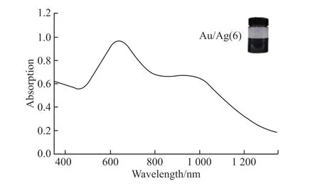
Au/Ag alloy nanostars ethanol solution showed dark blue color (Fig.3 inset).The absorption spectrum of the solution showed an almost unrecognized peak centered at 550 nm,a strong main peak center centered at 620 nm,and an infrared shoulder peak at 980 nm.The weak 550 nm peak could be attributed to the plasmonic resonance of the 60 nm sphere like core(Fig.2(b)),while the strong 620 nm peak and infrared shoulder peak originated from multiple modes[36],or the latitude and longitude propagation modes along the sharp tips[37],which was similar with infrared absorption from Au nanorods[38].The strong infrared absorption indicated that the synthesized Au-Ag alloy nanostars could be potentially applied for infrared SERS detection.

Fig.3 The absorption spectrum of synthesized Au-Ag nanostars,inset: digital image of the nanostructure ethanol solution
Much more intense maximum FDTD simulated electromagnetic fields were obtained from individual Au/Ag nanostar than from sphere nanoparticle with closed core diameter (Figs.4(a)-4(c)),under 633 nm excitation.The electric field intensity distribution on individual,colliding tip-top and intercrossed nanostars revealed that the strongest fields were at tip-top of the brunch,closed brunch tips gaps and the surface of crossed tips respectively,due to nanogap effect[39].The maximum electromagnetic field intensity could reach 38 V/m.The strong LSPR induced electromagnetic field and increased hot-spot area from nanostructures interacted surface implied possible better SERS performance of nanostars.
3.2 SERS performance of Au-Ag alloy nanostar based paper sensors
The SEM image of nanostar coated paper sensor shows dense and uniform distribution on paper fiber surface (Fig.5(a)).The area amplified nanostructure distribution images (Fig.5(b)) confirms nanostar geometry and near homogenous distribution on paper fiber surface,although there were several agglomeration clusters.Further amplified image (Fig.5(c)) shows individual,colliding tip-top,and crossed tips of nanostars on the paper surface,as the simulation conditions.
The dependency of coating layers on enhanced Raman intensities was investigated using 10-5M CV aqueous solution as analyte.Every dripped,permeated and dried 20 μL Au-Ag nanostar solution was counted as one layer.The Raman spectra showed five main scattering peaks at 1 619,1 390,1 305,1 172,920 cm-1,which could be attributed to ring C-C stretching vibration,N-phenyl stretching vibration,in-plane vibration of ring C-H,ring skeletal vibration of radical orientation,and out-of-plane vibration of ring C-H,respectively,from CV molecule[40],furtherly confirmed CV molecular detection.The intensities of 1 619 cm-1peak decreased with layer number increase from 5 to 25 with 5 layers interval (Figs.6(a),6(c)),probably because too high density nanostars decreased the gap space,and further reduced LSPR induced electromagnetic field strength.However,the 1 619 cm-1intensity increased at first,and then dropped with the layers increased from 3 to 8,with 1 layer interval (Figs.6(b),6(d)).The highest 1 619 cm-1peak intensity indicated optimized 5 layers coating paper sensor,for further Raman performance investigation.
The SERS performances of flexible paper sensors were characterized using different concentration CV aqueous solution,from 10-4to 10-11M.The Raman spectra shows characteristic scattering peaks from CV,and the intensities decreased with CV concentration decrease (Fig.7(a)).In fact,the peaks kept recognizable until the CV concentration was as low as 10-11M.The lowest detectable concentration or detection limit could reach 10-10M,following the signal noise ratio larger than three standard.This detection limit was similar to or lower than reported flexible fiber or paper-based SERS sensors[41].The better SERS performance probably resulted from strong LSPR induced electromagnetic field from star-shaped geometry and Au/Ag alloy components (Fig.4) and suitable Au-Ag nanostar distribution on paper fiber surface (Fig.5).The relationship between CV concentrations and 1 619 cm-1peak intensities showed well linear (Fig.7(b)),indicating potential quantitative analysis application of this flexible paper sensor.

Fig.4 FDTD simulated electromagnetic field distribution of Au-Ag alloy sphere (a),single nanostar (b),colliding tip-top nanostars,and intercrossed nanostars,under 633 nm laser excitation

Fig.5 SEM images of 5-layer Au-Ag nanostar coated paper SERS sensor (a) and amplified areas (b,c) for detailed nanostar distribution

Fig.6 The Raman spectra of CV molecules measured using paper sensor with different Au-Ag nanostar layers (a,b) and 1 619 cm-1 peak intensities change with layer numbers (c,d)

Fig.7 Raman spectra of CV molecules with different concentrations,using flexible paper sensor,under 633 nm laser excitation (a),and the relationship between CV concentrations and 1 619 cm-1 peak intensities
The SERS enhancement factor (EF) of the sensor could be calculated using the following equation by Gupta and others[42,43]
where,ISERSandIneatare the CV molecules Raman signal intensities from Au-Ag alloy SERS sensor and blank paper;NSERSandNneatare the molecules involved in Raman signal generation from flexible paper sensor and blank paper.The Raman intensities could be obtained from 10-9M CV using flexible paper sensor,and 0.01 M CV using blank paper,as shown in Fig.8.The 1 619 cm-1peak intensities were applied for the calculation.NSERSandNneatcould be simplified as CV concentrations,based on the monolayer assumption and identical Raman measurement parameters for SERS sensor and blank paper.The calculated EF was 4.795×10⁶,which was larger than other silver,gold nanostructures based SERS sensors[44,45],which was consistent with low detection limit and indicated better SERS performance of Au-Ag nanostar-based flexible paper sensor.Moreover,the sensor was low cost due to cheap paper supporting materials and simple fabrication process.

Fig.8 Raman spectra of 10-9 M CV using Au-Ag alloy nanostar based flexible paper sensor and 0.01 M CV using blank paper,under 633 nm laser excitation
4 Conclusions
In conclusion,Au-Ag alloy nanostars based flexible paper SERS sensors were fabricated,and their SERS performances were investigated in this research.Au-Ag alloy nanostars with sharp tips were synthesized by metal ions reduction method and UPD growth control,from 6 Au/Ag precursor molar ratio.The XRD pattern and crystal interplane distance from HR-TEM image indicated face-centered cubic Au or Ag crystals of nanostructure.TEM image confirmed nanostar shape,with~ 60 nm core and~30 nm tip length.The EDS element mappings revealed the Au and Ag components for alloy nanostar formation.LSPR induced absorption spectrum showed broad visible and near infrared absorption from sphere like core,latitude and longitude propagation of the tips,indicating potential applications for infrared SERS detection.The flexible paper SERS sensors with homogeneous nanostar distributions were fabricated by simple nanostructures solution dropping onto patterned regular office paper.Optimized 5-layer coated paper sensor were obtained from the relationship between layer numbers and Raman signal intensities.The detection limit could reach 10-10M for CV molecules.The relationship between CV analyte concentrations and Raman intensities showed well linear,for potential quantitative analysis.The enhancement factor could be 4.795×10⁶.Low detection limit and large enhancement factor were from the strong LSPR induced electromagnetic field,from nanostars.The low-cost flexible paper SERS sensors could be applied for trace molecule detection.
Conflict of interest
All authors declare that there are no competing interests.
杂志排行
Journal of Wuhan University of Technology(Materials Science Edition)的其它文章
- Fabrication of YAG: Ce3+ and YAG: Ce3+,Sc3+ Phosphors by Spark Plasma Sintering Technique
- Preparation of Modified UiO-66 Catalyst and Its Catalytic Performance for NH3-SCR Denitration
- Effect of Molecular Weight on Thermoelectric Performance of P3HT Analogues with 2-Propoxyethyl Side Chains
- Ultraviolet Photodetector based on Sr2Nb3O10 Perovskite Nanosheets
- Fabrication of Silane and Desulfurization Ash Composite Modified Polyurethane and Its Interfacial Binding Mechanism
- Bio-inspired Hydroxyapatite/Gelatin Transparent Nanocomposites
