New insights into astrocyte diversity from the lens of transcriptional regulation and their implications for neurodegenerative disease treatments
2024-03-05IbrahimOlabayodeSaliuGuoyanZhao
Ibrahim Olabayode Saliu ,Guoyan Zhao
Astrocytes are a major glial cell type in the central nervous system,and they provide trophic and metabolic support to neurons.In addition to these roles,they play crucial roles in modulating synaptic functions,development,and pruning (Brandebura et al.,2023).Αstrocytes become reactive (activated) by undergoing morphological,molecular,and functional alterations in response to neuropathology such as in injuries and neurodegenerative diseases (NDs)(Escartin et al.,2021).The pathological significance of reactive astrocytes has been implicated in the onset and progression of NDs,including Alzheimer’s disease (AD),Parkinson’s disease (PD),Lewy body disease,and Huntington’s disease.Heterogeneous responses of astrocytes have been seen to exert detrimental and/or beneficial effects on the brain depending on the stimuli.For example,astrocytes have been reported to initiate inflammation and inhibit axon regeneration or withstand neuronal insults and improve recovery (Brandebura et al.,2023).Reactive astrocytes can mitigate disease outcomes by releasing molecules that promote cell repair or exacerbate disease outcomes by releasing molecules that promote the formation of glial scars to prevent axonal regeneration or cause cell death (Brandebura et al.,2023).Therefore,it is critical to understand the molecular mechanisms that regulate the inherent cellular contribution of astrocytes to neuronal degeneration and how they can be manipulated to prevent degeneration or even facilitate neuronal regeneration in NDs.In this perspective,we discuss recent findings on the complexities in the heterogeneous response of astrocytes in NDs.We also emphasize that understanding astrocyte subtypes and their transcriptional profiles in different brain regions and different disease conditions is crucial for the development of effective therapies.
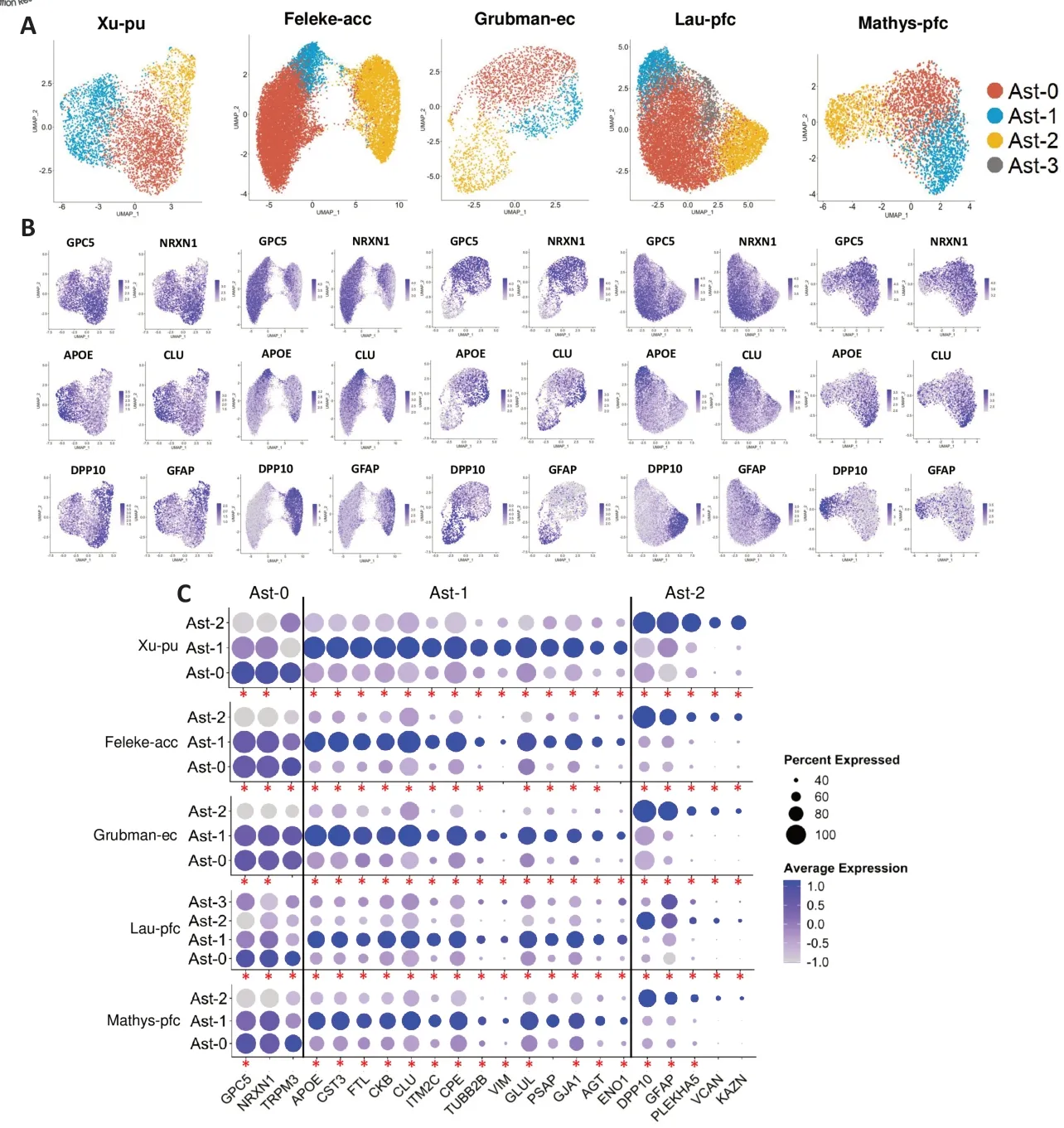
Astrocytes represent a diverse population of cells and display regional and local diversity in the adult brain (Endo et al.,2022).Recent developments in high-throughput single-cell or single-nucleus (snRNA-seq) RNA sequencing and spatial transcriptomic techniques have enabled the profound understanding of cell type heterogeneity and provide insights into their importance under both healthy and pathological conditions.However,the number of astrocyte subclusters/subpopulations in different regions of the brain,and how similar and different these subpopulations are in healthy and disease conditions are not clear.For example,Grubman et al.(2019) obtained 2171 astrocyte nuclei of the entorhinal cortex samples from the control and AD brains and clustered them into eight groups (a1–a8).Mathys et al.(2019) sampled 3392 astrocyte nuclei of the prefrontal cortex from the control and AD brains and identified four transcriptional subpopulations (a0–a3).The inconsistencies observed in the number of astrocyte subpopulations across multiple snRNA-seq studies may stem from many confounding factors.Sample differences such as postmortem intervals and cause of death;technical factors such as differences in single-cell extraction protocols,the expertise of the operators,and divergent sequencing depths;analytical factors such as analytical tools used for bioinformatic analysis,and variations in the analytical parameters are possible sources of variation observed among studies.However,the variation in the number of astrocyte subpopulations could also reflect biologically relevant differences in brain regions.To disentangle the biologicalversustechnical effects on astrocyte subpopulation structures,we performed independent data analysis on multiple snRNA-seq datasets with uniform parameters (Xu et al.,2023).The main distinguishing feature of our analysis is that each data set is independently analyzed using the same single-cell genomics tool Seurat with the same analysis algorithm and parameters.Surprisingly,we identified three evolutionarily conserved (between human and mouse) subpopulations of astrocytes that were shared in different ND conditions across multiple brain regions.Identification of shared populations enabled the comparison of molecular similarities and differences between orthologous subpopulations in different disease conditions and across different brain regions.
In our study,we identified three distinct subpopulations of astrocytes in the human putamen,characterized by the expression of unique marker genes shared by astrocytes from AD and PD subjects and cognitively normal controls (Xu et al.,2023).Αst-0 represents a homeostatic subpopulation with highlevel expression ofGPC5andNRXN1whereas Ast-1 and Αst-2 represent astrocytes at distinct activation states.Ast-1 expressed high levels ofAPOE,CLU,and many other conserved marker genes while Ast-2 expressed high levels ofGFAP,DPP10,and many other unique marker genes conserved across three conditions (Figure 1BandC).Moreover,RNAscopein situhybridization corroborated the presence of different astrocyte subpopulations in the putamen and white matter tissue of the internal capsule in all subject groups.Next,we performed an independent analysis of published snRNA-seq data from four cortical regions representing four NDs using the same analysis method and parameters.We found that the three astrocyte subpopulations in the putamen region and their transcriptome features were shared with astrocytes from all cortical regions including the prefrontal cortex,entorhinal cortex,and anterior cingulate cortex (Figure 1A–C).Furthermore,the diversity of astrocytes was critically explored in an ΑD animal model (Habib et al.,2020) and three subpopulations of astrocytes,Gfap-low,disease-associated astrocytes,Gfap-high astrocytes representing different activation states were identified,together with other astrocyte subpopulations at intermediate activation states.We found that the three subpopulations corresponded to the Ast-0,Ast-1,and Ast-2 subpopulations in human data with marker genes conserved between species,suggesting that the three astrocyte subpopulations are evolutionarily conserved between humans and mice.

Figure 1|Three astrocyte subpopulations shared across multiple brain regions and disease conditions.
Interestingly,even though the astrocyte subpopulations are shared across different brain regions,we observed regional differences in the transcriptional features between orthologous astrocyte subpopulations.Some conserved markers exhibited consistent expression patterns between the putamen and one cortical brain region but not the other.Kyoto Encyclopedia of Genes and Genomes and Gene Ontology enrichment analysis revealed that,out of all the compared brain regions,only putamen Ast-1 marker genes were highly enriched for pathways related to neurodegenerative disorders,amyloid fibril formation,tau protein binding,ferroptosis,regulation of inflammatory response and neuron death.These pathways consist of genes likeAPOEandPARK7,which are the top AD and PD risk genes.Αst-1 from putamen is additionally enriched in transcripts for proteins such asMT3,SOD1,andHSP90AB1,which enable astrocytes to protect neurons from amyloid-beta toxicity,dopamine quinone neurotoxicity,or oxidative stress,suggesting a potential neuroprotective role of Ast-1.Intriguingly,regional differences in astrocytes have been reported previously.For example,Chai et al.(2017) revealed significant differences in electrophysiological characteristics,Ca2+signaling,cellular morphology,gene and protein expression profiles,and proximity of astrocytes to synapse between striatal and hippocampal astrocytes.Morel et al.(2017) found that the expression of astroglialtranslating mRNΑs in major cortical and subcortical brain regions closely followed the dorsoventral axis,especially from the cortex/hippocampus to the thalamus/hypothalamus posteriorly.These studies,along with ours,illustrate the regional specialization of astrocytes in the brain,and region-specific astrocyte transcriptomes translate into neuralcircuit-based functional differences.
Moreover,NDs can differentially alter the brain microenvironment,which can influence the response of astrocytes to pathologies.NDs share a common feature known as “selective regional vulnerability”,in which the pathology initiates in one or two highly susceptible regions and then progressively and predictably affects more brain areas stereotypically.For example,amyloid plaques appear in the transentorhinal and entorhinal cortex,hippocampus,and prefrontal cortex early in AD and are present in non-demented older adults.In later stages,often after dementia symptoms have already manifested,these plaques develop in regions like the striatum and cerebellum.In PD,the deposition of alpha-synuclein (α-syn) starts in the dorsal motor nucleus of the vagus and spreads to the brainstem,resulting in the loss of dopaminergic neurons in the substantia nigra pars compacta.Αs the disease progresses,α-syn pathology predominantly spreads in a caudo-rostral route,ultimately impacting the neocortex.The factors underlying this regional selectivity remain unclear.In our study,we detected discordant transcriptome changes between cortical astrocytes and putamen astrocytes.We found genes differentially expressed in disease conditions overlap among cortical astrocyte subpopulations and are highly correlated with each other.However,putamen astrocyte genes differentially expressed were largely non-overlapping or showed expression changes in opposite directions compared to cortical astrocytes,including many AD and PD risk genes identified by genome-wide association studies.Furthermore,Kyoto Encyclopedia of Genes and Genomes and Gene Ontology analyses revealed differential regulation of multiple neurodegenerative disease-related pathways as well as amyloid-beta binding and amyloid precursor protein metabolic process pathways between cortical and subcortical astrocytes (Xu et al.,2023).Our findings suggest that regional differences in astrocyte population and transcriptomic changes may underly regional differences in disease vulnerability.
Due to the extensive transcriptional and proteinlevel alterations of astrocytes in NDs,targeting transcription factors (TFs) to modify the networks of RNA and protein expression in astrocyte reactivity may be a promising strategy for treating NDs (Endo et al.,2022).TFs have been shown to modulate astrocyte identity,reactivity,and disease pathogenesis in animal models.For example,modulating reactive astrocyte activity in APP/PS1 AD mouse model by selectively inhibitingSTAT3,a transcription factor that promotes astrogliosis,reduced amyloid-beta plaque burden and size,protected against learning and spatial memory decline,and triggered a phenotypical switch in reactive astrocytes (Reichenbach et al.,2019).Additionally,astrocyte-specific deletion ofBmal1,a potent regulator of astrocyte activation,prevents both α-syn and tau pathologies in P301S and α-syn preformed fibril injection mouse models.TheBmal1deletion also induces a distinct form of astrocyte reactivity,which has minimal impact on amyloid plaque formation but enhances the phagocytosis of tau and α-syn in ΑPP/PS1 mice (Sheehan et al.,2023).Α previous study systematically compared marker genes to distinguish astrocyte subpopulations in autoimmune encephalomyelitis,transient neuroinflammation,and spinal cord injury in mice and humans.They predicted TFs that differentially regulate more than 12,000 differentially expressed genes that are potentially associated with astrocyte reactivity in these conditions (Burda et al.,2022).Furthermore,by modulating astrocyte reactivity through transgenic knockout ofSmarca4orStat3in mice,they demonstrated that TFs can significantly modify disorder outcomes,indicating their potential as therapeutic targets.Analyses of the astrocyte region-enriched genes across 13 brain regions by Endo et al.(2022) revealed that differences and similarities in TFs may partly explain astrocyte interregional differences in mouse brains.Whether and how these regional differences in TF expression might contribute to the regional vulnerability of ND remains unknown.No systematic study on the heterogeneity of astrocytes and their transcriptional regulation in different human brain regions has been carried out.Therefore,probing the potential of TFs for the regulation of astrocyte reactivity in humans may provide insights into how transcriptomes and functions may be altered during astrocyte activation and how astrocyte activation may contribute to disease pathogenesis.Consequently,identifying TFs responsible for astrocyte inter-regional heterogeneity and reactivity in human,along with assessing their contribution to regional differences in gene expression and pathophysiology of NDs could offer new therapeutic avenues for the treatment of different NDs.
Finally,employing multimodal high-throughput technologies such as single-cell spatial transcriptomics,proteomics,and epigenomics to delineate the complex heterogeneity of astrocyte response to NDs will provide a holistic picture of the molecular pathophysiological mechanism of this group of related diseases.Future lineage tracing experiments are needed to determine whether astrocyte subtypes from different brain regions share the same or have different developmental paths.This is important as it remains unknown whether human astrocyte subtypes are genetically encoded or are just different activation states responding to different environmental stimuli.These investigations will fill the current knowledge gap on astrocyte heterogeneity,reactivity,and regulation and could provide novel therapeutic targets for the treatment of different NDs.
This work was supported in part by the R21AG077643,R01NS123571,1U19NS130607,and 5T U24 HG012070(to GZ);in part by Alzheimer Association Fellowship Award 23AARFD-1029969(to IOS).
This publication is solely the responsibility of the authors and does not necessarily represent the official view of the National Institutes of Health.
Ibrahim Olabayode Saliu*,Guoyan Zhao*
Department of Genetics,Washington University School of Medicine,St.Louis,MO,USA (Saliu IO,Zhao G)
Department of Neurology,Department of Pathology and Immunology,Washington University School of Medicine,St.Louis,MO,USA (Zhao G)
*Correspondence to:Guoyan Zhao,PhD,gzhao@wustl.edu;Ibrahim Olabayode Saliu,PhD,isaliu@wustl.edu.
https://orcid.org/0000-0001-5615-6774(Guoyan Zhao)
https://orcid.org/0000-0002-6182-5992(Ibrahim Olabayode Saliu)
Date of submission:October 31,2023
Date of decision:December 13,2023
Date of acceptance:December 30,2023
Date of web publication:March 8,2024
https://doi.org/10.4103/NRR.NRR-D-23-01790
How to cite this article:Saliu IO,Zhao G(2024)New insights into astrocyte diversity from the lens of transcriptional regulation and their implications for neurodegenerative disease treatments.Neural Regen Res 19(11):2335-2336.
Open access statement:This is an open accessjournal,and articles are distributed under the terms of the Creative Commons AttributionNonCommercial-ShareAlike 4.0 License,which allows others to remix,tweak,and build upon the work non-commercially,as long as appropriate credit is given and the new creations are licensed under the identical terms.
杂志排行
中国神经再生研究(英文版)的其它文章
- Notice of Retraction
- Next-generation regenerative therapy for ischemic stroke using peripheral blood mononuclear cells
- Ruxolitinib improves the inflammatory microenvironment,restores glutamate homeostasis,and promotes functional recovery after spinal cord injury
- OSMR is a potential driver of inflammation in amyotrophic lateral sclerosis
- Optimal transcorneal electrical stimulation parameters for preserving photoreceptors in a mouse model of retinitis pigmentosa
- Neuroprotective effects of G9a inhibition through modulation of peroxisome-proliferator activator receptor gamma-dependent pathways by miR-128
