Liver immunity,autoimmunity,and inborn errors of immunity
2023-03-18YavuzEmreParlarSefikaNurAyarDenizCagdasYaseminBalaban
Yavuz Emre Parlar,Sefika Nur Ayar,Deniz Cagdas,Yasemin H Balaban
Yavuz Emre Parlar,Yasemin H Balaban,Department of Gastroenterology,Hacettepe University Faculty of Medicine,Ankara 06100,Turkey
Sefika Nur Ayar,Department of Internal Medicine,Hacettepe University Faculty of Medicine,Ankara 06100,Turkey
Deniz Cagdas,Department of Pediatric Immunology,Hacettepe University Ihsan Dogramaci Children's Hospital,Ankara 06100,Turkey
Abstract The liver is the front line organ of the immune system.The liver contains the largest collection of phagocytic cells in the body that detect both pathogens that enter through the gut and endogenously produced antigens.This is possible by the highly developed differentiation capacity of the liver immune system between self-antigens or non-self-antigens,such as food antigens or pathogens.As an immune active organ,the liver functions as a gatekeeping barrier from the outside world,and it can create a rapid and strong immune response,under unfavorable conditions.However,the liver's assumed immune status is anti-inflammatory or immuno-tolerant.Dynamic interactions between the numerous populations of immune cells in the liver are key for maintaining the delicate balance between immune screening and immune tolerance.The anatomical structure of the liver can facilitate the preparation of lymphocytes,modulate the immune response against hepatotropic pathogens,and contribute to some of its unique immunological properties,particularly its capacity to induce antigen-specific tolerance.Since liver sinusoidal endothelial cell is fenestrated and lacks a basement membrane,circulating lymphocytes can closely contact with antigens,displayed by endothelial cells,Kupffer cells,and dendritic cells while passing through the sinusoids.Loss of immune tolerance,leading to an autoaggressive immune response in the liver,if not controlled,can lead to the induction of autoimmune or autoinflammatory diseases.This review mentions the unique features of liver immunity,and dysregulated immune responses in patients with autoimmune liver diseases who have a close association with inborn errors of immunity have also been the emphases.
Key Words: Liver immunity;Autoimmunity;Immune tolerance;Autoinflamation;Autoimmune liver diseases;Inborn errors of immunity
INTRODUCTION
The immune system is a complex cellular and molecular network that provides the body with defense against harmful and foreign substances.While the immune system provides defense against pathogens in healthy individuals,it also plays a role in clearing the body’s own dead cells and cell remnants to prevent tumoral cell formation.On the other hand,one of the main features of the immune system is"immune tolerance" which ensures that the body does not harm its own tissues and maintains tissue homeostasis while performing the aforementioned active immune screening of tissues and organs.
Conceptually,the elements of the immune system can be divided into two main groups: Innate and acquired (adaptive) immunity,which interact closely with each other.The innate immune system provides a pre-structured first response to a wide range of situations and stimuli,and thus constitutes an initial rapid response against immune insults.However,the adaptive immune system learns to recognize previously encountered stimuli and provides a specific immune response against them.Both types of immunity are mediated by both molecules and cells.The general characteristics of the innate and adaptive immune systems are summarized in Table 1[1].
Immune tolerance implies the inertia of the immune response towards self-antigen.Immune tolerance can occur in two ways.“Central tolerance” is acquired by training lymphocytes about autoantigens during their development in primary immune organs (e.g.,thymus),whereas “peripheral tolerance” defines the maintenance of tolerance towards self-antigens by lymphocytes at the target organ,such as the liver,which has previously completed their development and spread around the body.Central tolerance is achieved by apoptosis of self-reacting T lymphocytes in the thymus and by the loss of autoreactive feature of lymphocytes by changing their receptors in the bone marrow.Peripheral tolerance is provided by T regulatory (Treg) cells and co-stimulatory surface molecules that control antigen presentation by dendritic cells (DCs)[2].
Autoimmunity is the formation of a cellular or humoral immune response against the body’s own antigens due to defects in immune tolerance mechanisms.Autoimmune diseases are characterized by tissue damage resulting from a dysregulated immune reaction of an organism against its own antigens[3].The autoimmune reaction can be limited to an organ (e.g.,autoimmune hepatitis [AIH]) or systemic reactions involving several organ systems (e.g.,systemic lupus erythematosus).Although both the innate and adaptive immune systems contribute to the development of autoimmune disease,it is generally known that adaptive immunity plays a major role.Indeed,recent literature proposed to classify the disease with loss of immune tolerance as “autoinflammatory disease“ which is mainly associated with disorders in innate immunity and “autoimmune disease“ which is driven by pathological responses of the adaptive immune system[4].
Various factors and mechanisms can trigger autoimmunity.In general,the presence of underlying genetic predisposition factors,environmental factors,infection,inflammation,and apoptotic bodies triggers the development of autoimmunity[5].Although the pathogenesis of autoimmunity is still not fully understood and studies are ongoing,the currently known mechanisms that are thought to cause autoimmunity are summarized at the cellular and molecular levels in Table 2[6].
The prevalence of autoimmune diseases in the general population is around 3%-5%[7,8].They include a diverse group of diseases that can affect almost all organs and sometimes multiple systems,such as autoimmune thyroiditis and autoimmune hemolytic anemia,which can be organ-specific whereas systemic lupus erythematosus and vasculitides have systemic involvement.The presence of one autoimmune disease predisposes patients to other autoimmune diseases.In fact,autoimmune liver diseases (AILD) are organ-specific,namely,liver-restricted autoimmune diseases,and are commonly associated with autoimmune diseases of other organs[9].

Table 1 Characteristics of immune system components

Table 2 Mechanisms of autoimmunity
Dysregulated immune responses not only increase infection risk but also make individuals prone to autoimmune and malignant diseases.Inborn errors of immunity (IEI) were previously named “primary immunodeficiency diseases”.IEI is a heterogeneous group of diseases caused by one or more disorders in the innate or adaptive immune system,affecting the development or function of the immune system and increasing susceptibility to infections[10].Unlike secondary immune deficiencies,which develop due to various drugs and diseases,IEI is a genetic disorder.More than 350 genes involved in the etiology of IEI have been identified.While some IEIs are inherited by a single gene,other is polygenic.Except for selective IgA deficiency,all other forms are rare,occurring in approximately 1:10000 Live births.However,it is estimated that IEI is more common in consanguineous or genetically isolated populations[11].According to a classification updated in 2019,IEIs were grouped under ten headings as shown in Table 3[12,13].Most IEIs present with symptoms and are diagnosed in childhood;however,symptoms of some diseases,such as common variable immunodeficiency (CVID),may appear later in life.Diagnosis may be delayed because of the heterogeneous and indolent course of symptoms associated with IEI.The risks in these patients are not limited to susceptibility to bacterial,viral,or opportunistic infections but also include autoimmunity,malignancy,lymphoid proliferation,atopy,and granulomatous disease[12,14,15].The treatment method varies according to the type of IEI,such as prophylaxis for bacterial,fungal,and/or viral infections;intravenous or subcutaneous immunoglobulin;immunosuppressive or modulatory drugs;and hematopoietic stem cell transplantation.
ROLE OF THE LIVER IN IMMUNITY
The liver has been proposed as an “immunological organ”.Beginning with intrauterine life,the liver has several unique immunological features,including a high level of immune tolerance,powerful innate immunity,and over-reactive autoimmunity against a weak adaptive immune response.In addition,the liver has a dual arterial blood supply from the hepatic artery and portal vein;thus,it is a bridge between the two circulatory systems of the body,namely,the caval and portal systems.Oxygen-rich arterial blood enters the liverviathe hepatic artery,which supplies one-third of the liver’s blood flow.The portal vein carries most of the blood to the liver,which is rich in both nutrients and pathogen-derived molecules[16,17].After passing through a network of liver sinusoids,blood leaves the parenchymaviathe central hepatic veins.Various antigenic structures and cells from the gut and other organs mix within the liver sinusoids and are cleaned by hepatocytes.Approximately 30% of the total cardiac output passes through the liver every minute,and it carries approximately 108peripheral blood lymphocytes in 24 h[18].Decreased blood velocity in the feeding vessels of the liver,minimal increases in systemic venous pressure,and disturbances in sinusoidal flow result in stasis.This prolongs the contact time between lymphocytes and antigen-presenting cells (APCs) in the sinusoids and promotes lymphocyte extravasation.The sinusoids are lined with special liver sinusoidal endothelial cells (LSECs)containing multiple fenestrae that allow blood lymphocytes to reach the space of Disse between LSECs and hepatocytes,where they contact the extracellular matrix,stellate cells,and hepatocytes[19].
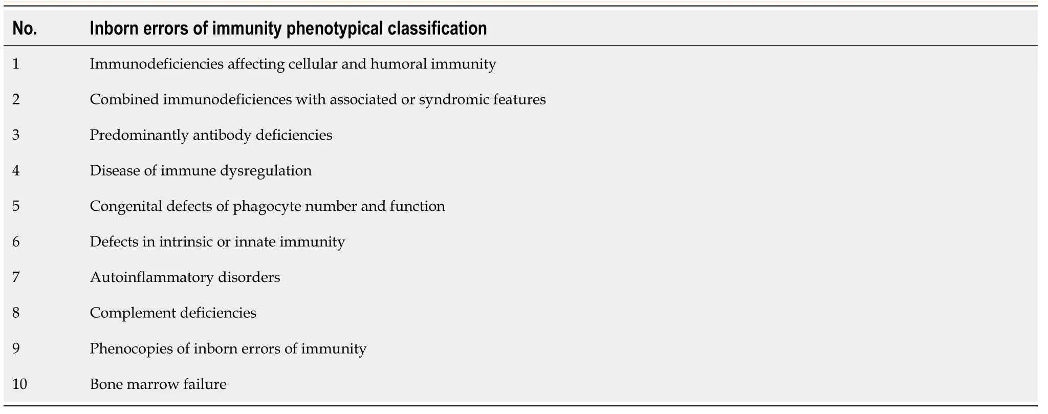
Table 3 Categories of inborn errors of immunity
The liver is considered to be one of the primary organs of the immune system,with its own microanatomy and lymphoid and non-lymphoid cells.Liver parenchymal cells are hepatocytes and cholangiocytes,which constitute 60%-80% of liver tissue (Figure 1) and function as part of the “liver immune system”.Non-parenchymal cells,namely,LSECs,hepatic satellite/into cells,Kupffer cells,neutrophils,mononuclear cells,T and B lymphocytes,natural killer (NK) cells,and NKT cells,also have immunological functions[18].Lymphocytes are scattered throughout the hepatic lobules and portal areas.The liver contains approximately 1010lymphocytes,including conventional and nonconventional lymphocyte subpopulations of the immune system.
Conventional T cells include clusters of differentiation (CD)8+and CD4+T cells.Both groups of T cells exhibit a diverse repertory of T cells that recognize antigens in the context of major histocompatibility complex (MHC) class I and II molecules.CD4+T cells are less in number than CD8+T cells in the liver.There are more memory cells in the liver than in blood.Unconventional T cells contain a variety of cell types and are categorized into two main populations based on NK cell marker presentation.Unconventional T cells presenting T cell markers are named NKT cells,and they bridge the gap between the adaptive and innate immune systems.NKT cells have a limited T-cell receptor (TCR) repertoire.They recognize and eliminate tumor and virus-infected cells.Unlike conventional T cells,NKT cells recognize glycolipid antigens that are presented by CD1d.NKT cells are further classified as “classical NKT cells”and “nonclassical NKT cells”.Classical NKT cells are divided into two groups: CD4-positive or CD4/CD8-double negative.Nonclassical NKT cells contain TCR αβ and TCR γδ T cells[20].Classical and non-classical NKT cells are found in higher proportions in the liver than in other organs and may constitute 30% of the intrahepatic lymphocyte population[21].
The liver comprises various types of resident APCs that can capture cell-associated released antigens,either passing through the liver or during the death of pathogen-infected hepatocytes.Resident APCs include Kupffer cells,LSECs,and DCs.Kupffer cells constitute the majority of the macrophage group in the body and constitute approximately 20% of the non-parenchymal cells in the liver[22].Kupffer cells originate from circulating monocytes and localize in the sinusoidal vascular space of the liver.Here,they settle perfectly to remove endotoxins from the blood and phagocytose residues and microorganisms.Their slow migration through hepatic sinusoids leads to temporary stasis,facilitating close contact with the passing lymphocytes[23].LSECs constitute the majority of non-parenchymal cells in the liver (50%).Their morphology forms a sieve-like fenestral endothelium.LSECs express molecules containing mannose and scavenger receptors,which facilitate antigen uptake.LSECs also include MHC class I and II and co-stimulatory molecules (CD40,CD80,and CD86) that facilitate antigen presentation[24].DCs are professional APCs that control immunity and tolerance.Hepatic DCs are derived from the bone marrow and are mostly found around the central veins and portal tracts of the liver[25].Hepatic DCs produce certain cytokines in response to signals from invading microbes and their cellular environment,support the adaptive immune system,and act as a bridge between innate and adaptive responses[26].
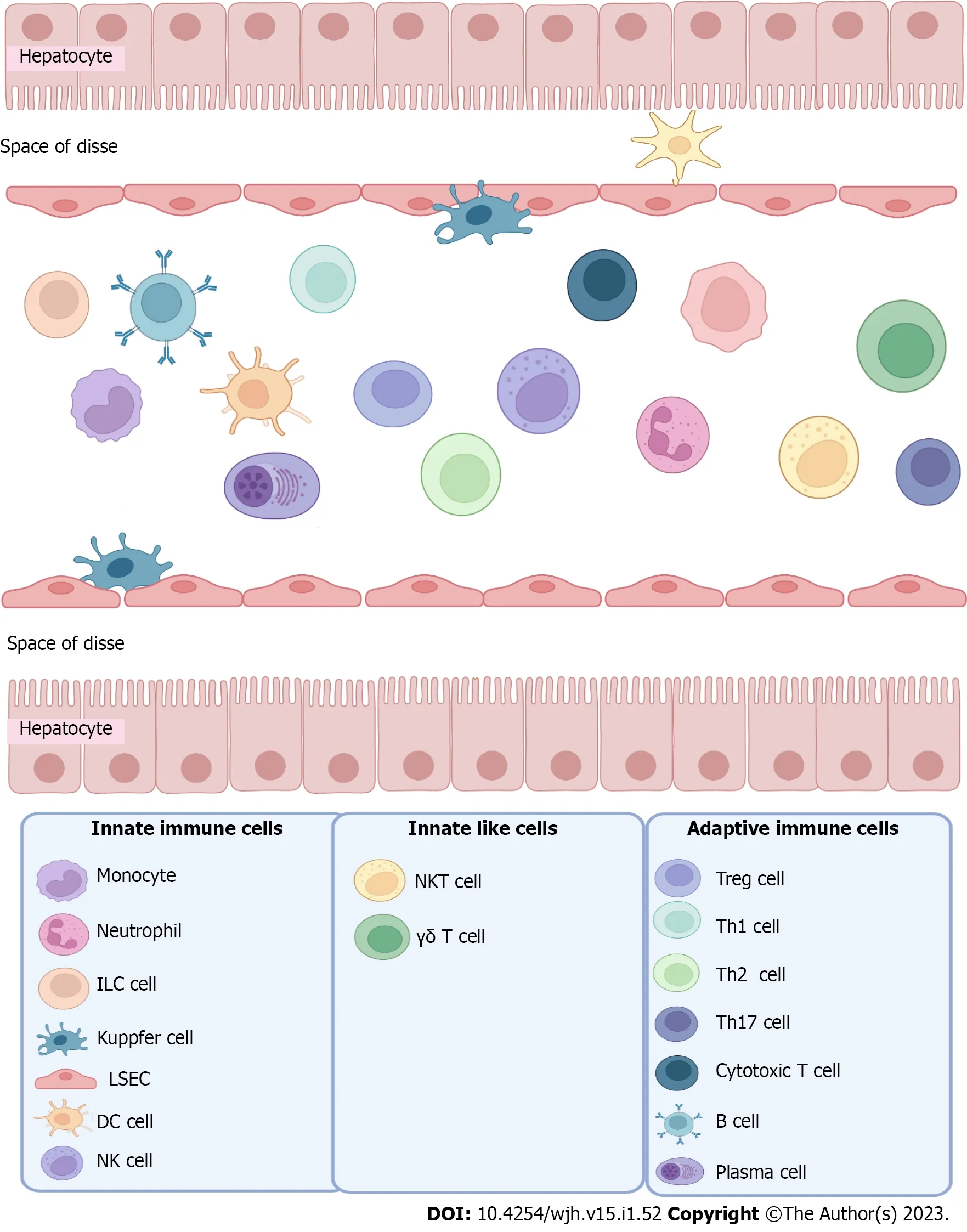
Figure 1 Cell composition of the healthy liver.
IMMUNE SYSTEM ELEMENTS IN THE LIVER
Innate immunity
The innate immune system is the first crucial defense against infections.It quickly reacts to possible pathogenic attacks.The innate immune system contains physical and chemical barriers,humoral factors,phagocytic cells,and lymphocytic cells (NK and NKT cells).Although innate immune responses kill pathogens non-specifically,recent studies suggest that innate immunity can detect specific infections through “pattern recognition receptors (PRRs)”.PRPs identify structures reflected by pathogens called pathogen-associated molecular patterns (PAMPs)[27].Among them,the best-defined PAMPs are lipopolysaccharides and peptidoglycans.
Hepatocytes play an important role in the control of systemic innate immunity by secreting PRRs and complementing plasma.Liver expression of genes encoding these proteins is governed by transcription factors such as hepatocyte nuclear factors (nuclear factor-1) and CCAAT-enhancer-binding protein.During the acute phase of the systemic inflammatory response,various pro-inflammatory cytokines[such as interleukin (IL)-6,IL-1,tumor necrosis factor α (TNF-α),and interferon-gamma (IFN-γ)]stimulate hepatocytes to produce high levels of complement and PRRs[28].
The complement system comprises plasma and membrane proteins that affect each other to protect against infection.In addition,it contributes to the pathogenesis of various liver disorders including fibrosis,alcoholic liver disease,and ischemic liver injury.There are three different ways to activate the complement system: Classical,lectin,and alternate pathways.After activation,the complement system mediates various biological activities,such as opsonization,and inflammatory and cytotoxic functions.The liver biosynthesizes the main complement components in the plasma,including C1r/s,C2,C4,Cbp,C3,mannan-binding lectin,factor B,mannan-binding lectin-associated serine proteases 1-3,and the terminal components of the complement system C5,C6,C8,and C9.Hepatocytes are also involved in the biosynthesis of certain regulatory proteins in the plasma,such as factor I,factor H,and C1 inhibitors[29,30].
The liver contains membrane-bound PRRs,such as Toll-like receptors (TLRs),which are a family of proteins that recognize PAMPs expressed by microorganisms.Diverged TLRs are expressed by liver cells.They have been shown to participate in liver injury and repair,and contribute to the pathogenesis of various liver diseases.Recently,cytoplasmic PRRs,including nucleotide-binding oligomerization domain-like receptors and retinoic acid-inducible gene (RIG)-like helicases,have been identified.RIG-1 serves as a pathogen receptor that regulates cellular transition to hepatitis C virus (HCV) replication[31].
Many studies have shown that hepatic NK cells play a significant role in innate immune responses against tumors,viruses,intracellular bacteria,and parasites.NK cells also contribute to innate defense against primary liver tumors and liver metastases in patients.This effect is achieved by direct killing of tumor cells and stimulation of tumor-specific immunity[32].Activation of NK cells is also involved in liver injury,fibrosis,and repair[33].Liver lymphocytes are enriched in Tγδ cells.Evidence suggests that Tγδ cells play an important role in innate defense against viral and bacterial infections and in tumor formation.The percentage of Tγδ cells is considerably increased in the livers of tumor-bearing mice and patients with viral hepatitis[34].
In addition to host defense against infection,innate immunity can detect signals from damaged hepatocytes during non-infectious liver injury.Acetaminophen hepatotoxicity and ischemic liver injury can cause liver damage by inducing sterile neutrophilic inflammation.Neutrophilic inflammation after partial hepatectomy can promote liver regeneration by triggering a local inflammatory response,leading to hepatocyte proliferation[35].IL-1 is an important mediator of sterile neutrophilic inflammation in liver injury.
All chronic liver diseases lead to liver fibrosis,which is characterized by the activation of hepatic satellite cells (HSCs) overproducing collagen,and eventually,its accumulation in the liver[36].HSCs are generally inactive in healthy livers,but become activated during liver injury and differentiate into myofibroblastic cells.Transforming growth factor β (TGF-β) and platelet-derived growth factor induce HSC transformation and proliferation.Evidence suggests that the innate immune system plays a key role in regulating HSC activation and liver fibrosis[37].The complement system is activated after liver damage.A recent study showed that C5 deficiency caused a decrease in liver fibrosis,whereas overexpression of the C5 gene caused an increase in liver fibrosis[38].TLRs likely play a significant role in the pathogenesis of liver fibrosis because various TLRs are expressed in liver cells,including HSCs[39].TLR9-deficient mice have been shown to be resistant to liver fibrosis because HSCs require TLR9 for DNA activation[40].Kupffer and NK cells have been shown to play significant roles in liver fibrosis[33].It is thought that Kupffer cells activate HSC by producing cytokines/growth factors such as TGF-β.NK cells have an inhibitory effect on liver fibrogenesis.Activated HSCs are directly killed by NK cells by expressing the NK cell-activated ligand retinoic acid early inducible gene 1 and tumor necrosis factorrelated apoptosis-inducing ligand receptors[41,42].
Adaptive immunity
The liver is a front-line filter for pathogens and PAMPs entering the body from the gutviathe portal vein,and is often one of the first points of contact with other antigens entering the body.Similar to lymphoid organs,the liver is involved in the development and function of the adaptive immune response.Despite the abundance of APCs in the liver and their ability to rapidly recruit diverse immune cell populations,establishing an integrated adaptive immune response in the liver is a complex process.The immune response in the liver must be in delicate balance between tolerance to non-threats and immunity to pathogens.
There is insufficient data on the functions of B cells in the liver.The scarcity of B cells in the healthy liver is the reason for not obtaining the intended information.In adaptive immunity in the liver,these T cell subsets are highly regulated in all stages of diverse disorders.The major T lymphocytes involved in adaptive immunity include CD4+T cells,CD8+T cells,and γδ cells.CD4+T cells have at least five functional subgroups,including helper T (Th),Th2,Th17,follicular helper T (Tfh),and T-regulatory(Treg) cells.The innate and adaptive immune responses in the liver are supported by Tfh cells,which are often suppressed by Treg cells.CD8+T cells are composed of two subgroups: Cytotoxic T (Tc) cells and CD8+Treg cells.Tc cells are the main killer cells in adaptive immunity,and CD8 Treg cells suppress immune responses to infection.Tγδ cells participate in both the innate and adaptive immune responses.
Adaptive immunity and viral hepatitis
Although hepatitis B virus (HBV) and HCV are both hepatotropic viruses,hepatocellular necrosis during infection primarily results from an adaptive immune response targeting virus-infected liver cells[43].Naive T cells specific to viral antigens can be locally activated in the liver.In the initial stage of adaptive immunity,antigen-specific naive T cells are usually prepared by APCs in the lymph nodes,differentiate into effector cells,and then migrate to the target (liver)[44].However,HBV-specific naïve T cells can exert their anti-HBV effects by directly entering the liver before maturation in lymphoid organs[45].Th17 cells can exacerbate liver lesions during HBV infection.In patients with HBV infection,the number of Th17 cells increases in the blood and liver,accompanied by high levels of IL-17 and IL-22 in the blood[46].In contrast,HBV-specific CD4+CD25+foxp3+Treg cells have immunosuppressive effects during HBV infection[47].Evidence demonstrates that HBV-specific CD8+T cells play a significant role in viral clearance and in the prognosis of HBV infection.When HBV-specific CD8+T cells are activated,they produce IFN-γ and TNF-α,which in turn inhibit HBV replication in infected hepatocytes and enable viral clearance.However,studies in mice infected with HBV have shown that HBV components also induce specific immune tolerance through clonal deletion,clonal ignorance,and clonal anergy[48].It has been reported that there are more CD11b+Gr-1+myeloid-derived suppressor cells (MDSCs) in the liver of patients with chronic hepatitis B.The suppressive role of MDSCs in T cells contributes to the dysfunction of HBV-specific CD8+T cells.Additionally,γδ-T cells may promote CD8+T-cell depletion in these patients by recruiting MDSCs to the liver[49].
Adaptive immunity and hepatocellular carcinoma
Most cases of hepatocellular carcinoma (HCC) occur in individuals with a history of HBV or HCV infection,with or without cirrhosis.Two main mechanisms explain the close association between viral infection and HCC: Immunosuppression due to viral infection,and viral gene integration.The occurrence and prognosis of HCC are closely related to T-cell-mediated immunity[50].It has been known that CD8+T cells are the essential cells of adaptive immunity that kill tumor cellsviahistocompatibility leukocyte antigen class I molecule limitation on the tumor cells.Several HCC tumorassociated antigen (TAA)-specific CD8 T cells have been identified.Alpha-fetoprotein (AFP) is the most common TAA in HCC patients.AFP has been reported to transform DCs into tolerogenic DCs,which inhibit the induction of tumor-specific CD8+T cells[51].Among the CD4+T-cell subsets in HCC,CD4+CD25+Foxp3+Treg cells play an important immunoregulatory role.As the number of infiltrating Treg cells increased,the number of CD8+T cells in the liver decreased.When the number of Treg cells is decreased by cyclophosphamide treatment in patients with HCC,the number of CD4+T cells that secrete IFN-γ increases[52].Evidence suggests that the number of MDSCs is increased in the peripheral lymphatic tissue and blood of patients with HCC,resulting in suppression of both innate and adaptive immunity.MDSCs suppress NK cells in HCCviacell-cell contact.Studies have suggested that MDSCs inhibit CD8+T cells through indirect pathways by producing inhibitory cytokines such as IL-10[53].It has been shown that programmed death 1 (PD-1) is highly expressed in T cells that are infiltrating the hepatic tumor,whereas PD-1 Ligand (PD-L1) is overexpressed on tumor cells.IFN-γ secreted by CD8+T cells with increased PD-1 expression induces high levels of PD-L1 expression in cancer cells.This may lead to the exhaustion of TAA-specific CD8+T cells in the tumor through tumor cell immune escape.Increased PD-L1 expression in HCC cells is inversely related to HCC prognosis[54].
IMMUNE TOLERANCE AND THE LIVER
Besides being an immunological organ,the liver is also an “immune tolerant” organ.Approximately 1.5 L of blood per minute comes to the liver from both the circulatory systems.This blood contains pathogenic antigens as well as harmless substances such as dietary antigens,intestinal microbiota products,and autoantigens.This necessitates advanced “immune tolerance mechanisms” that prevent untoward immune responses in the liver.The first observations on the immunotolerant effect of the liver are that rejection did not develop in liver transplant patients despite allograft major MHC incompatibility,and also that combined transplant patients (transplantation of other organs together with liver from the same donor) accepted non-hepatic allografts more easily even without immunosuppression[55].Therefore,considering the antigenic diversity to which the liver is exposed in its normal physiology,it is accepted that the liver is not an “immune reactive” but an “immune tolerogenic” organ[56].Immunosuppressive agents,including calcineurin inhibitors (cyclosporine and tacrolimus) and corticosteroids,which target the activation,expansion,and cytotoxicity of the recipient’s T lymphocytes,have led to advances in transplant surgeries since the 1970s,reducing the rate of acute rejection to less than 15%.However,the long-term use of immunosuppressants is associated with an increased risk of infection and malignancy.It has been observed that hepatic allografts can be accepted by MHCincompatible individuals for a short period of time without immunosuppressant treatment.Cellular and humoral alloimmune responses contribute to the rejection.It is also important to know that liver transplantation itself can induce inflammatory pathways,such as hepatic ischemia-reperfusion injury.The liver microenvironment is permeated by waves of pro-inflammatory and anti-inflammatory responses throughout life,and this regenerative profile,as well as the subtypes of secreted cytokines,is closely associated with the restoration of liver function and clinical outcomes after liver transplantation.
Immune cells in the liver have their own mechanisms that make the liver more immune tolerant than other organs.The key factor in ensuring immune tolerance is the anti-inflammatory effect of Treg cells(CD4+25+T lymphocytes) on other lymphocytes.Although Tγδ lymphocytes have cytotoxic effects against bacteria and tumors,they also play a role in limiting hepatic inflammation and fibrosis by releasing anti-inflammatory cytokines.Unlike other tissues,antigen-presenting DCs in the liver exhibit an “immature” phenotype that expresses low levels of MHC and costimulatory molecules (CD40,CD80,and CD86).DCs also contribute to immune tolerance by secreting IL-10,which activates Th2 rather than Th1,and by enabling the formation of Treg cells.In response to inflammation,PD-L1 upregulation occurs in hepatocytes and HSCs;thus,inflammation is suppressed.It is interesting that “autoimmunity”can also be seen in the liver,an organ where such different immune-tolerance mechanisms are at the forefront[57,58].
AUTOIMMUNITY AND LIVER DISEASE
AILD is a group of diseases,including AIH,primary biliary cholangitis (PBC),primary sclerosing cholangitis (PSC),and variant syndromes (AIH with PBC or PSC).Each AILD is heterogeneous in itself,and genetic and environmental factors play roles in the underlying pathogenesis.Although all of them affect the liver,the target cells for autoimmune damage,the pattern of inflammation,presenting clinical findings,and treatment options vary divergently within the AILD spectrum.
Primary biliary cholangitis
PBC is a typical organ-specific autoimmune disease,in which the biliary tract is the main target of destruction.Patients with PBC experience symptoms ranging from lymphocytic cholangitis associated with cholestasis and biliary fibrosis to progressive ductopenia.The presence of antimitochondrial antibodies (AMA) directed to pyruvate decarboxylase E2 (PDC-E2) is a diagnostic and serological feature of PBC.Anti-PDC-E2 antibodies primarily belong to the IgG3 subclass;however,IgM and IgA autoantibodies targeting this antigen may also be found.Anti-PDC-E2 antibodies have a potential pathogenic role,and immunohistochemical examinations of liver tissues from patients with PBC revealed predominantly CD4 and CD8 T cells of the bile ducts in the portal area[59].The innate and adaptive immune cell elements and cytokines involved in the PBC pathology are shown in Figure 2.
Adaptive immunity and PBC:Infiltration of mononuclear cells around the small- or medium-sized bile ducts in the hepatic portal area is one of the characteristic histopathological features of PBC.These infiltrating lymphocytes are adjacent to the biliary epithelial cells in the damaged bile ducts.Loss of tolerance to PDC-E2 is the initiating event leading to clinical biliary pathology,and PDC-E2-specific CD4+and CD8+T cells are highly enriched in the PBC liver[60].Among the T cells,CD8+T cells play a predominant role in the immunopathogenesis of PBC.In patients with PBC,CD8+T cells highly infiltrate the portal area.PDC-E2-specific CD8+T cells were detected in the peripheral blood at the early stages of PBC.In experimental models of PBC,liver lesions with extensive CD8+T-cell infiltration in the portal region,granuloma,and even fibrosis have been detected[61,62].Different subsets of CD4+T cells are also involved in the pathogenesis of PBC.In liver samples from patients with PBC,infiltration of CD4 T cells,including PDC-E2-specific CD4+T cells,is evident during inflammation in the portal areas[63].An increased number of CD4+T cells (Th17) have been observed in the portal tracts compared to the peripheral blood in PBC patients.The analysis showed that Th17 cells play a significant role in maintaining PBC immunopathology,which is mediated by Th1 cells at an early stage[64].
IL-12 and IL-23 are pleiotropic cytokines with proinflammatory effects that play an important role in various autoimmune diseases.Additionally,genome-wide association studies identified the important elements of the IL-12/Th1 signaling pathway,IL-12A,IL-12Rβ2,and STAT4,as susceptibility gene loci for PBC[65].Although there was a low amount of Treg cells in the serum of patients,they were detected in lymphocyte aggregates located in the portal area.Studies have shown that Treg cells from patients with PBC significantly increase IFN-γ secretion in response to low-dose IL-12 stimulation.This effect was achieved by rapid and potent phosphorylation of STAT4 on Treg cells in these patients[66].
Innate immunity and PBC:The role of innate immunity in the immunopathogenesis of PBC has been supported by numerous studies,demonstrating the ability of cholangiocytes to express various TLRs,cellular activators of innate immunity,and other PPRs.Peroxisome proliferator-activated receptor γ(PPARγ) is constitutively expressed in biliary epithelial cells of small intrahepatic bile ducts.PPARγ appears to be downregulated in the bile ducts of PBC patients.PBC is characterized by the upregulation of TLR4 and TLR9 in cholangiocytes,and TLR3 and type I IFN-γ signaling pathways in the portal tracts[67].Evidence suggests that IL-17-positive cells accumulate around the damaged bile ducts.Biliary epithelial cells can produce Th17-inducible cytokines,such as IL-6 and IL-1β,as a result of the innate immune response.These results suggest that periductal IL-17-secreting cells facilitate the migration of inflammatory cells around the bile ducts in PBC,which may worsen chronic cholangitis[68].
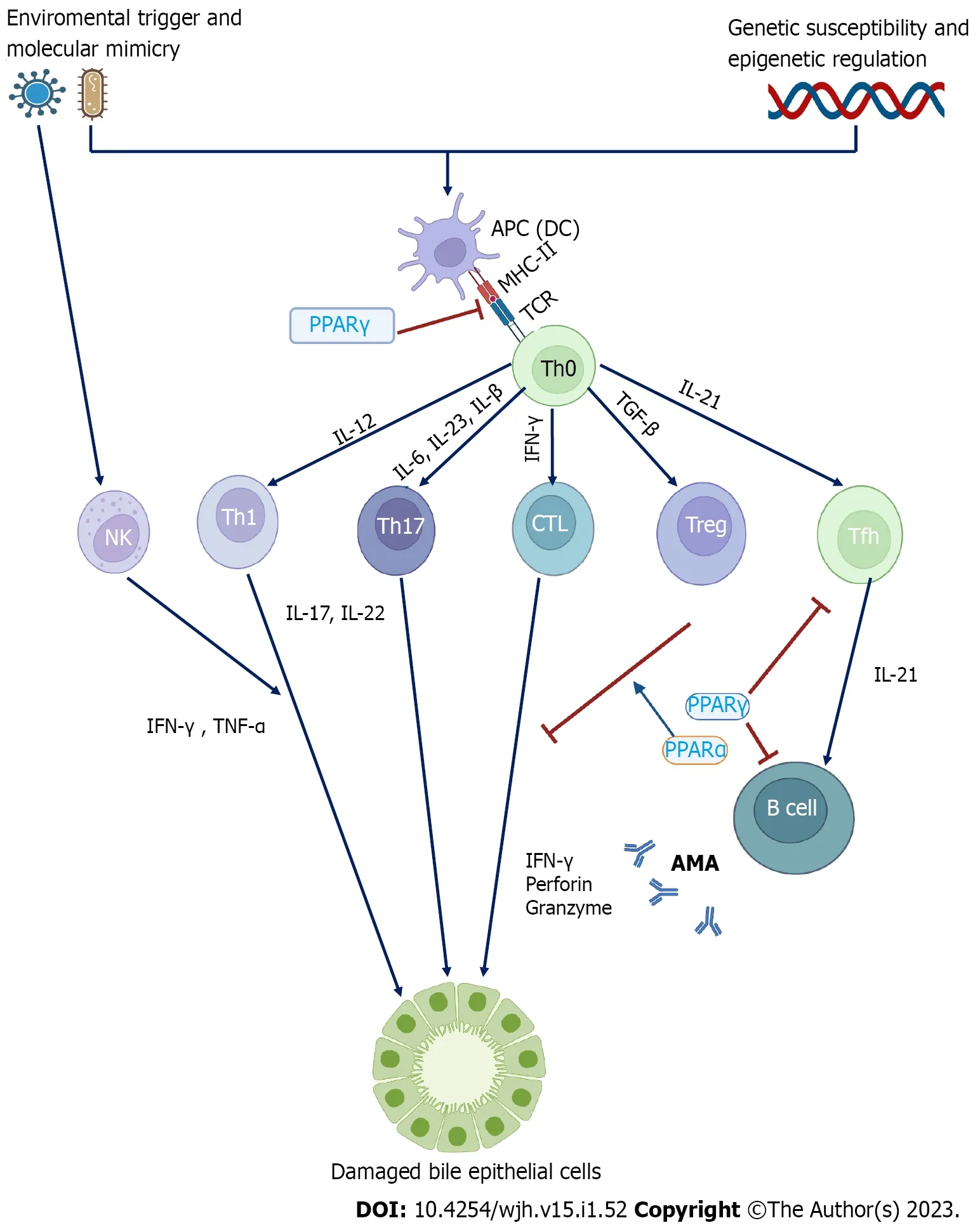
Figure 2 Model of pathogenic mechanisms in primary biliary cholangitis.
Autoimmune hepatitis
AIH is an autoimmune chronic inflammatory liver disease characterized by the presence of multiple autoantibodies,elevated serum aminotransferase levels,and excessive hepatic lymphoplasmacytic infiltration.However,the exact pathogenesis of AIH remains unclear.Although autoantibody positivity is asine qua nonof AIH,T cells rather than B cells are the major mediators of AIH immunopathogenesis.Current evidence suggests that T cells are immune regulators,and multiple autoantibodies are also important participants[69].
The frequency of infiltrating CD4+T cells is histopathologically higher than that of CD8+T cells in the early stages of AIH.Spontaneous apoptosis of CD4+T cells is markedly reduced in AIH[70].The ratio of liver CD8+/CD4+T cells (Tc/Th) increases with disease activity in patients with AIH.CXCR3 and CCR6 are highly expressed in CD8+T-cells.This shows that the ligands CXCL9 and CCL20 are highly expressed in the inflamed liver,thus facilitating the uptake of CD8+T cells into the liver[71].Emperipolesis is defined as the presence of an intact,viable cell (lymphocyte) within the cytoplasm of another cell (hepatocyte),and is one of the histopathological and diagnostic features of AIH.Emperipolesis is predominantly mediated by CD8+T cells and is correlated with severe necroinflammation and fibrosis[71].
Every morning, when she saw me come, she liked the Chinese children seen their parents come home from shopping. They so excited, because their parents have bought their favourites.
Different subsets of CD4+T (Th) cells,particularly Treg cells,have been found to exert remarkable effects in AIH.Treg cells in patients with AIH suppress autoimmunity by direct contact with CD4+CD25-T cells and secretion of regulatory cytokines,such as IL-4,IL-10,and TGF-β[72].Treg cells mediate immune suppression through the expression of CD39 and CD73.Treg cells in AIH exhibit reduced NTPDase-1 activity as well as a reduced ability to inhibit IL-17 secretion from Th17 cells in AIH,which contributes to autoimmunity.Circulating and intrahepatic IL-17 Levels were significantly higher in AIH patients than in healthy controls.Hepatic expression of IL-17 is associated with inflammation and fibrosis in the liver[73].Studies have shown that the interaction between Gal-9 on Treg cells and Tim-3 on Th cells may be an important mechanism for direct contact suppression mediated by Treg cells.Although some studies have reported a decrease in the number of Treg cells in AIH,others have shown that Treg cells do not decrease in AIH[74,75].These results suggest that the role of Treg cells in AIH immunopathology remains controversial.
In addition to Treg cells,Thf cells are associated with adaptive cell immunity in AIH.CD8 T cells have been shown to be activated by IL-21,secreted by Tfh cells.Tfh cells are widely recognized as a subset of CD4+T cells that aid in B-cell development[76].The number of T γδ cells was increased in patients with AIH.T γδ cells secrete higher levels of IFN-γ and granzyme B than healthy controls,which may contribute to autoimmune damage in AIH patients.
Studies have shown that B cells inhibit CD4+T cells in animal models of AIH.Its suppressive function is dependent on the expression of CD11b in B cells.IL-10 is mainly secreted by CD4+T cells and increases CD11b expression.This means that CD4+T cells and B cells can regulate each other in AIH[77].The possible immune cells and mediator cytokines involved in the autoimmune hepatitis pathogenic pathway are shown in Figure 3.
AUTOIMMUNITY AND IEI
With a simplistic approach,autoimmunity and IEI can be thought of as “over” and “insufficient”functioning of the immune system,respectively.In other words,autoimmunity and IEI might be accepted as opposites in the spectrum of immune system functioning.However,with the accumulation of knowledge and experience in both disease groups,this simple distinction disappeared,and it was revealed that the immune system was “dysregulated” in both groups.
The coexistence of autoimmunity and IEI is a well-known entity[78].An analysis conducted in France showed that 26.2% of patients with IEI had one or more autoimmune or autoinflammatory symptoms during their lifetime[79].In a two-center prevalence study in Turkey including 1435 patients with IEI,autoimmunity was reported at a rate of 2.2%[80],although antibody deficiencies take the first place among immunodeficiencies.According to this study,the most common type of immunodeficiency associated with autoimmune diseases is CVID,and the most common accompanying autoimmune diseases include vasculitis,autoimmune hemolytic anemia,and autoimmune thrombocytopenia.In a national data-based study conducted in France,Fischeret al[79] found that autoimmunity is mostly associated with T cell-related diseases and CVID.The cumulative incidence graph of lifelong autoimmune development in patients with IEI increased almost linearly after 8-10 years of age,and 40%of patients developed autoimmune disease by the age of 50 years.The most common accompanying autoimmune diseases were cytopenia and gastrointestinal,skin,rheumatological,and endocrine diseases.Therefore,it is important for all physicians dealing with autoimmune diseases or immunodeficiencies to keep in mind that various autoimmune diseases can accompany almost all types of IEI syndrome,either as the first finding or during their course.
Pathophysiology of autoimmunity developing on the background of IEI
It is thought that there are common genetic and pathophysiological mechanisms for IEI and autoimmune diseases based on the frequent occurrence of their association and the increased incidence of autoimmunity in the families of individuals with IEI.The leading cause of autoimmunity in IEI is loss of immune tolerance.In Autoimmune Polyendocrinopathy-Candidiasis-Ectodermal Dystrophy (APECED)and DiGeorge syndrome,T cell development and function are impaired,resulting in “loss of central tolerance”,and the development of autoreactive T cells triggers autoimmunity[81].The “peripheral tolerance loss” is lost in patients with Immune Dysregulation,Polyendocrinopathy,Enteropathy,Xlinked (IPEX) syndrome,hyper immunoglobulin-M (HIGM) syndrome,and CVID,and autoreactive B cells play a role in the emergence of autoimmunity in these patients[82].Autoimmunity can also occur with a disorder in signaling pathways in the immune system,and one of the best examples is Wiskott-Aldrich Syndrome (WAS).Loss of the WAS protein,a regulatory protein that plays a key role in signaling from TCR to the cytoskeleton in WAS,results in impaired number and function of Treg lymphocytes,which triggers autoimmunity[83].Autoimmunity may develop as a result of the failure of autoreactive lymphocytes to be cleared by apoptosis in autoimmune lymphoproliferative syndrome(ALPS) and some combined immunodeficiencies[84].Autoimmunity develops in partial IgA (PIgA)deficiency and complement disorders due to impaired antigen clearance and increased exposure to antigens[85].X-linked chronic granulomatous disease causes an abnormal immune response against cellular wastes,and this is blamed for the pathogenesis of SLE developing in one-third of female carriers of this disease[86].
Autoimmunity should also be considered as a warning sign in terms of the IEI.On the one hand,the hypogammaglobulinemic state and cellular deficiency affect the results of serology tests and biopsies,creating diagnostic difficulties for autoimmune diseases in patients with IEI.Therefore,the interpretation of diagnostic tests in these patients should be done very carefully,and even weak autoantibody positivity,which is normally ignored,should be taken into account.
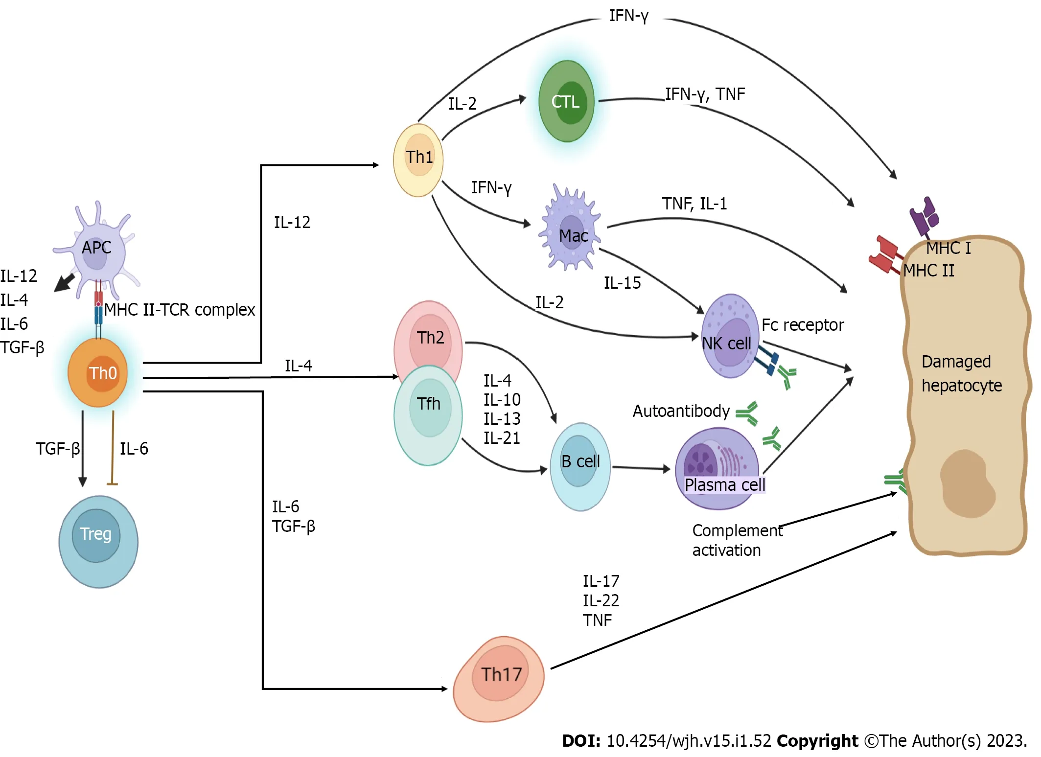
Figure 3 Pathogenic pathways of autoimmune hepatitis.
Association of AILD and IEI
APECED syndrome is an IEI characterized by the predominance of autoimmunity,and AIH can occur in up to 43% of cases[91].In MHC II disorders,autoimmunity may develop against hepatocytes and cholangiocytes in the liver[92].There is a case report of an association between mucocutaneous candidiasis and AIH in a child with a STAT-1 gain-of-function mutation[93].In a case series of 274 individuals with a STAT-1 gain-of-function mutation,AIH was reported in six (2%) patients[94].A high titer positivity for AMA autoantibodies,indicating a predisposition to the development of PBC,has been reported in a case of IPEX syndrome[95].In a series of 11 patients with hyperimmunoglobulin M syndrome,PSC developed in five (45%) patients.SinceCryptosporidium parvumwas detected in the stool of four of them,it was thought to play a role in the pathogenesis of PSC[96].In a series of 90 patients with ALPS,seronegative AIH was detected in three (3.3%) patients (83).A case report of a five-year-old boy with IL-2 receptor alpha (CD25) deficiency provided important information about the pathogenesis of AILD in IEI[97].He was diagnosed with PBC,a disease that is not normally expected to be observed in this age and sex.It was shown that he had an increase in autoreactive T cells due to a decrease in CD4+CD25+Treg cells.After allogeneic bone marrow transplantation,AMA/PDC-E2 positivity disappeared,and PBC findings improved,along with improved T cell composition.
CONCLUSION
The liver has a unique anatomical design to protect the host from potential pathogens passing from the intestine to the portal circulation,while maintaining a general state of immune hyposensitivity.The liver is the main organ of the innate and adaptive immune systems.As the mechanisms of antigen capture,presentation,and recognition in the liver will be understood,the biological mechanisms of immune tolerance in the liver will become clearer.The balance between immune tolerance and effective immune screening is maintained by interactions between numerous immune cells that are present in and recruited into the liver.This is necessary for normal functioning of the liver.If an inappropriate immune response disturbs this delicate balance,autoimmune liver pathologies can develop.In addition,failure to initiate an effective immune response results in chronic viral infections or failure to clear cancer cells.This function of the liver in maintaining immune responses and tolerance demonstrates the importance of the liver as a vital immune organ.
FOOTNOTES
Author contributions:Parlar YE and Balaban YH contributed equally in collecting the data and writing the paper;Ayar SN and Cagdas D edited the manuscript and contributed opinions on liver immunity;all authors have read and approved the final manuscript.
Conflict-of-interest statement:There are no conflicts of interest to report.
Open-Access:This article is an open-access article that was selected by an in-house editor and fully peer-reviewed by external reviewers.It is distributed in accordance with the Creative Commons Attribution NonCommercial (CC BYNC 4.0) license,which permits others to distribute,remix,adapt,build upon this work non-commercially,and license their derivative works on different terms,provided the original work is properly cited and the use is noncommercial.See: https://creativecommons.org/Licenses/by-nc/4.0/
Country/Territory of origin:Turkey
ORCID number:Yavuz Emre Parlar 0000000273498415;Sefika Nur Ayar 0000-0002-7772-0968;Deniz Cagdas 0000000322134627;Yasemin H Balaban 0000-0002-0901-9192.
S-Editor:Chen YL
L-Editor:Wang TQ
P-Editor:Chen YL
杂志排行
World Journal of Hepatology的其它文章
- Clinical characteristics and outcomes of COVID-19 in patients with autoimmune hepatitis: A population-based matched cohort study
- Influence of non-alcoholic fatty liver disease on non-variceal upper gastrointestinal bleeding: A nationwide analysis
- Rising incidence,progression and changing patterns of liver disease in Wales 1999-2019
- Prognostic role of ring finger and WD repeat domain 3 and immune cell infiltration in hepatocellular carcinoma
- Detection of colorectal adenomas using artificial intelligence models in patients with chronic hepatitis C
- Acute-on-chronic liver failure in patients with severe acute respiratory syndrome coronavirus 2 infection
