Qingchi San (青赤散) treats ulcerative colitis in mice by inhibiting the nuclear factor-kappa B signaling pathway and Nucleotidebinding oligomerization domain,leucine-rich repeat and pyrin domain-containing 3 inflammasome formation
2023-02-15ZHOUZhenghuaJIJianbinWANGHongxiaYANLinKANGHongchang
ZHOU Zhenghua,JI Jianbin,WANG Hongxia,YAN Lin,KANG Hongchang
ZHOU Zhenghua,JI Jianbin,WANG Hongxia,YAN Lin,KANG Hongchang,Department of Gastroenterology,First Teaching Hospital of Tianjin University of Traditional Chinese Medicine,Tianjin 300381,China
ZHOU Zhenghua,JI Jianbin,WANG Hongxia,YAN Lin,KANG Hongchang,National Clinical Research Center for Chinese Medicine Acupuncture and Moxibustion,Tianjin 300381,China
Abstract OBJECTIVE: To investigate the efficacy of Qingchi San(青赤散,QCS),a preparation of Traditional Chinese Medicine,on ulcerative colitis (UC)in mice by inhibiting the nuclearfactor-kappa B (NF-κB) signaling pathway and nucleotide-binding oligomerization domain,leucine-rich repeat and pyrin domain-containing 3 (NLRP3) inflammasome formation .METHODS: The UC model was established with maleC57BL/6J as the animal model.Bodyweight,Disease Activity Index (DAI),colon length and weight were detected.Furthermore,colonic histology was performed by hematoxylin-eosin (HE) staining.interleukin-1β (IL-1β),interleukin-6 (IL-6),tumor necrosis factor-α (TNF-α),myeloperoxidase (MPO) and superoxide dismutase(SOD) were performed by enzyme-linked immunosorbent assay.Cyclooxygenase 2 (COX2) and inducible nitric oxide synthase (iNOS) mRNA expression wereconducted by real-time quantitative polymerase chain reaction (RT-qPCR).NF-κB,inhibitor of NF-κBα (iκBα),Phosphorylated inhibitor of NF-κBα (p-iκBα),caspase-1,NLRP3 and Apoptosis-associated speck-like protein containing a caspase recruitment domain (ASC) protein expression were conducted by Western blotting.RESULTS: Compared with UC model group,Bodyweight was significantly increased in QCS treatment.At the same time,DAI was significantly decreased in QCStreatment.Colon length and weight and colonic histology were significantly improved in QCS treatment.Furthermore,the expression of IL-1β,IL-6,TNF-α,MPO,SOD,COX2,and iNOS were significantly decreased in QCS treatment.Finally,the expression of NF-κB signaling pathway-related proteins NF-κB,iκBα,p-iκBα,and the expression of NLRP3 inflammasome related proteins caspase-1,NLRP3 and ASC were significantly decreased in QCS treatment.CONCLUSION: Traditional Chinese drug QCS could treat UC by inhibiting the NF-κB signaling pathway and NLRP3 inflammasome formation in mice.
Keywords: colitis ulcerative;NF-kappa B;disease activity index;NLR family;pyrin domain-containing 3 protein;inflammasomes;Qingchi San
1.INTRODUCTION
Ulcerative colitis (UC),also known as chronic nonspecific ulcerative colitis,is mainly inflammatory and ulcerative lesions in the rectum,colonic mucosa and submucosa,and in serious cases can invade the whole colon and terminal ileum.1Together with Crohn disease(CD),both of these diseases are known as inflammatory bowel disease (IBD).The disease often manifests as recurrent attacks,which severely reduces the quality of life of patients and brings huge economic burdens to patients.2The disease is characterized by a relapsing remission process,which leads to considerable disability and high direct and indirect costs.3Epidemiological data showed that there was no significant gender advantage in the incidence of UC,and the incidence of UC is higher between 30 and 40 years old.4UC in many regions is also rising.In recent years,the incidence and prevalence of UC around the world have been increasing year by year.In addition,studies have also shown that immigrant children from countries with low incidence to countries with high incidence have a similar risk of ulcerative colitis as non-immigrant children.5These studies also indicate that the incidence of UC is closely related to the living environment,dietary habits,and other factors.In China,statistics show that as the level of urbanization increases,the incidence of UC in many regions is also rising.5In addition,there are significant differences in the incidence of UC in different regions worldwide,which may be closely related to the differences in the local industrialization level,economic level,and medical level.6As the specific etiology of UC is still unclear,the specific pathogenesis of UC is not well known.In general,the pathogenesis of UC is closely related to the imbalance of intestinal flora,colonic mucosal barrier damage and abnormal immune function.7So far,previous studies have shown that the interaction between the host and the intestinal flora is one of the key factors in the pathogenesis of UC.8,9Generally,the host’s innate or adaptive immunity can tolerate normal bacteria and prevent the invasion of harmful bacteria.However,the imbalance of intestinal flora will cause intestinal inflammation,because the change of dominant bacteria will also lead to a rapid increase of harmful bacteria in the intestine.The proliferation of harmful bacteria can directly invade and damage the intestinal epithelial cells,causing damage to the intestinal mucosal barrier.10
Damage of intestinal mucosal barrier will undoubtedly lead to intestinal microbial invasion,and the body can sense the presence of invaders through the process of identifying pathogen-associated molecular patterns(PAMPs).11Both pattern-recognition receptor signals activate the production of nuclear transcription factor nuclear factor-kappa B (NF-κB) and inflammatory mediators.NF-κB signaling pathway plays an important role in the development of UC and is also one of the research hotspots.12Transcription factor NF-κB is mediated by a regulatory subunit IκB kinase (IKKg) and the catalytic IκB kinase complex consisting of a subunit IκB kinase α (IKKa) and an inhibitor of NF-κB (IκB)kinase β (IKKb).When upstream kinase is activated,the IKK complex phosphorylates the NF-κB inhibitor IκBa,causing its degradation and subsequent release of RelA/p50 dimer.The RelA/p50 dimer is rapidly transferred to the nucleus to activate transcription of multiple inflammatory mediators,such as cyclooxygenase 2 (COX2),inducible nitric oxide synthase (iNOS),tumor necrosis factor-α (TNF-α),interleukin-1 (IL-1),and interleukin-6 (IL-6).13,14TNF-α is a small molecular protein secreted by mononuclear macrophages,which can improve the phagocytosis of neutrophils,promote the adhesion of neutrophils to endothelial cells,and stimulate a local inflammatory response.IL-1 and IL-6 are closely related to the pathogenesis of UC.COX2 is induced by a variety of damaging factors and participates in inflammatory responses by catalyzing prostaglandin synthesis.15Under the action of stimulating factors,iNOS induced expression is increased and a large amount of NO is synthesized,mediating the inflammatory response.Several studies have confirmed that COX2 and iNOS are closely related to the incidence of UC.14,16,17
Nucleotide-binding oligomerization domain,leucinerich repeat and pyrin domain-containing 3 (NLRP3) is mainly expressed in the cytoplasm of neutrophils,monocytes,dendritic cells,T and B lymphocytes,epithelial cells,and osteoblasts.NLRP3 and apoptosisassociated speck-like protein containing a caspase recruitment domain (ASC) and pro-caspase-1 are assembled into a large molecular multi-protein complex,namely NLRP3 inflammasome.NLRP3 inflammasome can be activated by a variety of pathogenic or injuryrelated molecular modes,which play an extremely important role in the immune function of the innate immune system.The activation process of NLRP3 inflammasome consists of two steps: firstly,TLR activation promotes the transcription and translation of various molecules of NLRP3 inflammasome through NF-κB.Then NLRP3 inflammasome activation under the action of the inflammasome activator.Inactive procaspase-1 can be transformed to active caspase-1 by NLRP3 inflammasome.Active caspase-1 not only activates the interleukin-1β (IL-1β) and interleukin-18(IL-18) molecules,IL-1β and IL-18 cause a severe inflammatory reaction of the body,eventually promote the occurrence and development of a variety of inflammatory diseases,but also caspase-1 can make special cell apoptosis,named as pyroptosis.Currently,it is believed that NLRP3 inflammasome plays an important role in the recognition of intestinal flora,maintenance of intestinal environment stability,regulation of intestinal inflammatory response,and its disorder plays a certain role in the pathogenesis of UC.Both dampness and heat and stagnation ofQican cause stasis,which further blocksQiand blood.Stagnant blood is not only the pathological product of UC but also an important pathogenic factor of UC,so the blockage of collaterals by stagnant blood runs throughout UC.In the treatment of UC,Traditional Chinese Medicine enema also follows the principle of "treating its standard when it is urgent,treating its root when it is slow".18It is advisable to treat the disease according to its location and stage.At the same time,the use of Traditional Chinese Medicine enema in addition to dialectical treatment should also be based on the extent and degree of lesions to determine whether combined with oral Chinese medicine.Similar to western medicine,UC has lower expectations for the therapeutic effect of the whole colon than Traditional Chinese Medicine enema combined with oral Chinese medicine.19Qingchi San (青赤散,QCS) is a Traditional Chinese Medicine enema prescription summarized by the Department of Spleen and Stomach of the First Affiliated Hospital of Tianjin University of Chinese Medicine for the treatment of UC on the basis of many years of clinical practice.The recipe is composed of Chishizhi(Halloysitum Rubrum),Qingdai (Indigo Naturalis),Huangbai (Cortex Phellodendri Amurensis),Luganshi(Calamina),Kushen (Radix Sophorae Flavescentis),Ercha (Catechu),Sanqifen (Radix Notoginseng Powder),and Baiji (Rhizoma Bletillae Striatae).Because it contains two drugs "Qingdai (Indigo Naturalis)" and"Chishizhi (Halloysitum Rubrum)",it is also called"Qing Chi San".QCS is mainly used with Huangbai(Cortex Phellodendri Amurensis) and Sanqifen (Radix Notoginseng Powder),Huangbai (Cortex Phellodendri Amurensis) heats dry wet with thick intestines and stomach,matrine heats dry wet check flow field.QCS has a good therapeutic effect on dysentery and blood in feces.The two drugs together can eliminate damp heat,so QCS plays an important role in treating UC and regulating and protecting gastrointestinal function.However,its specific molecular mechanism for the treatment of UC remains unknown.In order to reveal the key to solving this problem,in this study,we designed an experiment to determine the therapeutic action mode and molecular mechanism of QCS on dextran sodium sulfate(DSS)-induced UC in animal models.Interestingly,we found that QCS alleviated the UC by inhibiting the NFκB pathway and NLRP3 inflammasome formation in mice.
2.MATERIALS AND METHODS
2.1.Preparation method of QCS
QSC is composed of Chishizhi (Halloysitum Rubrum),Qingdai (Indigo Naturalis),Huangbai (Cortex Phellodendri Amurensis),Kushen (Radix Sophorae Flavescentis),Luganshi (Calamina),Ercha (Catechu),Sanqifen (Radix Notoginseng powder) and the ratio is 6∶1∶3∶3∶3∶1∶1∶1,respectively.Provided by the pharmacy of the First Affiliated Hospital of University of Chinese Medicine (Tianjin,China).First put half of the Chishizhi (Halloysitum Rubrum),Luganshi(Calamina),Huangbai (Cortex Phellodendri Amurensis),and Kushen (Radix Sophorae Flavescentis) in a 2000 mL flask,and soak the medicine in sterile water (composition∶water=1∶10 w/v) lasts for 1 h.The condensing tube is fixed on the flask with an iron stand.The condensing tube relates to a rubber tube to condense the circulating water.It is heated by a digital display temperature control heating mantle.The water is boiled and decocted for 1 h.The suspension is filtered with gauze.The Sanqifen (Radix Notoginseng powder),Ercha(Catechu) and 5 kinds of granules are mixed into a decoction,using a rotary evaporator to concentrate the medicine,and finally the liquid is autoclaved and stored at -20 ℃ until use.
2.2.Animals and groups
The male C57BL/6J mice (6-8 weeks old,22-24 g) were purchased from Beijing Vital River Laboratory Animal Technology Co.,Ltd.,and randomly divided into 6 groups: control group,UC model group,mesalazinepositive treatment group,QCS low dose (QCS-L) group,QCS medium dose (QCS-M) group and QCS high dose(QCS-H) group,with 8 mice in each group.
2.3.Mouse UC model and drugs dosage
The mice were free to drink sterile water for 0-11 d,and new sterile water was replaced every day in the control group.At the same time,sterile water was given by enema twice a day,and mice were killed on the 11th day.In the model group,sterile water was freely consumed for the mice for the first 3 d,and new sterile water was replaced every day.On the fourth day,3% DSS solution was freely consumed with mice,and new DSS solution was replaced every day.Meanwhile,sterile water was given by enema twice a day for 0-11 d,and mice were killed on the 11th day.The mice were free to drink sterile water for the first 3 d and replaced with new sterile water every day in the mesalazine positive group.On day 4,3%DSS solution was used to replace sterile water for free drinking,and new DSS solution was replaced every day.At the same time,mesalazine solution was used for enema twice a day for 0-11 d,and mice were killed on day 11.The preparation of mesalazine solution was referred to the literature,and the equivalent dose ratio of adult to mouse was 12.3,and the average dose of mice was 0.82 g·kg-1·d-1.According to the average body weight of mice of 25 g,methalazine was calculated.The average enema volume of each mouse was about 0.30 mL/d,and the enema volume was 0.15 mL each time for two times a day.In the low-dose QCS (QCS-L) group,sterile water was freely consumed for the mice for the first 3 d,and new sterile water was replaced every day.On the fourth day,3% DSS solution was freely con-sumed with mice,and new DSS solution was replaced every day.At the same time,QCS-L solution was used for enema twice a day for 0-11 d,and mice were killed on the 11th day.Convert the equivalent dose ratio of adult to mouse to 12.3,the crude drug dosage of QCS low-dose group was 19.50 g·kg-1·day-1.When the drug concentration was 2.5 g/mL,the average enema volume of each mouse was 0.2 mL/d,and the enema was given twice a day,0.1 mL each time.In the medium-dose QCS (QCS-M)group,sterile water was freely consumed for the mice for the first 3 d,and new sterile water was replaced every day.On the fourth day,3% DSS solution was freely consumed with mice,and new DSS solution was replaced every day.At the same time,QCS-M solution was used for enema twice a day for 0-11 d,and mice were killed on the 11th day.Convert the equivalent dose ratio of adult to mouse to 12.3,the crude drug dosage of QCS middle-dose group was 29.25 g·kg-1·d-1.When the drug concentration was 2.5 g/mL,the average enema volume of each mouse was 0.29 mL/d,and the enema was given twice a day,0.15 mL each time.In the highdose QCS (QCS-H) group,sterile water was freely consumed for the mice for the first 3 d,and new sterile water was replaced every day.On the fourth day,3%DSS solution was freely consumed with mice,and new DSS solution was replaced every day.At the same time,QCS-H solution was used for enema twice a day for 0-11 d,and mice were killed on the 11th day.Convert the equivalent dose ratio of adult to mouse to 12.3,the crude drug dosage of QCS low-dose group was 38.99 g·kg-1·d-1.When the drug concentration was 2.5 g/mL,the average enema volume of each mouse was 0.39 mL/d,and the enema was given twice a day,0.19 mL each time.
2.4.Enema administration treatment
Taking a prone position placed in mice,with 1 mL syringe from each group the required number of drugs,with 20 G trocar inserting from 4 cm colon,anus by trocar fixed 1 mL syringe slowly to inject liquid junction within the lumen,knead gently with the palm mice abdomen 1-2 min,so homogeneous solution contact with colonic mucosa in mice,mice and then head down about 45° inclined body,still placed 20 min,and then flat in mice.During the enema administration,the mice have continuously inhaled isoflurane,so that the mice were always in the state of anesthesia,and the liquid was kept in the colon for at least 20 min.After 20 min,the mice stopped inhaling isoflurane,and the mice regained consciousness about 30 s later.
2.5.Weight measurement of mice
Starting from the first day of the experiment,the weight of mice in each group was weighed at a fixed time every day,taking the weight of mice on the first day as the base.
2.6.Assessment of disease activity index (DAI)
Starting from the first day of the experiment,the weight,fecal traits and hematemesis of mice in each group were observed at a fixed time every day.The mice were scored according to the following criteria,and the weight,fecal traits and hematemesis scores of mice were added together,namely the DAI score of mice,to evaluate the severity of UC.The scoring criteria were as follows: (a)Weight change scoring criteria (0-4 points): mice with weight loss of 0-1% were rated as 0 point;a drop of 1%-5% is rated as 1point;a drop of 5%-10% is rated as 2 points;a drop of 10%-20% is rated 3 points;a drop of more than 20% is rated 4 points.(b) feces character score standard (0-4 points): normal feces 0 point;Soft stool 1 points;Loose bowel movement 2 points;diarrhea is 4 points.(c) blood stool scoring standard (0-4 points):normal feces 0 points;2 points of blood on the surface of feces;Severe bleeding 4 points.The average of the above 3 scores together to obtain the score of DAI.
2.7.Colonic length and weight measurement of mice
After anesthetized mice in each group were killed,the colon and anus were separated at the end of the rectum,the colon and mesentery were carefully separated with forceps to remove the complete colon tissue,and the remaining mesentery was cleaned as much as possible.Colonic length and weight were measured by electronic vernier caliper and electronic balance respectively.
2.8.Hematoxylin-eosin (HE) staining
The colon tissues were immersed in 4% paraformaldehyde for 4 h and transferred to 70% ethanol.Individual lobes of colonbiopsy material were placed in processing cassettes,dehydrated through a serial alcohol gradient,and embedded in paraffin wax blocks.Before immunostaining,5-um-thick colon tissue sections were dewaxed in xylene,rehydrated through decreasing concentrations of ethanol,and washed in PBS.And then stained with HE.After staining,sections were dehydrated through increasingconcentrations of ethanol and xylene.The pictures were obtained using a bioelectronic microscope.
2.9.RNA extraction and real-time quantitative polymerase chain reaction (RT-qPCR)
Total tissue RNA was extracted using RNA Fast kit(Solarbio,Beijing,China).The cDNA synthesis was performed by HiScript®II Reverse Kit (Vazyme,Nanjing,China).The RT-qPCR was performed using SYBR Green PCR Mix (Biosahrp,Hefei,China) on IQ5 PCR System (Biorad,Los Angeles,CA,USA).The reaction procedures: 94 ℃ for 1min,94 ℃ for 20 s,58 ℃for 30 s,72 ℃ for 30 s,with 40 cycles.The primers used are listed in Table 1.The relative expression of target genes was calculated with 2-ΔΔCt.GAPDH was used as the internal control for RT-qPCR.
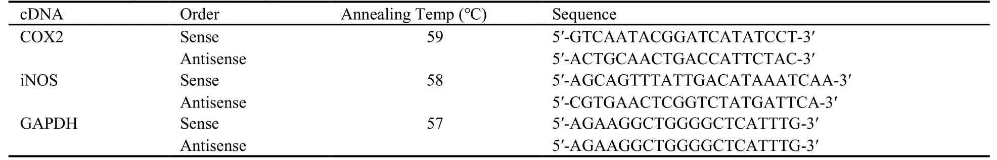
Table 1 Primer sequence used in RT-qPCR
2.10.Western blotting
The protein of colon tissues was collected and resuspended in 0.5 mL RIPA buffer (Solarbio,Beijing,China).Tissue lysates were placed on ice for 30 min.After centrifugation at 100 00gfor 20 min,the protein concentration was determined using the BCA kit(Thermo Scientific,Los Angeles,CA,USA).20 μg samples were electrophoresedviaSodium dodecyl sulfate polyacrylamide gel electrophoresis and transferred onto a PVDF membrane.Samples were blocked with 5% non-fat milk in TBST solution at room temperature for 2 h and then probed with NF-κB,inhibitor of NF-κBα (IκBα),phosphorylated inhibitor of NF-κBα (p-IκBα),caspase-1,NLRP3,apoptosisassociated speck-like protein containing a CARD (ASC)and GAPDH primary antibodies (Boster,Wuhan,China,dilution: 1 ∶2000) at 4 ℃ overnight.Secondary antibodies used for detection included HRP-conjugated anti-rabbit IgG,and the protein expression was detected using an ECL chemiluminescence detection kit (Boster,Wuhan,China).
2.11.Enzyme-linked immunosorbent assay (ELISA)assay
The mice serum samples were obtained by collecting the serum and centrifuging at 3000 rpm for 15 min before the samples were stored at-20℃ for IL-1β,IL-6,TNF-α.MPO and SOD activities in colon tissues were determined using MPO and SOD ELISA kits (Boster,Wuhan,China).
2.12.Statistical analysis
The classic scientific software GraphPad Prism 8.0 (State of New York,New York,USA) was used for data statistical analysis.The data were expressed as mean ±standard deviation.Thet-test was used for comparison between the two groups and one-way analysis of variance analysis of variance was used among the multiple groups.P <0.05 was considered statistically significant.
3.RESULTS
3.1.QCS inhibits the weight loss and reduces the DAI in UC mice
None of the six groups died during the experiment.The bodyweight of mice in the control group increased steadily.Compared with the control group,the bodyweight of mice in the UC model group decreased significantly after day 3,and the difference in bodyweight continued to increase compared with the control group.On day 10,the bodyweight of mice decreased to the lowest value,about 30% (P <0.05).Compared with the UC model group,the weight-loss trend of mice in the mesalazine positive treatment group and QCS dose groups was slower and the weight loss amplitude was significantly lower than that of the UC model group (P <0.05).In addition,the effect of QCS on the bodyweight of mice was dose-dependent,and the body weight trend of mice in the QCS-H group was close to that of the mesalazine positive treatment group (Figure 1A).Furthermore,there was no enteritis lesion in the control group,and the disease score was always 0.Compared with the control group,the enteritis score in the UC model group increased continuously from the second day after DSS treatment and reached a peak of about 2 points on day 10 (P <0.05).Compared with the UC model group,the enteritis score of the mesalazine positive treatment group and the QCS dose groups with different doses increased continuously from the second day after DSS treatment,but the scores were always lower than that of the UC model group (P <0.05).In addition,The DAI score of mice in the QCS-H group was superior to that of the mesalazine positive treatment group (Figure 1B).The results showed that QCS could significantly inhibit the weight loss and enteritis disease score of UC mice.
3.2.QCS can significantly repair colonic tissue injury in UC mice
Compared with the control group,the colonic length and weight of UC model mice were significantly shortened(P <0.05).Compared with the UC model group,colonic length and weight of mice in the mesalazine positive treatment group and QCS dose groups with different doses were significantly restored (P <0.05).In addition,the therapeutic effect of QCS was dose-dependent,the colonic length and weight of mice in the QCS-H group were close to that of the mesalazine positive group(Figure 1C,1D).The results showed that QCS could significantly alleviate the abnormal phenomenon of shortened colon length and weight loss in UC mice.
At the same time,HE staining results showed that mice colon in the blank group the layers of structure is clear,rich intestinal mucous membrane layer,rules,submucosa and muscular are sawed no obvious abnormalities and lesions.And mice UC model group local intestinal dot columnar epithelium cells necrosis,nucleus pycnosis,reduce the lamina propria adenomas disappeared,replaced by granulation tissue,accompanied by more inflammatory cells infiltration,damage and submucosa,there were also seen in the submucosa inflammatory cells.The mesalazine positive treatment group and QCS dose groups of different doses of colon tissue inflammation in mice were improved to a certain extent,is an almost complete structure,small inflammatory cell infiltration was occasionally observed in the mucosal layer,while no obvious changes or abnormalities were observed in the rest.Moreover,the colonic tissue structure of mice in the QCS-H group was superior to that of the mesalazine positive treatment group (Figure S1).In summary,QCS can obviously repair DSS-induced colon tissue damage in UC mice.
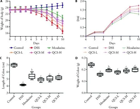
Figure 1 QCS significantly inhibited weight loss,decreased DAI score,and inhibited abnormal lesions in the colon in UC mice
3.3.QCS effectively inhibits the expression of inflammatory factors
Compared with the control group,the expression of inflammatory factors TNF-α,IL-1β and IL-6 in the UC model group were significantly up-regulated (P <0.05),and compared with the UC model group,the expression of inflammatory factors TNF-α,IL-1β and IL-6 were significantly inhibited in the mesalazine positive treatment group and QCS dose groups at different doses(P <0.05).In addition,the inhibitory effect of QCS was dose-dependent,and the expression of inflammatory factors TNF-α,IL-1β and IL-6 in the QCS-H group were superior to those in the mesalazine positive group (Figure 2A-2C).The results showed that QCS could effectively inhibit the expression of inflammatory factors in UC mice.
3.4.QCS reasonably regulates oxidative stress factors in UC mice
The results are similar to those of the inflammatory factors mentioned above.Compared with the control group,the mRNA expression of oxidative stress factors COX2 and iNOS and the contents of MPO and SOD in the UC model group was significantly up-regulated (P <0.05),and compared with the UC model group,the mRNA expression of oxidative stress factors COX2 and iNOS and the contents of MPO and SOD were significantly inhibited in the mesalazine positive treatment group and QCS dose groups at different doses(P <0.05).In addition,the inhibitory effect of QCS was dose-dependent,and the mRNA expression of oxidative stress factors COX2 and iNOS and the contents of MPO and SOD in the QCS-H group were superior to those in the mesalazine positive group (Figure 2D,2E,S2).The results showed that QCS could reasonably regulate oxidative stress factors in UC mice.
3.5.QCS inhibits NF-κB signaling pathway in UC mice
Compared with the Control group,the protein expression of NF-κB,iκBα,and p-iκBα significantly increased in the UC model group (P <0.05).However,compared with those in the UC model group,the NF-κB,iκBα,and p-iκBα expression levels decreased significantly in the mesalazine positive treatment group and QCS dose groups at different doses (P <0.05),especially the QCSH group (Figure 3).
Interestingly,compared with those in the UC model group,the protein expression of NF-κB signaling pathway-related pro-apoptotic factors caspase-1,NLRP3 and ASC in the mesalazine positive treatment group and QCS dose groups at different doses were also significantly reduced,as shown in Figure 4.The results showed that QCS could alleviate inflammation and tissue necrosis and apoptosis of UC by inhibiting the NF-κB signaling pathway in the UC mice.

Figure 2 ELISA assay of TNF-α,IL-1β,IL-6,MPO and SOD

Figure 3 Protein relative expression of NF-κB,IκBα and p-IκBα

Figure 4 Protein relative expression of caspase-1,NLRP3 and ASC
4.DISCUSSION
There have been reports on the use of Traditional Chinese Medicine to treat inflammatory bowel disease decades ago.20However,due to the limited technological level and experimental methods in the past,the molecular mechanism of the anti-ulcer effect of Traditional Chinese Medicine has not yet been revealed.This study is different from previous studies and used different UC model establishment methods: This study adopted the DSS water feeding method to establish a mouse acute UC model.The DSS-induced mouse UC model is an internationally recognized classic experimental animal model for UC research.21The clinical and pathological features of the model are most similar to clinical UC.22In addition,the drug composition ratio of QCS used in this study was significantly different from that of Traditional Chinese Medicine.Moreover,given the improvement of clinical symptoms of UC and the histological recovery of the colon at present,it is an important indicator to evaluate the efficacy of drug therapy for UC.23Therefore,this study comprehensively evaluated the therapeutic effect of QCS on DSS-induced UC in mice by quantitative scoring of the clinical manifestations of mice,combined with the detection of colon histopathology and physiological and biochemical parameters.Besides,revealed the mechanism of QCS in the treatment of UC in mice at the molecular level.
In some ways,UC is considered an autoimmune disease,along with rheumatoid arthritis,psoriasis,and systemic lupus erythematosus.24Existing data have shown that both in US patients and in animal models of colitis,there are significant inflammatory and immune dysfunction,which is mainly manifested by an imbalance in the secretion of pro-inflammatory cytokines.25,26Proinflammatory cytokines such as TNF-α,IL-1β and IL-6,as products of early inflammatory reaction,have a positive promoting effect in the formation of inflammation.They not only promote endothelial cell expression of adhesion molecules,to assist the neutrophil to migrate on but also can increase the ability of macrophage cells and induce the release of inflammatory mediators,to further enlarge activation and inflammatory signaling cascade,inducing the body to produce inflammation to resist heterogeneous intrusion.Proinflammatory cytokines are an important part of the body's nonspecific immune.27However,excessive secretion of pro-inflammatory cytokines such as TNF-α,IL-1β and IL-6 often causes some damage to the body.Therefore,regulating and maintaining the balance of proinflammatory cytokines and restoring the normal immune function of the body is crucial for the treatment of UC.28The experimental results showed that the serum levels of TNF-α,IL-1β and IL-6 in the model group were significantly increased,indicating that the inflammatory response in the model group was significant,which was consistent with the results previously reported.16As expected,QCS treatment significantly reduced plasma TNF-α,IL-1β and IL-6 levels in mice,indicating that QCS had significant anti-inflammatory effects and inhibited abnormal immune responses.
Studies have shown that in addition to inflammation,there is significant oxidative stress in UC patients and colitis mice.25,29In fact,oxidative stress plays a crucial role in the pathogenesis of many diseases,such as IBD,stroke,atherosclerosis and Alzheimer's disease.30,31Therefore,antioxidant treatment strategies are also of great promise in these diseases.This study confirmed the significant oxidative stress in UC mice induced by DSS through the detection of oxidative stress parameters in colonic tissues of mice.QCS intervention can significantly reduce the expression levels of COX2 and iNOS mRNA in the colon tissues of UC mice,and regulate the content of MPO and SOD,indicating that the therapeutic effect of QCS on UC is also related to its antioxidant effect.
In order to further explore the molecular mechanism of QCS,the key proteins of the NF-κB signaling pathway in mouse colon tissue were detected in this study.NF-κB signaling pathway plays an important role in the inflammatory process.32,33Studies have shown that the NF-κB signaling pathway is abnormally activated in both IBD patients and DSS-induced UC mouse models,which is closely related to the formation of UC.34,35Free NF-κB released by IκB translocated into the nucleus.Within the cell nucleus,the NF-κB with a combination of specific DNA sequences.Then,many genes encoding inflammatory response protein transcription of the cell,such as TNF-α,IL-1β and IL-6,to promote inflammation and the deterioration of UC.36,37In our study,compared with the control group,the expression levels of NF-κB,IκBα and p-IκBα were significantly increased in the UC model group.This illustrates that the UC model group significantly activates NF-κB signaling pathways.And the QCS treatment group can significantly reduce the expression levels of these proteins and has significant anti-inflammatory effects,which is consistent with the previous histological analysis of the QCS treatment group for UC treatment.Therefore,we believe that QCS treatment for UC is through the inhibition of NF-κB signaling pathway.
The innate immune system of the intestine is the first line of defense against various bacterial antigens in the body's immunity.38The weakened innate immune system of UC patients causes the accumulation of bacterial antigens,which then stimulates the waterfall inflammatory response of the acquired immune system.NLRP3 inflammasome plays a role in the relationship between the immune system and intestinal flora.NLRP3 inflammasome can maintain intestinal homeostasis and has a protective effect on experimental colitis,and its functional deficiency may lead to susceptibility to UC.NLRP3 inflammasome plays a pro-inflammatory role in the pathogenesis of colitis.Treatment of mice with FC11a-2,an inhibitor of pro-caspase-1,IL-1β and IL-18 that is dependent on NLRP3,can alleviate UC.In addition,NLRP3 inflammasomes were found to have effects on TNF-α,IFN-γ,IL-10,TGF-β,interferoninduced protein,myeloperoxidase,β-antimicrobial and intestinal flora.NLRP3 inflammasome is highly expressed not only in the colon tissues of acute and chronic colitis mice but also in the colon tissues of ulcerated lesions of UC patients.Results from human and experimental animals indicate that NLRP3 inflammasome can initiate and promote the inflammatory response of the colon.In our study,compared with the control group,the expression levels of caspase-1,NLRP3 and ASC were significantly increased in the UC model group,illustrating the UC model group significantly activates NLRP3 inflammasome formation and pyroptosis,and the QCS treatment group can significantly reduce the expression levels of these proteins and has significant antiinflammatory effects,which is consistent with the analysis of NF-κB signaling pathway of the QCS treatment group for UC treatment.Therefore,we believe that the core molecular mechanism of QCS treatment for UC is through the inhibition of NLRP3 inflammasome formation.
In conclusion,this study showed that QCS had a good therapeutic effect on DSS-induced UC in mice,and the mechanism of the therapeutic effect was related to the inhibition of the NF-κB signaling pathway.Moreover,the efficacy of high-dose QCS in the treatment of UC was superior to that of standard-dose mesalazine.Meanwhile,we propose to conduct clinical trials of QCS to evaluate the efficacy and safety of high-dose QCS in the treatment of patients with UC,to provide the oretical guidance and clinical evidence for the rational clinical application of this Chinese medicine.As the core of a series of inflammatory responses,the NLRP3 inflammasome plays an important role in the pathogenesis of UC.If the expression of NLRP3 inflammasome in patients with UC can be further studied and the interaction between NLRP3 inflammasome and related inflammatory factors can be clarified,QCS is expected to become a new target for UC treatment.
杂志排行

Journal of Traditional Chinese Medicine的其它文章
- Potential of natural medicines for treatment of osteoporosis: a narrative review
- Outlook on cultural inheritance and development
- Upholding fundamental principles and breaking new ground: ensuring positive efficacy and reaching new consensus
- General principle of high-quality academic development of traditional chinese medicine: “carrying on the essence,while pursuing innovations”
- Effects of moderate-intensity aerobic exercise combined with acupuncture on attention function of mentally-retarded adolescents: a randomised controlled trial
- Reveal the mechanisms of prescriptions for liver cancer' treatment based on two illustrious senior TCM physicians