Visual perception alterations in COVID-19: a preliminary study
2023-02-11MaraBegoaCocoMartnLuisLealVegaIreneAlcocebaHerreroAinhoaMolinaMartnDoloresdeFezMaraJosLuqueCarlosDueasGutirrezJuanFranciscoArenillasLaraDavidPiero
Mara Begoa Coco-Martn, Luis Leal-Vega, Irene Alcoceba-Herrero, Ainhoa Molina-Martn,Dolores de-Fez, Mara Jos Luque, Carlos Dueas-Gutirrez, Juan Francisco Arenillas-Lara,5,David P. Piero,6
1Group of Applied Clinical Neurosciences and Advanced Data Analysis, Department of Medicine, Dermatology and Toxicology, University of Valladolid, Valladolid 47005, Spain
2Group of Optics and Visual Perception, Department of Optics,Pharmacology and Anatomy, University of Alicante, Alicante 03690, Spain
3Department of Optics, and Optometry and Vision Sciences,University of Valencia, Valencia 46100, Spain
4COVID‐19 Unit, Department of Internal Medicine, University Clinical Hospital of Valladolid, Valladolid 47003, Spain
5Stroke Unit, Department of Neurology, University Clinical Hospital of Valladolid, Valladolid 47003, Spain
6Clinical Optometry Unit, Department of Ophthalmology,Vithas Medimar International Hospital, Alicante 03016,Spain
Abstract
● KEYWORDS: SARS-CoV-2; COVID-19; visual perception;contrast sensitivity function; color vision
INTRODUCTION
Severe acute respiratory syndrome coronavirus 2 (SARS‐CoV‐2) is the microbiological agent responsible for the coronavirus disease 2019 (COVID‐19) that has caused an unprecedented social and health crisis worldwide.Since the onset of the COVID‐19 pandemic, several papers have been published reporting on the clinical features of COVID‐19, but not much has been published with respect to ocular manifestations and their pathogenesis.
The most common ocular manifestation reported so far is conjunctival congestion (0.8%) due to direct infection of the ocular surface[1‐3], followed by a few cases of retinal vein occlusions presumably due to COVID‐19‐induced hypercoagulable state[4‐6]and other ocular manifestations such as dry eye, blurred vision, foreign body sensation, chemosis,epiphora, photophobia, and eye movement abnormalities[7‐12].
Reduced visual acuity (VA), visual field defects, and decreased retinal sensitivity have also been highlighted in various reports[13‐15], which seem to be related to the infectious capacity of SARS‐CoV‐2 and its unknown effect on the optic nerve and higher structures involved in the vision process[16]. However,despite this evidence, visual disturbances resulting from visual pathway impairment in these patients go unnoticed, as they are not life‐threatening conditions in view of the severity of other systemic signs and symptoms.
Currently, the prevalence of such visual disturbances in patients with COVID‐19 remains unknown, and due to the lack of visual assessment of these patients, the consequences of these disturbances are unpredictable.
Impairment of visual pathways may lead to alterations in visual perception. The contrast sensitivity function (CSF) is a variable that indicates the amount of contrast that a visual stimulus must have to be perceived, making it possible to detect whether there is a loss of information along the visual pathways and, depending on the type of stimuli for which this loss occurs, in which information channel the damage is located[17].
Another indirect consequence of visual pathway involvement is impaired color vision[18], defined as the ability to detect and differentiate stimuli with variations in tone.
For this reason, we set out to assess visual perception based on monocular measurement of color and chromatic‐achromatic contrast vision to inform on possible neurological involvement in patients with acute COVID‐19 in a rapid, simple and non‐invasive way, thus providing new data on the visual consequences of COVID‐19.
SUBJECTS AND METHODS
Ethical ApprovalThis study followed the principles of the Declaration of Helsinki and its protocol was approved by the Valladolid East Health Area Research Ethics Committee on 18 February 2021 (PI 21‐2177). In addition, all participants signed an informed consent form prior to the collection of their personal or clinical data.
Study DesignThis is a prospective cohort study to characterize the color and chromatic‐achromatic contrast vision of patients with COVID‐19 using a rapid, non‐invasive method (Optopad test) on a portable electronic display device(Apple iPad 6thGen A1893), comparing the results obtained with those of a sample of healthy controls matched for sex and age.
ParticipantsA convenience sampling of all patients admitted to the COVID‐19 Unit of the University Clinical Hospital of Valladolid during March 2021 was performed. Patients aged 16–65y with a confirmed diagnosis of COVID‐19 by polymerase chain reaction (PCR) or rapid antigen test, with near VA≥0.5 decimal, with no clinical history of ocular surgeries or eye diseases, in adequate physical or mental condition to undergo the test and who agreed to sign the informed consent form required for participation were included in the study. On the other hand, patients younger than 16y, with VA<0.5 decimal, with severe COVID‐19 or cognitive impairment and receiving antipsychotic or neuroleptic medications were excluded from the study. Due to hospital overbooking at the time and the inherent danger of handling such patients, refraction could not be assessed in the COVID‐19 group, but VA was measured with its most recent habitual correction to ensure that refractive errors did not affect test performance. A VA cut‐off of 0.5 decimal was defined as a selection criterion as this is the minimum value that does not affect the spatial frequencies assessed in the Optopad test. In the control group, the same procedure was followed.
Test DesignThe Optopad test battery allows the measurement of color (Optopad‐Color) and chromatic‐achromatic CSF(Optopad‐CSF) discrimination thresholds of the visual system using an iPad device as a display screen[19].Figure 1 shows as an example some plates of the Optopad test. The first plate is part of the Optopad‐Color test, while the remaining three plates correspond to a single spatial frequency for the measurement of achromatic and chromatic CSF(Optopad‐CSF test).
The iPad retina screen has a display resolution of 2048×1536 pixels at 267 pixels per inch, an 8‐bit resolution and a screen size of 9.7 inches, with a measured maximum screen luminance of 460 cd/m2. In order to ensure the correct reproduction of the spatial and colorimetric characteristics of the designed visual stimuli in this device[20], it has been previously characterized colorimetrically using 3DLUT method[21].
Optopad-ColorThis test contains two sets of plates: protan‐deutan‐tritan (PDT) plate, with stimuli along the cone‐isolating directions to detect congenital protan, deutan and tritan defects;and red‐green blue‐yellow (RG‐BY) plate, with stimuli along the opponent red‐green and blue‐yellow directions to detect acquired color defects.

Figure 1 Examples of some plates of the Optopad-Color and Optopad-CSF tests PDT plate (A), achromatic CSF 1.5 cpd (B),red-green CSF 1 cpd (C), blue-yellow CSF 1 cpd (D). CSF: Contrast sensitivity function; PDT: Protan-deutan-tritan.
PDT plate consists in a 3×10 grid of randomly oriented colored Landolt rings against a 60 cd/m2square achromatic surround subtending 1.7°, in an achromatic background with the device’s maximum generable luminance. Each row (protan: P, deutan:D and tritan: T) contains a decreasing color contrast series of stimuli isolating a single cone response in Smith and Pokorny cone space[22]. The luminance of surround and background has been chosen to reduce the sensitivity of the achromatic mechanism[23].
The chromaticity of the optotype on the RG‐BY plate is modulated along the cardinal directions of the opposite modulation Derrington‐Krauskopf‐Lennie (DKL) color space[24].The plate contains 4 rows: rows R and G, explore, respectively,the positive and negative directions of the red‐green axis,and rows Y and B, the positive and negative directions of the cardinal yellow‐blue axis. For more details, see de Fezet al[19].
Optopad-Contrast Sensitivity FunctionThe design of this test explores spatial CSF in the achromatic and chromatic mechanisms. The test contains plates for the measurement of the achromatic (CSF‐A), red‐green (CSF‐T) and blue‐yellow (CSF‐D) spatial contrast sensitivity along the cardinal directions of the DKL space. Each CSF is measured by means of 5 plates, one for each spatial frequency evaluated. Each plate contains a decreasing achromatic or chromatic contrast series of sinusoidal gratings in 2‐degree circular windows,arranged in a 4×4 grid against an achromatic background with the device’s maximum generable luminance.
The spatial frequencies are 1.5, 3, 6, 12, and 24 cycles per degree (cpd) for the achromatic CSF and 1, 2, 4, 8 y 12 cpd for the two chromatic CSFs. Grating orientation is randomly chosen among 3 possibilities (-15°, 0°, and 15°). Grating surround is, again, the device’s achromatic stimulus with 60 cd/m2. To minimize intrusions of the achromatic mechanism,chromatic gratings include random achromatic noise.
Both the Optopad‐Color and the Optopad‐CSF tests assess the patient’s discrimination thresholds. In other words, these tests are used to determine the minimum contrast (color contrast or chromatic‐achromatic contrast) needed to see the stimuli.For simplicity, these parameters will be referred to as color threshold and contrast threshold, respectively. The lower the threshold measured, the better the detection ability of the patient. In turn, sensitivity can be calculated as the inverse of the value of the discrimination threshold. Therefore, the lower the threshold, the better the discrimination ability and,consequently, the better the sensitivity.
Measurement ProcedureDuring the Optopad‐Color test,patients were asked to indicate the direction of the opening of the Landolt Cs in each row of each plate, while during the Optopad‐CSF test, the patient had to indicate the direction of the sinusoidal grating in the different rows of each plate.In both tests, the patient started with the first, most visible stimulus and carried on until the first error. The discrimination threshold was calculated as the mean between the first error and the last hit.
All measurements were performed in dark conditions,monocularly and at a viewing distance of 50 cm, with the iPad device connected to the mains and the screen brightness set to maximum. The eye to be tested in each patient was randomly selected and its near VA was assessed with a standard logMAR ETDRS test chart prior to testing. In the control group, the same measurement procedure was followed in a room set out for this purpose at the Department of Medicine, Dermatology and Toxicology of the University of Valladolid. Measures were repeated after 6mo in the same eye and in the same dedicated room, following the same measurement procedure in all patients able to attend a second visit. The medical records of the patients in the control group were carefully reviewed by trained research staff to ensure that none of them suffered from COVID‐19 between the first and the second visit.
Statistical AnalysisSPSS v.22.0 for Windows (IBM,Armonk, NY, USA) was used to perform the statistical analysis of the data. First, the Kolmogorov‐Smirnov test was used to confirm that the data samples were not normally distributed.Mann‐WhitneyUtest was used to analyze the significance of differences between groups. Statistically significant differences between visits in each group were assessed using the Wilcoxon signed‐rank test. All statistical tests were 2‐tailed, andP‐values less than 0.05 were considered statistically significant. Contrast and color threshold results from Optopad‐CSF (A: achromatic,T: red‐green; D: blue‐yellow contrast threshold) and Optopad‐Color (color threshold along Protan, Deutan, Tritan directions and RG‐BY color mechanisms) were obtained and analyzed as outcomes. Mean and standard deviation were used to represent color and contrast threshold results. CSF, obtained as 1/contrast threshold, was calculated for explanatory purposes line with the conventional clinical representation.
RESULTS
A total of 25 patients (9 females and 16 males) with COVID‐19 who were hospitalized for this cause in the COVID‐19 Unit of the University Clinical Hospital of Valladolid were included.Mean hospitalization time was 9.36±7.93d (from 2 to 31d).Mean time from the diagnosis (based on PCR or rapid antigen test date) until the performance of the visual examination was 14±12d (from 1 to 42d). Mean age in the COVID‐19 group was 54±10y (from 27 to 64y). Control group consisted of 16 healthy subjects (5 women and 11 men) with a mean age of 50±6y (from 44 to 62y). There were no statistically significant differences in age between groups (P=0.11). Mean VA was 0.78±0.18 decimal (from 0.50 to 1) and 0.91±0.13 decimal (from 0.63 to 1) in the COVID‐19 and control groups,respectively. There were statistically significant differences between groups in VA (P=0.02), with slightly higher values in the control group. Follow‐up at 6mo after inclusion could be performed in 75.6% of the participants (31/41): 64% (16/25) in the COVID‐19 group and 94% (15/16) in the control group.
Optopad-Contrast Sensitivity FunctionResults obtained with the achromatic Optopad‐CSF test (CSF‐A) in both study groups at baseline and at 6mo are summarized in Table 1.The achromatic contrast threshold results in the COVID‐19 group were significantly higher (i.e., worse sensitivity) than those obtained in the control group for all spatial frequencies studied (P<0.01 in all cases), being 1.6 times worse for 1.5 cpd, 1.5 times worse for 3 cpd, 2.7 times worse for 6 cpd,3.3 times worse for 12 cpd, and 2.7 times worse for 24 cpd. At 6mo, the contrast threshold results obtained in the COVID‐19 group remained significantly higher (i.e., worse sensitivity)than those obtained in the control group for almost all spatial frequencies studied (P<0.02), with the exception of 1.5 cpd(P=0.22). To summarize, the achromatic CSF values (CSF‐A)at baseline and at 6mo are plotted in Figure 2.
Results obtained with the chromatic Optopad‐CSF test for the red‐green mechanism (CSF‐T) in both study groups at baseline and at 6mo are summarized in Table 2. The chromatic contrast threshold results for the red‐green mechanism in the COVID‐19 group were significantly higher (i.e., worse sensitivity) than those obtained in the control group for all the spatial frequencies studied (P<0.02 in all cases), being 2.7 times worse for 1 cpd, 2.3 times worse for 2 cpd, 1.5 times worse for 4 cpd, 1.7 times worse for 6 cpd, and 1.7 times worse for 12 cpd. At 6mo, the contrast threshold results obtained in the COVID‐19 group remained significantly higher (i.e.,worse sensitivity) than those obtained in the control group for all spatial frequencies studied (P<0.01). To summarize, the chromatic CSF values for the red‐green mechanism (CSF‐T)obtained in each study group (Figure 3).
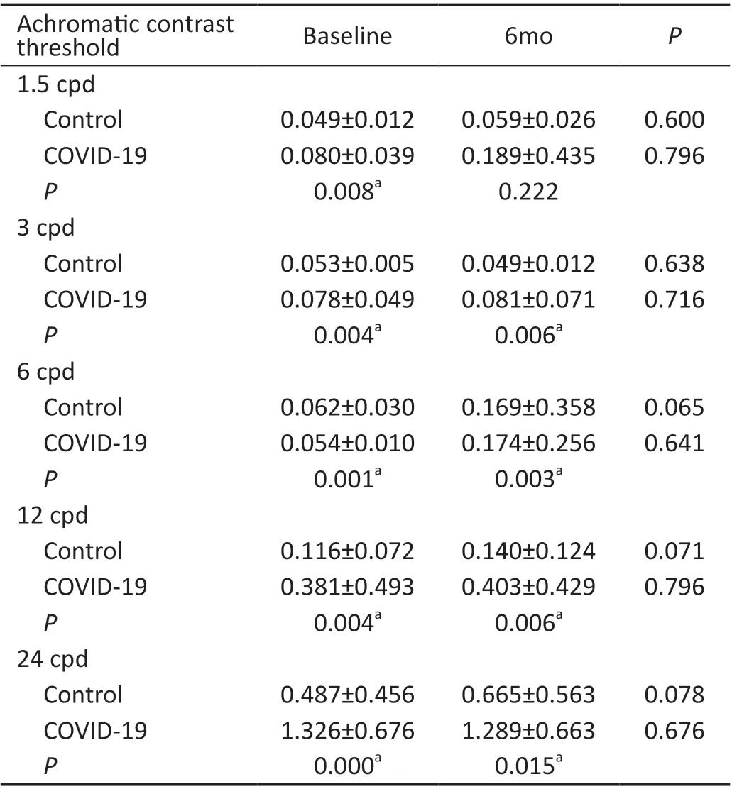
Table 1 Mean achromatic contrast threshold±standard deviation obtained in each study group for the spatial frequencies studied at baseline and at 6mo
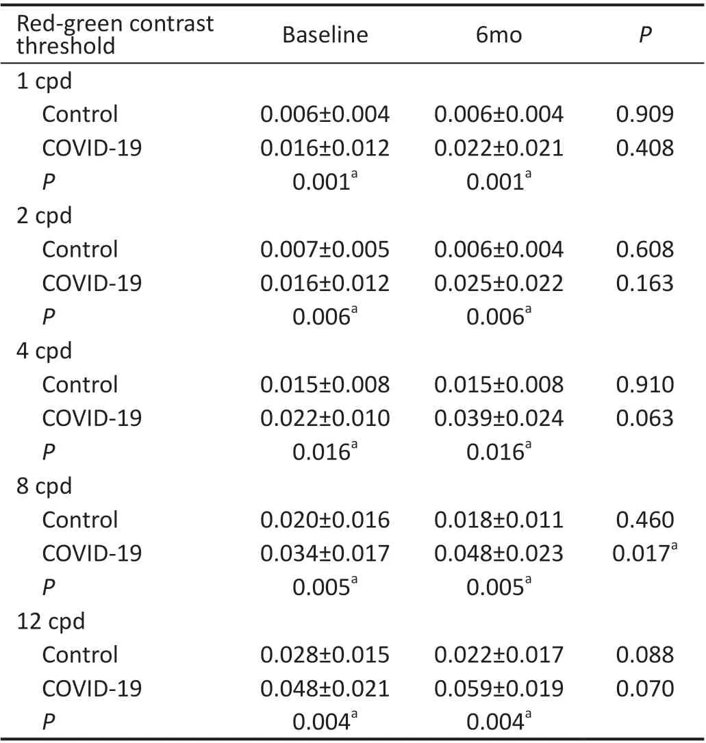
Table 2 Mean contrast threshold±standard deviation for the chromatic red-green mechanism obtained in each study group for the spatial frequencies studied at baseline and at 6mo

Figure 2 CSF (inverse of contrast threshold) values by frequencies for the achromatic mechanism (CSF-A) obtained in each study group Results obtained at baseline are represented by bars, while results obtained at 6mo are represented by lines. aBaseline visit, statistically significant; b6-month visit, statistically significant. CSF: Contrast sensitivity function; CSF-A: Achromatic contrast sensitivity function.
Results obtained with the chromatic Optopad‐CSF test for the blue‐yellow mechanism (CSF‐D) in both study groups at baseline and at 6mo are summarized in Table 3. The chromatic contrast threshold results for the blue‐yellow mechanism in the COVID‐19 group were higher (i.e., worse sensitivity) than those obtained in the control group for all spatial frequencies studied, being 2.1 times worse for 1 cpd, 2.1 times worse for 2 cpd, 1.1 times worse for 4 cpd, 1.2 times worse for 6 cpd, and 1.2 times worse for 12 cpd. These differences were statistically significant for almost all spatial frequencies studied (P<0.04),with the exception of 4 cpd (P=0.277). At 6mo, the contrast threshold results obtained in the COVID‐19 group remained slightly higher (i.e., worse sensitivity) in some spatial frequencies, but without statistically significant differences compared to controls (P>0.12 in all cases). To summarize, the chromatic CSF values for the blue‐yellow mechanism (CSF‐D)at baseline and at 6mo are plotted in Figure 4.
Optopad-ColorResults obtained with the Optopad‐Color test in both study groups at baseline and at 6mo are summarized in Table 4. Color threshold results in the COVID‐19 group were higher (i.e., worse sensitivity) than those obtained in the control group for all chromatic mechanisms studied, being 2.3 times worse for P direction, 2.0 times worse for D, 3.1 times worse for T, 2.3 times for R cardinal direction, 1.9 times worse for G; 2.7 times worse for B, and 1.7 times worse for Y. These differences were statistically significant for P, D and T channels (P<0.02 in all cases), and R and G channels(P<0.02 in both cases), but not for B and Y channels (P=0.079 and 0.234, respectively). At 6mo, the color threshold results obtained in the COVID‐19 group remained slightly higher (i.e.,worse sensitivity) for some channels than in the control group,but these differences were only statistically significant for P,D, and T channels (P<0.05 in all three cases). To summarize,color sensitivity values (inverse of color threshold) at baseline and at 6mo are plotted in Figure 5.

Figure 4 CSF (inverse of contrast threshold) values by frequencies for the chromatic blue-yellow mechanism (CSF-D) obtained in each study group Results obtained at baseline are represented by bars,while results obtained at 6mo are represented by lines. aBaseline visit, statistically significant. CSF: Contrast sensitivity function; CSF-D:Blue-yellow contrast sensitivity function.
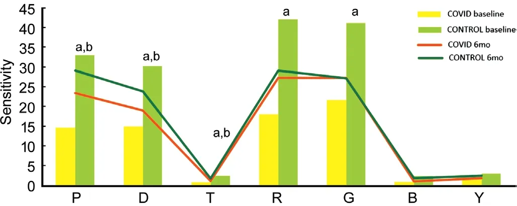
Figure 5 Color sensitivity (inverse color threshold) values obtained in each study group for P: L cone-isolating direction; D: M-isolating direction; T: S-isolating direction; R: Red cardinal direction; G: Green cardinal direction; B: Blue cardinal direction; Y: Yellow cardinal direction Results obtained at baseline are represented by bars, while results obtained at 6mo are represented by lines. aBaseline visit,statistically significant; b6-month visit, statistically significant.
Longitudinal AnalysisRegarding the Optopad‐CSF test results, in the control group, there were no statistically significant differences between visits for achromatic contrast threshold results at 1.5, 3, 6, 12, and 24 cpd (P≥0.065).Additionally, there were no statistically significant differences between visits for chromatic contrast threshold results at 1, 2, 4, 8, and 12 cpd (P≥0.088). On the other hand, in the COVID‐19 group, there were no statistically significant differences between visits for achromatic contrast threshold results at 1, 3, 6, 12, and 24 cpd (P≥0.641). Additionally, there were no statistically significant differences between visits for chromatic contrast threshold results (red‐green and blue‐yellow,respectively) with some exceptions: 1 cpd (P=0.408 and 0.013), 2 cpd (P=0.163 and 0.063), 4 cpd (P=0.063 and 0.214),8 cpd (P=0.017 and 0.499), and 12 cpd (P=0.070 and 0.958).Regarding the Optopad‐Color test results, in the control group,there were no statistically significant differences betweenvisits for color threshold results for the P, D, T, R, G, B, and Y channels (P≥0.256). On the other hand, in the COVID‐19 group, there were no statistically significant differences between visits for color threshold results for the P, D, T, R, G,B, and Y channels (P≥0.379).
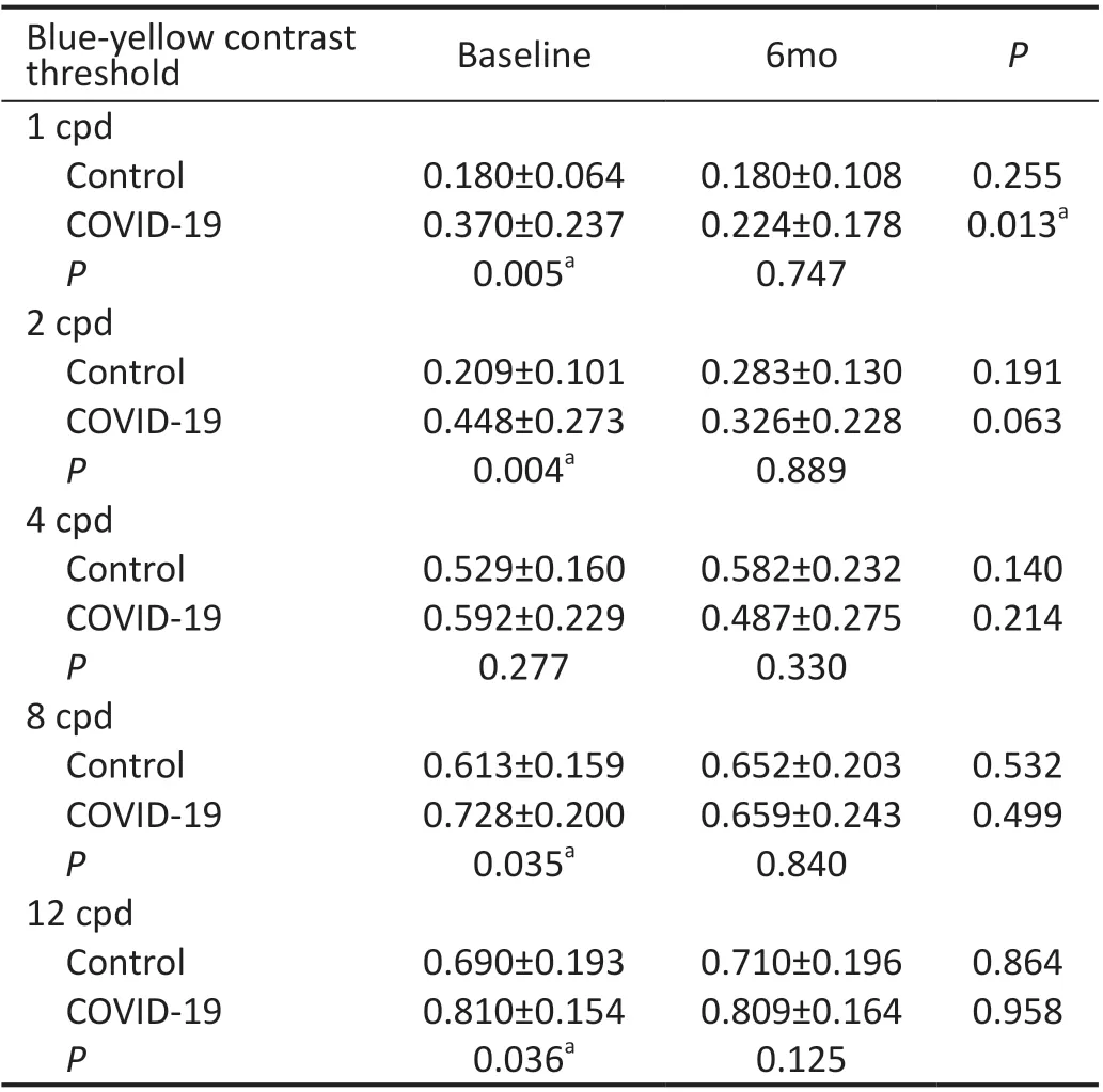
Table 3 Mean contrast threshold±standard deviation for the chromatic blue-yellow mechanism obtained in each study group for the spatial frequencies studied at baseline and at 6mo
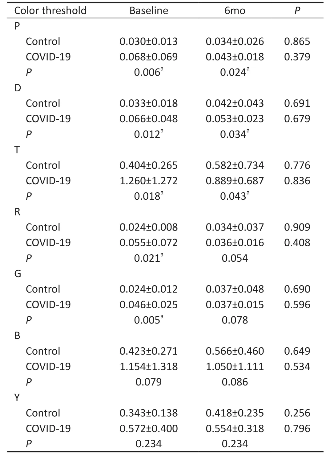
Table 4 Mean color threshold±standard deviation for the different channels obtained in each study group at baseline and at 6mo.
DISCUSSION
Although there are several studies providing evidence on the impact of COVID‐19 on the eye[1‐12,25‐28], few of them focus on its impact on visual perception[13‐15]. The present study has shown that there appears to be an impairment of color and achromatic‐chromatic contrast vision in hospitalized patients with COVID‐19. These disturbances were detected using an iPad‐based test that allowed trained research staff to perform the measurements during the patient’s hospitalization while maintaining all required safety measures[29‐30].
The visual disturbances found in COVID‐19 patients may be due to retinal diseases, ocular surface problems or neurological disorders affecting the visual pathway. Unfortunately, the presence of these associated problems could not be confirmed due to limitations in the management of these patients in our centre (they could not be transferred from one unit to another unless it was extremely necessary to avoid the spread of infection). In any case, none of the patients evaluated reported a significant or limiting vision loss requiring urgent ophthalmological examination. To partially overcome this limitation, the results obtained in the COVID‐19 group were compared with those obtained in another group of healthy patients of similar age in whom there were no pathological problems. This comparison revealed the presence of statistically significant differences for almost all color and contrast thresholds measured for different spatial frequencies,with worse results in the COVID‐19 group. Another possible factor that may have contributed to these results is the ocular irritation and dryness that the patient may experience as a consequence of wearing the surgery mask during the hospital stay.
Ocular surface problems in patients with COVID‐19 are well reported in the literature[1‐3]and may result in reduced CSF and color vision due to the dryness, tearing, or blurred vision that they can cause. Similarly, different reports have highlighted retinal problems associated with COVID‐19 that may lead to reduced VA, such as neuroretinopathy, central retinal vein occlusion, disc edema or retinitis[4‐6,14,31‐36]. All these studies report uncorrected and distance‐corrected VA loss, but none of them specifically describe a reduction in CSF or any assessment of visual resolution In this sense, it is worth noting that patients with neuroretinopathy have a markedly reduced CSF[37].
Likewise, optic neuritis, which is a condition that has been shown to significantly degrade CSF[38], has also been reported to be associated with COVID‐19[39‐41]. Specifically,optic neuritis results in large losses of sensitivity trough the magnocellular pathway and losses of contrast gain in the inferred parvocellular pathway[38]. Therefore, the worse achromatic and chromatic CSF pattern in patients with COVID‐19 compared to healthy patients of similar age may be due to the possible neuroretinopathy and optic neuritis that can be found in these patients. However, studies evaluating retinal structure in patients with COVID‐19 have shown limited changes, with no signs of active inflammation or optic nerve involvement[42‐43]. Turkeret al[42]found that the density of retinal capillary plexus vessels was reduced in COVID‐19 patients, posing a risk of retinal vascular complications.
In addition to these problems, other types of visual disturbances have been described in patients with COVID‐19 suggesting involvement of the visual pathway and not just the retina[15,44‐45]. For example, Sharmaet al[15]described the case of a 22‐year‐old woman with an absolute lower scotoma in her right eye for 4d and a history of fever and sore throat for 10d who was eventually diagnosed with COVID‐19.Similarly, Selvarajet al[44]described the case of a patient presenting with dysosmia, dysgeusia and loss of peripheral monocular vision after being diagnosed with COVID‐19. Even an acute reduction in VA after receiving the second dose of the Pfizer‐BioNTech COVID‐10 vaccine has been reported in the literature[46]. Therefore, visual pathway disorders cannot be ruled out as a potential etiological factor for the CSF disturbances detected in the present study.
In the cohort studied, color vision alterations were also detected in COVID‐19 patients compared to age‐ and sex‐matched healthy subjects, with significantly higher color threshold results (i.e., worse sensitivity) in the COVID‐19 group for almost all channels studied, except for the blue (B)and yellow (Y) cardinal directions. To our knowledge, this is the first study reporting color vision anomalies in patients with COVID‐19. It should be noted that predominantly red‐green color vision anomalies have been described in a wide variety of retinal and neurological disorders, such as acute macular neuroretinopathy or optic neuritis[18]. However, it should be considered that retinal signals carrying color information are transmitted through the lateral geniculate nucleus of the thalamus to the primary visual cortex (V1) and processed by the secondary visual cortex (V2) and then by cells located in subcompartments within the posterior inferior temporal cortex[47]. Therefore, any alteration in any of these areas involved in the processing of color information may lead to anomalies in color vision, which is a possibility considering the potential involvement of the central nervous system by COVID‐19[48]. Further research is needed to confirm the level of alteration that COVID‐19 may have on all these structures.Lastly, no significant changes were found in the visual parameters assessed in the control group during the 6‐month follow‐up. Surprisingly, this trend was also observed in the COVID‐19 group, suggesting that the reduction in the sensitivity of color and chromatic‐achromatic contrast vision does not fully recover after the end of the disease, at least after the first 6mo. Only a significant recovery was observed in the sample tested in the following parameters: CSF‐T (red‐green)for 8 cpd and CSF‐D (blue‐yellow) for 1 cpd. This minimal trend towards improvement is consistent with studies reporting a recovery of VA loss after overcoming COVID‐19 infection,with improvements also in associated optical coherence tomography findings[32‐34]. Long term studies are needed to confirm whether full recovery of color and chromatic‐achromatic contrast vision is achieved over time.
This study has some important limitations that must be acknowledged. The main limitation is that it was not possible to perform a complete ophthalmological examination in COVID‐19 patients due to the strict protocols of management of these patients to avoid the spread of infection. In any case, no patient reported significant or limiting visual loss requiring urgent ophthalmological examination. In addition,it was necessary to include an age and sex‐matched control group to confirm that the sensitivity of color and chromatic‐achromatic contrast vision measured in patients with COVID‐19 were significantly worse than that measurable in healthy individuals of similar sex and age. Likewise, a functional magnetic resonance imaging study would also have been useful to define the actual involvement of the visual pathway disturbance detected in the present cohort of patients.The sample size used can be considered another limitation,as it did not allow us to stratify according to the degree of severity of COVID‐19. Future studies should consider this stratification to allow clinicians to characterise how visual function deteriorates with disease progression. Finally, the use of validated subjective questionnaires to analyze how patients perceive the degradation of visual function and how this affects the performance of activities of daily living (ADLs) in these patients would also have been useful.
Visual perception disturbances (color and chromatic‐achromatic contrast vision) seem to be present in patients hospitalized with COVID‐19 and may be a consequence of involvement of the ocular surface, the retina, the visual pathways, or a combination of all of these. To the best of our knowledge, these alterations are present while the infection is active and most of them remain after a period of at least 6mo.However, further research is needed to better understand how these visual disturbances occur, why they are more pronounced in some patients than in others, and whether they are sustained for periods of time longer than 6mo after infection.
ACKNOWLEDGEMENTS
Foundation:Supported by the Institute of Health Carlos III(No.COV20/00539).
Conflicts of Interest: Coco-Martín MB, None; Leal-Vega L,None;Alcoceba-Herrero I,None;Molina-Martín A,None;de-Fez D,None;Luque MJ,None;Dueñas-Gutiérrez C,None;Arenillas-Lara JF,None;Piñero DP,None.
杂志排行
International Journal of Ophthalmology的其它文章
- COVID-19 pandemic impact on ocular trauma in a tertiary hospital
- Apolipoprotein A1 suppresses the hypoxia-induced angiogenesis of human retinal endothelial cells by targeting PlGF
- Comparison of vegetable oils on the uptake of lutein and zeaxanthin by ARPE-19 cells
- Identifying a novel frameshift pathogenic variant in a Chinese family with neurofibromatosis type 1 and review of literature
- Recurrence risk factors of intravitreal ranibizumab monotherapy in retinopathy of prematurity: a retrospective study at one center
- Changes of optic nerve head microcirculation in high myopia
