Clinical implications and mechanism of histopathological growth pattern in colorectal cancer liver metastases
2022-07-30KongBTFanQSWangXMZhangZhangGL
Kong BT, Fan QS, Wang XM, Zhang Q, Zhang GL
Abstract Liver is the most common site of metastases of colorectal cancer, and liver metastases present with distinct histopathological growth patterns (HGPs),including desmoplastic, pushing and replacement HGPs and two rare HGPs.HGP is a miniature of tumor-host reaction and reflects tumor biology and pathological features as well as host immune dynamics. Many studies have revealed the association of HGPs with carcinogenesis, angiogenesis, and clinical outcomes and indicates HGP functions as bond between microscopic characteristics and clinical implications. These findings make HGP a candidate marker in risk stratification and guiding treatment decision-making, and a target of imaging observation for patient screening. Of note, it is crucial to determine the underlying mechanism shaping HGP, for instance, immune infiltration and extracellular matrix remodeling in desmoplastic HGP, and aggressive characteristics and special vascularization in replacement HGP (rHGP). We highlight the importance of aggressive features, vascularization, host immune and organ structure in formation of HGP, hence propose a novel "advance under camouflage" hypothesis to explain the formation of rHGP.
Key Words: Colorectal cancer liver metastases; Histopathological growth pattern;Desmoplastic histopathological growth pattern; Replacement histopathological growth pattern; Prognostic value; Vessel co-option
lNTRODUCTlON
Colorectal cancer (CRC) is the third most frequent tumor worldwide and the second most common in Europe. In diagnosed CRC, 20%-25% of patients are classified as stage IV and 15%-25% of those develop liver metastases[1]. For these whose metastases are confined to the liver, the only opportunity to cure is radical surgical resection. However, not all patients are fit for surgery and there is still a high rate of intrahepatic recurrence after curative resection. Therefore, the attempt on seeking for more comprehensive prognostic and stratification markers is of utmost importance. Deriving from this aim,the invasive margin as one pathological variable was selected to construct a prognostic system for rectal cancer patients[2]. There, tumor margin was classified into expanding type (pushing or well circumscribed) and infiltrating type. Based on these studies, the histopathological growth pattern (HGP)initially came into shape.
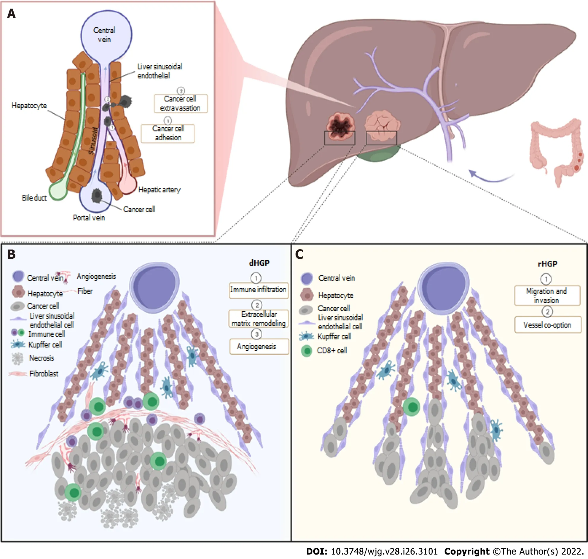
In 2016, international consensus guidelines[3] for scoring HGPs of liver metastasis were produced.Three common HGPs,i.e., desmoplastic HGP (dHGP), pushing HGP (pHGP) and replacement HGP(rHGP), and two rare HGPs,i.e., sinusoidal HGP and portal HGP are described in these consensus guidelines. In principle, HGPs could be distinguished according to the character of the invasive margin and morphology of the tumor, which is usually observed in light microscopy on standard hematoxylin and eosin (H&E)-stained tissue sections. Distinct biological and invasive patterns are presented in different HGPs. The key histopathological characteristic of dHGP is that there is a broad desmoplastic rim at the tumor periphery, which separates tumor cells from normal liver tissue. rHGP tumor cells mimic the liver architecture and replace the hepatocytes within liver plates, and the tumor displays an infiltrative border and irregular contours. pHGP tumor expands in a pushing way and the adjacent liver is compressed. Its interface is as sharp as that of dHGP but without a desmoplastic rim[3]. Figurative knowledge of dHGP and rHGP is presented in Figure 1.
PROGNOSTlC VALUE OF HGPS
Frequently, HGP predicts overall survival and recurrence in patients resected for CRC liver metastasis(CRCLM). HGPs have been extensively characterized not only in liver metastases but also in the primary cancer and metastases in lung, brain and skin; therefore, there are different categorization about HGPs in different tissues. They are ordinarily classified into dHGP and non-dHGP or expanding and infiltrative HGP[2] when used as prognostic biomarkers. In this fashion, dHGP refers to pure dHGP with a 100% desmoplastic interface on every section, while non-dHGP actually includes pushing,replacement and a mixed (pushing-replacement) pattern. As the major form in most cases, rHGP represents non-dHGPs and infiltrative HGP. One of the most important findings from studies on predictive value of HGP is that CRCLM patients with dHGP, especially pure dHGP, are prone to longer overall survival. dHGP is verified as an independent factor for superior overall survival (OS) and progression-free survival (PFS)[4-8], while non-dHGPs are strong prognostic indicators of worse survival[8-14]. More than in CRCLM, a similar trend has been observed in liver metastases from cutaneous melanoma[15], which implies that the prognostic value of HGP is generally applied in various liver metastases.
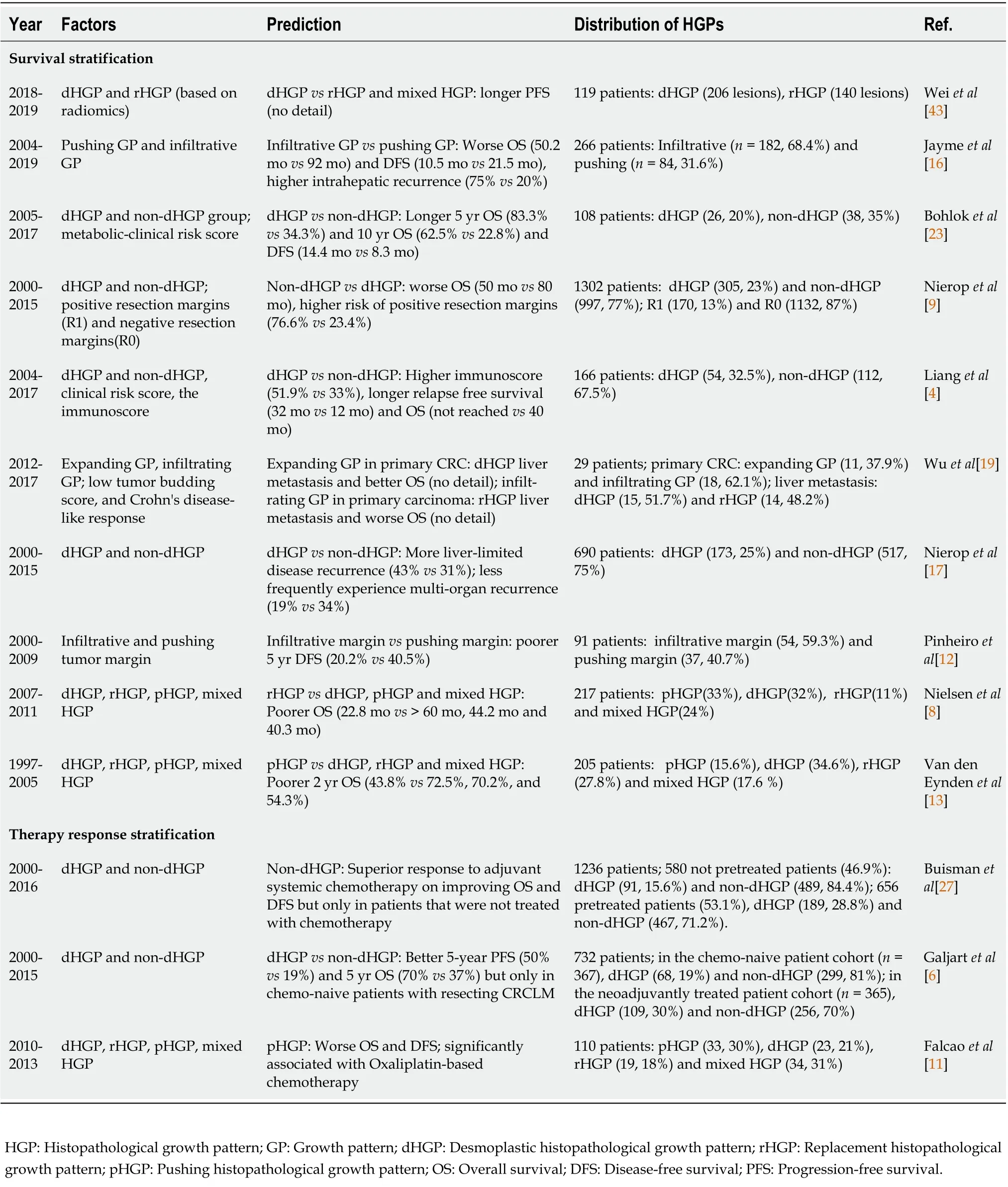
For CRCLM patients who have undergone hepatectomy, high recurrence rate may cause repeat resection and impair their survival. Non-dHGP might be responsible for this damage from intrahepatic as well as overall recurrences[12]. Compared with dHGP, rHGP and pHGP more frequently experience multiorgan recurrence, while dHGP has more liver-limited disease recurrence[16-18]. Evidence supports that dHGP predicts good outcomes, but the infiltrative pattern or rHGP indicates poorer outcomes and higher recurrence. In this context, we provide an overview of studies focusing on prognostic and stratification value of HGPs in Table 1.

Figure 1 Formation and mechanism of desmoplastic histopathological growth pattern and replacement histopathological growth pattern.A: Cancer cells originating from colorectal cancer arrive in the liver via portal vein, adhere to lumen of liver sinusoid and migrate with extravasation through fenestrae on liver sinusoidal endothelial cells (LSECs); B: There is a desmoplastic rim in interface of tumor with desmoplastic histopathological growth pattern, with tumor cells destroying liver plate, causing immune infiltration and extracellular matrix remodeling induced by activated fibroblasts and deposited fiber. Both angiogenesis and necrosis are presented in the tumor; C: In replacement histopathological growth pattern, tumor cells with highly migration and invasion replace hepatocytes and coopt LSECs but without disturbing the liver structure and extensive immune infiltration. dHGP: Desmoplastic histopathological growth pattern; rHGP: Replacement histopathological growth pattern.
Apart from predicting survival, the growth pattern of primary CRC was also found to predict the HGP of liver metastases. The primary CRCs with expanding growth pattern significantly tend to form dHGP liver metastasis, while CRC patients presenting with infiltrating growth pattern are more likely to have rHGP liver metastasis[19]. It seems that some invasive characteristics are inherited from primary tumor to secondary metastases. However, there are few reports about this. Wuet al[19] found several specific gene mutations, among which representative gene mutationPIK3CAappearing in 40% of primary CRC patients with dHGP liver metastasis was speculated to mediate vascular development and angiogenesis through the vascular endothelial growth factor (VEGF) signaling pathway, further supporting dHGP. Nevertheless, these findings have not revealed the genomic correlation between growth pattern in primary CRC and HGP in liver metastases.
Some relevant factors such as surgical margin, immunoscore[20] and glucose uptake[21,22] were also widely investigated as predictor in outcomes of CRC. The combination of HGP with resection margin,parameters of immune status, genes and metabolism also paves the way for creating comprehensive and accurate predictive models. Surgical margin in tumorectomy is a critical but controversial issue asbiologic factors driving margin-based differences may lead to the need for larger surgical margins,which indicate less chance of residual tumor cells and recurrence. Nieropet al[9] found that patients with non-dHGP are at higher risk of positive resection margins. It seems to be necessary to enlarge resection margin size when tumors present more aggressive borders or worse HGPs. Partly answering this question, Jaymeet al[16] found that those with infiltrative-type borders indeed presented with worse overall and disease-free survival, and had a 2.32 higher risk of hepatic recurrence than patients with pushing borders. However, a larger resection margin (> 10 mm) in patients with infiltrative borders did not affect the prognosis. It could be pointed out that what really counts is tumor biology rather than tumor size. In parallel with HGPs, high preoperative glucose uptake representing high metabolism rate is associated with poor prognosis in CRCLM[21,22]. When integrating preoperative metabolic parameters with HGPs, the postoperative prognostic value could be improved[23]. As expected, similar results appeared in studies on correlation between HGPs and immunophenotype and genomic mutation[19,24].

Table 1 Studies on stratification value of histopathological growth pattern in colorectal cancer liver metastasis
HGPS AND THERAPlES
HGP is also a useful tool to stratify patients for their response to therapy. For both resectable and unresectable patients, additional chemotherapy and angiogenesis inhibitors (AIs) are regarded as essential adjuvant therapy, however, their timing of administration and effect remains a matter of debate. Previous studies showed perioperative chemotherapy was not that beneficial as expected[25,26]. Retrospective analysis on long-term outcomes of 236 resectable CRCLMs suggested that there were no measurable differences between groups receiving adjuvant and perioperative chemotherapy[26].Whereas an interesting phenomenon is that the effectiveness of adjuvant therapies varies with different HGP subgroups. For patients who were not pretreated with chemotherapy, non-dHGP subgroup have longer OS and disease-free survival (DFS) after adjuvant chemotherapy[27]. dHGP and pHGP are more sensitive to triplets + cetuximab and triplets + bevacizumab, respectively, while rHGP has poor response to the both therapies[28]. In other words, HGPs may determine the sensitivity to chemotherapy and AIs. Besides, chemotherapy somehow changes the component of HGPs. One study found more than half (55%) of chemonaive resected CRCLMs showed rHGP, while patients with neoadjuvant therapy presented the opposite phenomenon, in which dHGP comprised the major proportion (66%)[6]. Chemotherapy as an independent relevant factor induced an HGP phenotypic change with an increase of dHGP. A similar trend was shown in another study in which Nieropet al[29]evaluated the effect of preoperative systemic chemotherapy on the HGPs of CRCLM, and obtained multiple verification in three independent cohorts. Conclusively, HGP could be a stratification factor when considering the effectiveness of adjuvant chemotherapy after resection.
The remarkable results with AIs in preclinical studies brought hope to patients; however, with passing decades, AIs have failed to demonstrate a survival advantage. Increasing findings strongly imply that rHGP is insensitive to AIs and increasing proportion of rHGP furtherly drives reactive resistance after AIs therapy, all of which is mainly due to vessel co-option being as predominant vascularization. Serial clinical studies in patients with liver metastases verified that rHGP is prevalent in post-treatment patients and rHGP subgroup poorly responds to AI therapy[30]. Whereas, supplementary inhibition of rHGP through suppressing cancer cell motility and migration facilitated effectiveness of AIsin vivo. Other direct evidence supporting vessel co-option (rHGP) as a mechanism of acquiring resistance to AI therapy was obtained from an orthotopic human hepatocellular carcinoma(HCC) model[31]. The researchers paid more attention to vascularization within the tumor instead of the interface between the tumor and liver, and found the number of co-option vessels was elevated from 23.3% in untreated controls to 75% in resistant tumors, along with a shift from angiogenesis (dHGP) to vessel co-option (rHGP)[31]. These observations make it clear that rHGP induces resistance to AI therapy inin vivoandin vitro.
Collectively, HGPs determine the sensitivity to therapy, and both chemotherapy and AIs somehow change the components of HGPs. Chemotherapy tends to the change with increase of dHGP, while AIs make a shift from angiogenesis (dHGP) to vessel co-option (rHGP). Considering prognostic significance and drug response of HGP in liver metastases, HGP could be used to guide treatment for CRCLM patients, select out ideal subgroup, design precision therapy for individual patient, even develop approaches to transform HGP of drug-resistant group to one sensitive type so that they can benefit from current therapy.
HGPS AND VASCULARlZATlON
Tumors adopt various forms of vascularization, among which, angiogenesis occupies a dominant position. Yet, increasing evidence shows that vessel co-option is a crucially alternative nonangiogenic strategy by which tumor cells hijack the pre-existing vessels instead of creating new vessels and gain access to nutrients to support tumor survival, growth and metastasis. Different HGPs correspond with distinct vascularization patterns, which also vary with metastatic organs. In CRCLM, rHGP and dHGP obtain blood supplyviadifferent routes,i.e., the former is vessel co-option while the latter is sprouting angiogenesis. This difference of vascularization contributes to distinct therapy response.
For a long time, dHGP has been known to represent an angiogenic growth pattern. Different from rHGP, tumors with dHGP completely destroy liver architecture and form neovascularization.Chemotherapy and antiangiogenic agents caused 100% necrosis and few surviving carcinoma cells left under the desmoplastic rim[32]. It is believed that leakiness of immature vascular allows cytotoxic agents sufficient interaction with malignant cells contributing to more efficient of chemotherapy on dHGP. In the process of sprouting angiogenesis, with extracellular matrix (ECM) and basement membrane degrading, endothelial cells and pericytes proliferate, migrate and eventually form immature vasculature[33,34]. Like a double-edged sword, abnormal basement and loose pericytes of new vessels lead to leakiness and heavily impair blood supply to the whole tumor which causes hypoxia and acidosis[35]. This leakiness may affect drug delivery and only after normalizing the abnormal tumor vasculature can chemotherapy as well as oxygen efficiently penetrate throughout tumor[35]. AIs has been verified with the ability to prune proliferating vessels, render tumor vasculature normalized and enhance the delivery and efficacy of cytotoxic agents[36,37]. Collectively, sprouting angiogenic vascularization contributes to favorable effect of chemotherapy on dHGP.
In contrast, rHGP tumors expand in a “conventional way” by replacing liver cells, attaching liver sinusoidal endothelial cells (LSECs) and co-opting liver vessels with liver structure preserved. The data from Frentzas and colleagues[30] provide direct evidence supporting cancer cells utilizing pre-existing sinusoidal blood vessels in rHGP liver tumors. They found invading tumor cells followed the “RR” rule that tumor cells replaced hepatocytes in the liver parenchyma but respected the sinusoidal blood vessels, leaving the sinusoidal space complete. Sinusoidal vessels with one end embedded in the tumors were frequently observed, with the other end originating from normal liver. Different from dHGP, there were more mature vessels in continuity with the sinusoidal network in rHGP. rHGP lesions preserve more viable carcinoma cells, more microvascular density (MVD) and showed no necrosis after chemotherapy and antiangiogenic agents[32].
These vascularization characteristics could partly explain why rHGP tumors initiate less response to chemotherapy and antiangiogenic therapy than dHGP tumors do, and even resistance to antiangiogenic therapy. Tumor with rHGP lacks proliferating vasculature, while the co-opted liver sinusoidal capillary network as a mature and endogenous vascular network does not respond to AIs. Current studies on vascularization of HGPs extensively pay attention to tumor periphery other than central region, and particularly co-option vessels were only observed in interface. In this case, what the vascularization in central region of rHGP tumor requires further investigation.
lDENTlFYlNG HGPS lN NONlNVASlVE METHODS
In spite of strong prognostic and stratification value, HGP has not been put into clinical decisionmaking, which is mainly because of its limitation of requiring histopathological assessment of surgical resection specimens. Thus, a noninvasive method identifying HGP is urgently needed, especially when selecting optimal therapies for patients with untreated and unresectable liver metastasis and predicting their survival for long-term healthcare. Rim enhancement as a hotspot topic has attracted much attention. Radiomics stands out with a remarkable performance in identifying HGPs and predicting outcomes and several novel predictive models combining morphological score or not have superior ability to conventional response evaluation criteria.
Over the years, several noninvasive approaches, such as computed tomography (CT), magnetic resonance imaging (MRI) and ultrasound, were used to identify HGP and explore their correlation with response to therapy and even survival forecasting. There are some common imaging features of HGPs,e.g., a significantly enhanced rim and clear tumor-liver interface in dHGP, compared with an indefinite margin and no rim enhancement in rHGP. Earlier studies on the correlation between radiology images and morphological features concentrated on rim enhancement since knowledge on HGP was insufficient. In the 1990s, scientists studied the enhancing rim through comparing radiology images with histopathological slides, attributed this phenomenon to blood flow and perfusion and believed that the morphological substrate of rim enhancement of colorectal metastases seen by CT was compressed liver parenchyma[38]. The compressed liver parenchyma lacked a portal blood supply but was compensated by an increase in the arterial blood supply, and the rim enhancement of metastases could only be seen during hepatic arteriography. Similarly, Terayamaet al[39] found two-way blood flow between the tumor and the adjacent liver tissues contributed to peritumoral enhancement. This abnormal blood circulation and occlusion in the tumoral-peritumoral area caused sinusoidal congestion, the thickness of which reflected thickness the perilesional hyperintense rim[40]. The findings from Semelkaet al[41] implied that the level of compression of hepatic parenchyma was not positively correlated with the degree of perilesional enhancement. The concomitant reactions such as peritumoral desmoplastic reaction, vascular proliferation and peritumoral inflammation contributed a lot to the increase in rim enhancement. Above all, these findings on the mechanism of rim enhancement focusing on inflammatory infiltration and reactive vascularization step forward to comprehensive knowledge on HGP.
Nevertheless, in some cases, it is difficult to recognize some minor differences in rim enhancement because distinct vascularization for HGPs cannot be exactly and quantitatively identified visually.Current studies have focused on the correlation between HGPs and radiomics. Radiomics is a promising tool to predict HGPs but still faces great challenges. Radiomics containing pre- and post-contrast(arterial and portal venous phase) multidetector CT images were demonstrated to improve distinguishing accuracy on HGPs in the training cohort[42]. Importantly, the performance predicting HGPs of radiomics models alone did not differ from combining clinical and qualitative imaging factors[42].Given the diagnostic performance of this model with area under the curve > 0.9, another study verified its potential for predicting survival[43]. In unresected patients additionally treated with bevacizumabcontaining chemotherapy, the dHGP subgroup had > 1-year PFS, which was in line with HGP prediction on resected specimens[43].
CT criteria based on morphological forecast outcomes superior to Response Evaluation Criteria In Solid Tumors (RECIST) based on tumor size and number[44,45]. A radiomics model based on tumorliver interface exhibited better predictive value compared with a model based on tumor zone;nevertheless, combination of the two models was superior to any single one, even clinical model[46].Some evaluative and predictive models,e.g., SPECTRA-score[47] based on a radiomic nomogram,displayed superior sensitivity and accuracy to standard evaluation with RECIST 1.1. Even though CT,MRI and positron emission tomography/CT have comparative diagnostic value in detecting CRCLM,gadoxetate-disodium-enhanced MRI was found to have greater accuracy in a systemic review[48]. These imply that the combined prediction model of morphological characteristics and imaging studies perform better than the mono-model.
UNDERLYlNG MECHANlSM OF HGPS
dHGP: Immune infiltration and ECM remodeling
Immune infiltration: The tumor microenvironment not only carries out major tumor-relevant activities including antitumor response and stromal remodeling but also involves in formation of distinct pathological phenotypes. The immunoscore of patients with dHGP remains high, which implies that dHGP is an abundant immune status. As unique marker of dHGP tumor, the desmoplastic rim usually indicates active antitumor response meanwhile rim itself is a product of matrix deposition and immune infiltration. Here, adaptive lymphocytes participate in both cytotoxic and immunosuppressive effects. In dHGP, there was an increase of CD3+T cells, CD8+T cells, CD20+B cells and CD8/CD4 ratio; all of which enhance antitumor immunity, while decrease of CD4+T cells[4,5]. That this phenomenon appeared in peritumoral and intratumoral regions and even distant tumor-free liver but not peripheral blood suggested local immune situation instead of systemic immunity drove and determined the HGP phenotype. In dHGP, Eefsenet al[49] observed macrophages accumulating at the tumor border in patients given neoadjuvant chemotherapy and higher level of urokinase-type plasminogen activation receptor in chemonaive patients. As part of the plasminogen activation system, urokinase-type plasminogen activation receptor is mainly expressed in macrophages and myofibroblasts and some cancer cells[50], and induces changes in the macrophages during tumor invasion. Therefore, higher urokinase-type plasminogen activation receptor expression in desmoplastic metastases may be a secondary reaction of the tumor cells to the desmoplasia.
ECM remodeling: Desmoplasia is also a host-specific reaction to protect against malignant cells invasion of adjacent parenchyma and plays a protective role in tumor progression. On formation of desmoplasia, cancer-associated fibroblasts (CAFs) play a pivotal role through producing collagen and fiber, coupled with the role of self-produced cytokines and growth factors, thereby remodeling the ECM[51]. When stromal content automatically decreases within tumor microenvironment or inducing stromal depletion with medication,e.g., type I collagen, the tumor will become more aggressive, with lower tissue stiffness and rapid growth, leading to poorer overall survival of the host[52]. This desmoplastic capsule mainly consists of collagen fibers originating from activated myofibroblasts.Capsule formation typically occurs when there is a high level of infiltration by CD4+, CD45RO+and CD8+T cells in the near stromal region[53]. However, the distant stromal area with low density of immune cells hardly ever forms a capsule. The desmoplastic rim may be a complicated collection of several components,e.g., fibers, blood vessels, fibroblasts and immune cells. This rim originates from the antitumor response, and functions as a barrier separating malignant cells from normal tissues.
rHGP: Aggressive characteristics and special vascularization
Metastatic capability: Cell motility, invasion and migration: To our knowledge, rHGP was initially described as infiltrating growth pattern[2] other than replacement growth pattern, implying that this type of tumor has aggressive biology and is prone to adopt an infiltrative growth patten. Tumor cells with rHGP present a highly motile, invasive and adhesive phenotype, which accelerates infiltration of the adjacent liver and utilizes the pre-existing vasculature.
For molecules involved in cancer cell motility supporting rHGP, the actin-related protein 2/3(ARP2/3) complex is indispensable. It has been demonstrated ARP2/3 mediates the nucleation of actin filaments at the frontier cells to drive cell movement and facilitate the motility and invasion of breast and colorectal cancer cells[54]. ARP2/3 subunit ARPC3 is highly expressed in rHGP human CRCLM specimens, and its knockdown results in a significant decrease of rHGP in animal models[30].In vitrodata have shown that ARPC3 silencing suppresses migration other than proliferation of CRC cells.Apart from ARP2/3, Runt-related transcription factor (RUNX)1 also regulates motility and epithelial-mesenchymal transition (EMT) in cancer cells. As a key upstream transcriptional regulator of ARP2/3, RUNX1 is overexpressed in rHGP CRCLM lesions[55], and its inhibition could suppress tumor cell motility and EMT. Downstream proteins ARP2/3 and thrombospondin 1 are also reported to facilitate cancer cell motility; however, whether RUNX1 support of rHGP is ARP2/3 dependent needs more investigation.
Tumor with strong adhesion to hepatocytes has potential to develop rHGP. Claudins as critical components of tight-junctional complexes that modulate carcinogenesis and metastasis[56]. Claudin-2 acts as an essential determinant in the formation of rHGP liver metastases from either CRC or breast cancer[57]. During tumor metastasis to the liver, there is functional shift in claudin-2 from tightjunctional complex to adhesion molecule between cancer cells and hepatocytes[58,59]. Claudin-2 promotes tumor cell adhesion to hepatocytes and is specifically expressed at high levels in rHGP; at the same time, claudin-8 is specifically expressed at high levels in dHGP[57].
Co-opting vessels: Different HGPs are associated with different types of tumor vascularization. In rHGP, tumor cells replace hepatocytes to spread and co-opt liver vessels to obtain blood. Since rHGP corresponds with vessel co-option, those regulating vascular factors should be paid attention.Angiopoietin (Ang)1 and Ang2 are vascular growth factors that act as agonists to active their ligand Tie2, which then together induce endothelial cell (EC) formation, survival, proliferation and migration.Ang1 has a distinct vascular-relevant effect, protects against EC apoptosis, and mediates vessel maturation by enhancing pericyte recruitment. Ang2 has a proangiogenic function through mediating pericyte detachment and blood vessel destabilization[60]. In human resected CRCLM specimens with rHGP, Ang1 supporting vessel co-option displays higher expression in the liver adjacent to tumor[61].To demonstrate the critical role of Ang1, an animal model of Ang1 knockout was established and showed a change from rHGP to dHGP along with a decrease of liver metastases[61]. This verified that high expression of Ang1 in the host liver supported rHGP. Additionally, some inflammatory molecules partly participated in rHGP. RNA sequencing showed that two genes,CXCL6andLOXL4, were significantly upregulated in rHGP tumorsvsdHGP tumors, and were involved in cell migration and wound healing[62]. Neutrophils expressingLOXL4concentrated at the tumor-liver interface and in areas of inflammation in rHGP lesions, and circulating neutrophil expression of LOXL4 protein is increased in CRCLM patients. It indicates that the chemoattraction and subsequent activation of neutrophils may be vital in promoting rHGP in CRCLM[62].
Hypothesis for HGPs
Liver injury hypothesis: When coping with exogenous or endogenous injuries, the liver has both the potential for healthy regeneration following acute injury and the potential for repair toward fibrosis under persistent damage[63]. Given malignant cells proliferate within the liver, causing injury and activating host reactions, van Damet al[64] proposed the hypothesis that HGPs represent the response patterns of the liver to injury.
Development of liver metastases has similar pathological changes to those in liver injury. In liver fibrosis, portal fibroblasts and hepatic stellate cells transform to activated myofibroblasts, binding with components of ECM (crosslinking collagen), and together induce fibrogenic activation and support fibrogenic units based on activation and reorganization of cholangiocytes, accounting for fibrotic progression[65]. For dHGP, the peripheral desmoplastic rim shares one common ECM remodeling and ductular reaction with liver fibrosis, among which the activated cholangiocytes proliferate and form small nonfunctional bile ducts. Differently, rHGP adopts an analogous pattern with liver regeneration where the process of new cells replacing older hepatocytes takes place. In the context of vascularization,in liver regeneration, regenerating liver cells co-opt pre-existing sinusoidal capillaries instead of sprouting angiogenesis which is akin to rHGP. Even though it is far from reaching a certain conclusion that dHGP and rHGP exemplify liver fibrosis and regeneration, but there is still similarity between formation of liver metastasis and liver development and regeneration; knowledge of which would enable us to determine the underlying mechanism of HGPs.
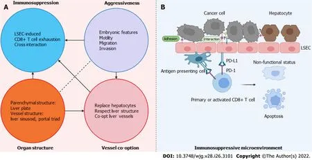
Advance under camouflage hypothesis: Researchers have detailed knowledge of the mechanism of dHGP, but there is little ideal explanation for the formation of rHGP. Except for some tumoral phenotypic and molecular drivers observed in rHGP, there is still no systematic model to support the explanation. We put forward a hypothesis that malignant cells benumb and educate the immune system, and advance by an unknown path under camouflage (Figure 2).

Figure 2 “Advance under camouflage” hypothesis for replacement histopathological growth pattern. A: “Advance under camouflage” hypothesis of replacement histopathological growth pattern (rHGP) includes four elements. Embryonic features, motility, migration and adhesion contributes to aggressiveness of tumor, which drives tumor progression as an intrinsic factor. Tumor cells interact with liver sinusoidal endothelial cells (LSECs) and hepatocytes, so that tumor cells educate these normal liver cells and remodel the immune microenvironment into a tolerance state via CD8+ T cells. In this manner, under camouflage of LSECs,tumor cells are able to survive and slowly progress. Co-opting LSECs enable the tumor to obtain sufficient blood supply and less chance of exposure to immune system. With its unique parenchymal and vessel structure, organ architecture of liver supports the whole advance process; B: Immunosuppressive microenvironment in tumor with rHGP. Under cross interaction of cancer cells and LSECs and other immune cells (unknown mechanism), programmed death (PD) ligand 1 on LSECs and antigen presenting cells binds to PD-1 on T cells making CD8+ T cells activated to a dynamic but nonfunctional type which fails to produce effector cytokines and has decreased cellular cytotoxicity, or is apoptostic. LSEC: Liver sinusoidal endothelial cell; PD: Programmed death.
The tumor cells with an infiltrative pattern paralyze or tame the local immune systemviainteraction with LSECs and induce immune tolerance. LSECs are a major group of hepatic cells that specialize in detection and capture of pathogens from the blood. However, in some cases, this group also downregulates T-cell response through crosstalk with immune cell subsets, leading to immune escape[64,66].Through programmed death (PD) ligand 1 on T cells binding to PD-1 on LSECs, CD8+T cells are activated to a dynamic but nonlicensed type, which fails to produce effector cytokines (e.g., interferon-γ)and have decreased cellular cytotoxicity[67]. Similar suppressive immune activity takes place with mediation of another specific surface protein LSECtin, through which LSECs directly identify activated T cells and inhibit immune-response-mediated T cells[68]. These strongly imply that LSECs are important players in immune tolerance, which enhances invasive and metastatic potential of tumor cells. Taking advantage of this point, tumor cells obtain sufficient interaction with LSECs and hepatocytes, so that tumor cells educate these normal liver cells and remodel the immune microenvironment into a state of tolerance. In this manner, under camouflage of LSECs, tumor cells are able to survive and slowly advance. To expand further, the leading cells at the tumor border also initiate adaptive changes,e.g., enhanced metastatic capacity including cell motility, adhesion and migration.Many of them are driven by some embryonic features that are relevant to co-option-type metastases[69,70]. As a whole, embryonic characteristics, interaction between tumor and normal liver cells, and immunological inertia allows efficient and safe advance of tumor cells. Two other factors must be paid attention: (1) Co-opting normal hepatic vasculature reduces the chance of direct exposure of the tumor to the immune system and achieves normalization of the blood supply. In this case, the intratumoral microvessel keep their normal structure and evenly perfuse the whole nodule, which reduces hypoxia and necrosis and is conducive to overall development of the tumor; and (2) Organ structure is another essential aspect. In an organ with a clear and distinct architecture such as the lungs and liver, the metastatic foci can often expand and develop along with its structure[71,72], which implies that organ architecture sometimes directs how tumor cells expand. We propose a complex axis that ‘embryonic characteristics playing a primitive driving role - tumor hepatic sinusoidal endothelial cell interaction as the mediator - organ structure as the support’ and advance under camouflage hypothesis to explain the underlying mechanism of rHGP.
CONCLUSlON
We proposed a novel hypothesis to explain the mechanism of rHGP formation. We denoted four elements,i.e., intrinsic features of cancer cells, tumor vascularization, immunosuppressive microenvironment and host organ structure in the HGP formation. Vascularization and tumor microenvironment have been emphasized in previous studies while the pivotal role of organ structure is addressed for the first time in this review. Common features in angiogenic HGP, for instance, desmoplastic reaction,immune infiltration and sprouting angiogenesis, have been shown. Organ specific morphology was only observed in non-angiogenic HGP, exhibiting angio-tropism and structure-dependent properties. It was observed that non-angiogenic HGP metastases in brain[73] and skin[74] adopted pericytic mimicry and extravascular migratory behavior to get access to the blood. Similarly, metastatic cells remained the mesenchymal structure (pulmonary alveoli and hepatic plate) and presented with alveolar HGP,interstitial HGP and perivascular cuffing HGP in the lung[75], as well as rHGP in the liver. Thus, itcould be hypothesized that the organ architecture provides metastatic tumor cells with attachment and support for their growth. We also highlight the specific interaction of tumor cells with LSECs in rHGP which might indirectly contribute to tumor progress. However, it is still a pure conceptual idea and it remains to be verified.

Table 2 Characteristics of desmoplastic histopathological growth pattern and replacement histopathological growth pattern in colorectal cancer liver metastasis
Clinical implications of HGP are as follows: (1) The association of HGP with clinical outcomes suggests that HGP can be used to stratify patients by survival risk. Early risk stratification helps provide individualized care and guide long-term follow-up; (2) Patients with dHGP are more likely to benefit from systematic therapy, those with rHGP are prone to acquire resistance to AIs and those with nondHGP are at high risk of positive surgical margin, indicating that HGP can serve as a biomarker for therapy. Based on pre-treatment prediction of HGP, stratification of patients may help clinicians in treatment decision-making and surgical planning for CRCLM patients. The patients with non-dHGP tumor are at higher risk of hepatic recurrence, therefore radical surgery may be of utmost importance.Furthermore, combination therapy would be an inevitable choice for the subgroup with rHGP.Nevertheless, the above benefit from HGP is on the premise of identifying HGP by a non-interventional method. In this regard, radiomics aiming to distinguish HGP in combination with other markers may be a powerful tool in classification of patients; and (3) Studies of mechanism of HGP favor development of therapeutic approaches, and it is encouraging that there have been relevant preliminary trails. For instance, vessel co-option was found to be inhibited through suppressing cancer cell motility and migration in rHGP tumor, and inhibitors targeting both angiogenesis and vessel co-option were more effectivein vivo[30]. Considering the enrichment of fibrotic and angiogenic reaction of dHGP tumors,anti-angiogenic and anti-fibrotic therapies may be effective for these tumors. However, even though both anti-fibrotic[76,77] and anti-VEGF[78] treatment could restore the immune response, these treatments should be used with caution due to the unsatisfying results shown in trials[79].
In addition, we put forward our perspectives on some hot topics. HGP is a miniature of tumor-host reaction and reflects tumor biology and pathological features as well as host immune dynamics. In this sense, HGP builds a bridge between microscopic characteristics and clinical implications. Is HGP plastic? With existence of spatial and temporal heterogeneity, tumors utilize different vascularization at different stages, and their HGP also changes with development of tumor, but knowledge about this is still lacking. What is the key motivation shaping HGP? In addition to motility, invasion and migration giving rise to formation of HGP, the association between HGP and other biological characteristics such as embryonic features, stemness, and spontaneous mutation should be explored. In summary, HGP is a paradox involving several dimensions: malignant and normal cells, central and peripheral sites,angiogenesis and non-angiogenesis, and aggressive and mild characteristics (the summary on characteristics of dHGP and rHGP is listed out in Table 2). Combination of AIs with immune checkpoint inhibitors and AIs with vessel co-option inhibitors showed better effects, suggesting that complex targeted treatment would be a direction for the precision therapy in the future.
FOOTNOTES
Author contributions:Kong BT performed the majority of the writing, and prepared the figures and tables; Fan QS,Wang XM and Zhang Q conceptualized the manuscript; Zhang GL revised the manuscript, provided guidance on the overall concept and execution of the manuscript; and All authors have read and approved the final manuscript.
Supported byNational Nature Science Foundation, No. 81873111, No. 82174454, and No. 82074182; Natural Science Foundation of Beijing, No. 7202066.
Conflict-of-interest statement:All authors report no relevant conflicts of interest for this article.
Open-Access:This article is an open-access article that was selected by an in-house editor and fully peer-reviewed by external reviewers. It is distributed in accordance with the Creative Commons Attribution NonCommercial (CC BYNC 4.0) license, which permits others to distribute, remix, adapt, build upon this work non-commercially, and license their derivative works on different terms, provided the original work is properly cited and the use is noncommercial. See: https://creativecommons.org/Licenses/by-nc/4.0/
Country/Territory of origin:China
ORClD number:Bing-Tan Kong 0000-0001-5352-1406; Qing-Sheng Fan 0000-0002-5648-2833; Xiao-Min Wang 0000-0003-
1274-224X; Qing Zhang 0000-0003-3713-4785; Gan-Lin Zhang 0000-0003-3674-3060.
S-Editor:Ma YJ
L-Editor:A
P-Editor:Ma YJ
杂志排行
World Journal of Gastroenterology的其它文章
- Involvement of Met receptor pathway in aggressive behavior of colorectal cancer cells induced by parathyroid hormone-related peptide
- Role of gadoxetic acid-enhanced liver magnetic resonance imaging in the evaluation of hepatocellular carcinoma after locoregional treatment
- Bifidobacterium infantis regulates the programmed cell death 1 pathway and immune response in mice with inflammatory bowel disease
- Tumor-feeding artery diameter reduction is associated with improved short-term effect of hepatic arterial infusion chemotherapy plus lenvatinib treatment
- Impact of sodium glucose cotransporter-2 inhibitors on liver steatosis/fibrosis/inflammation and redox balance in non-alcoholic fatty liver disease
- Endoscopic techniques for diagnosis and treatment of gastro-entero-pancreatic neuroendocrine neoplasms:Where we are
