Periodontal-orthodontic interdisciplinary management of a“periodontally hopeless” maxillary central incisor with severe mobility: A case report and review of literature
2022-06-23KeJiangLiShanJiangHouXuanLiLangLei
lNTRODUCTlON
Periodontitis, an inflammatory response to invading periodontal pathogens, may lead to the destruction of periodontal supporting tissues[1]. Tooth mobility, a common symptom of advanced periodontitis, is a major reason patients seek dental consultation and a main factor of clinical decision-making by dentists[2]. Loss of supportive periodontal tissue height and widening of the periodontal ligament are the underlying mechanisms of increased tooth mobility[3]. Occlusal trauma-induced tooth mobility, a result of periodontal membrane widening rather than inadequate bone support, can be caused by acute periapical periodontitis, orthodontic treatment, or prosthesis implantation[4]. Malocclusion-associated occlusal trauma is well recognized in patients with a deep impinging overbite or anterior dental crossbite[5,6].
Teeth with significant supportive periodontal tissue loss are especially prone to occlusal trauma, which has been defined as secondary occlusal trauma[7,8]. Primary occlusal trauma mainly refers to occlusal trauma occurring before the presence of periodontal disease and is regarded as a synergistic factor of periodontal disease. Despite consensus on the definitions of primary and secondary occlusal trauma, specific criteria of reduced periodontal support that leads to a clinical diagnosis of secondary occlusal trauma have not been identified clearly. Since both periodontal inflammation and occlusal trauma can result in an increase in the tooth mobility, any clinical decision should be made only after periodontal inflammation is well controlled and occlusal trauma is clearly alleviated[9]. Clinical diagnosis that occlusal trauma has occurred or is occurring may include progressive tooth mobility, fremitus, occlusal discrepancies, tooth migration, root resorption, etc. Once alveolar bone absorption exceeds 70% of total bone support, secondary occlusal trauma and severe tooth mobility may ensue, leading to a possible diagnosis of “periodontally hopeless teeth”[10].
Orthodontic treatment after the careful periodontal treatment of periodontally compromised teeth with pathological tooth migration has been reported in some cases[11-13], and the prerequisites for orthodontic intervention are controlled periodontal inflammation and reduced mobility after strict periodontal treatment[14]. Occlusal adjustment and periodontal splinting have been advocated to stabilize periodontally questionable and hopeless teeth[15,16]. Traditionally, severe tooth mobility may affect the prognosis after orthodontic tooth movement[14], since orthodontic treatment has been reported to increase tooth mobility[17]. However, there is no consensus concerning whether orthodontic tooth movement can be conducted for “periodontally hopeless teeth” with class III mobility.
Extraction of teeth with severe attachment loss has been a common practice in the past because it is expensive to preserve such teeth and the prognosis is usually poor. In addition, in the absence of periodontal treatment, the retention of "hopeless" teeth has a destructive effect on the periodontium of adjacent teeth[18,19]. Although orthodontic treatment in subjects with aggressive periodontitis have been reported[14,20], orthodontic-periodontal interdisciplinary treatment in periodontally hopeless teeth has seldom been reported. In this case report, we describe the periodontal-orthodontic interdisciplinary treatment of a “periodontally hopeless” upper central incisor with class III mobility with a good prognosis over a follow-up period of 3 years.
CASE PRESENTATlON
Chief complaints
A 27-year-old female had a chief complaint of severe tooth mobility and discomfort of the maxillary incisor.
History of present illness
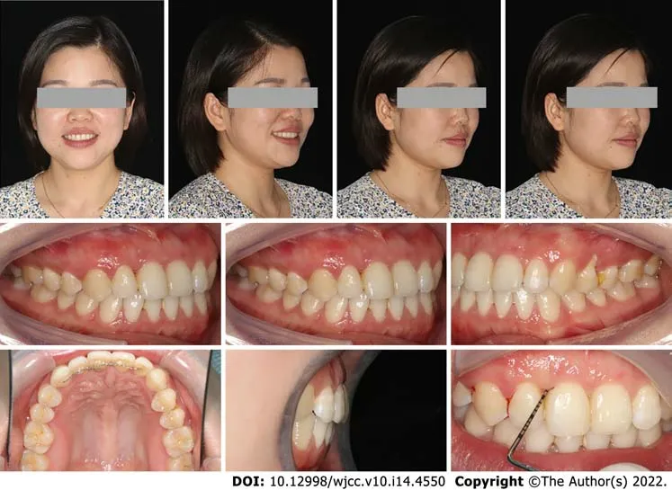
The patient was referred by the periodontist for orthodontic advice. Despite strict periodontal maintenance for half a year, periodontal treatment failed to reduce tooth mobility. The patient had a site of edge-to-edge occlusion with a tendency for a crossbite in the incisor region. Angle class I molar relationships were observed on both sides. Her maxillary central incisors were rotated, especially the upper right central incisor (Figures 1 and 2). The upper right central incisor displayed class III mobility.


History of past illness
Biofilm-induced periodontal infection, occlusal trauma-associated sterile inflammation, and orthodontic tooth movement-related inflammation can all contribute to periodontal membrane widening and tooth mobility[4]. Our present case report demonstrates that despite class III tooth mobility and vertical bone absorption to the apex, a “periodontally hopeless tooth” can be retained after comprehensive periodontal treatment and precise orthodontic treatment.
May hundreds of colossal74 shells, some of a deep red, others of a grass green, stood on each side in rows, with blue fire in them, which lighted up the whole saloon, and shone through the walls, so that the sea was also illuminated
The Porcelain Maiden and the Golden Blackbird know you too? Yes, replied the youth, and the Porcelain Maiden can tell you the whole truth, if she only will
Personal and family history
The patient had a non-contributory personal and family history.
Physical examination
The clinical decision of fixed appliances or removable clear aligners is important for the prognosis of periodontally hopeless teeth[35]. Despite its convenience of oral hygiene, clear aligners may produce large instantaneous stress on the periodontal tissue during repeated removal and wear[36]. Considering the extreme tooth mobility in this case, mechanic force during wearing aligners may aggravate periodontal status. Therefore, fixed appliance is a better choice for teeth with severe tooth mobility.
Apprentice-boys, children of the poor, and even the poor peoplethemselves, stood before the house, watching the lighted windows;and the watchman might easily fancy he was giving a party also,there were so many people in the streets. There was quite an air offestivity about it, and the house was full of it; for Mr. Alfred,the sculptor, was there. He talked and told anecdotes3, and every onelistened to him with pleasure, not unmingled with awe4; but none feltso much respect for him as did the elderly widow of a naval5 officer.She seemed, so far as Mr. Alfred was concerned, to be like a pieceof fresh blotting-paper that absorbed all he said and asked formore. She was very appreciative6, and incredibly ignorant- a kind offemale Gaspar Hauser.
Laboratory examinations
THEY stopped at a little hut; it was very mean looking; the roof sloped nearly down to the ground, and the door was so low that the family had to creep in on their hands and knees, when they went in and out
Imaging examinations
In the region of the maxillary right central incisor on the apical radiograph, less than 25% of remaining alveolar bone was observed, and extensive but relatively mild alveolar bone resorption could be seen on the panoramic radiograph in other regions (Figure 3). The upper right central incisor displayed class III mobility. Cephalometric analysis showed that the patient showed a skeletal class III tendency and an average angle with proclined upper incisors and retroclined lower incisors (Table 1).


FlNAL DlAGNOSlS
Based on the information, the patient was diagnosed with skeletal class I malocclusion and anterior crossbite. When the patient first visited the periodontal department, she was diagnosed with severe aggressive periodontitis (classification 2018: stage IV grade C). The primary aim of periodontal and orthodontic treatment was to reduce tooth mobility and preserve the upper right central incisor. The long-term goal was to establish stable occlusion and maintain periodontal tissue health to prevent tooth extraction. The treatment plan was as follows: (1) Initial periodontal treatment (full-mouth scaling and root planning, tooth brushing instruction); (2) Periodontal surgery (guided tissue regeneration, GTR); (3) Orthodontic treatment; and (4) Supportive periodontal therapy.
TREATMENT
The overall treatment process is shown in Figure 4. After initial periodontal therapy, a splint was placed on the maxillary front teeth to avoid further occlusal trauma and facilitate periodontal repair. Periodontal surgery (guided tissue regeneration) was performed to augment the alveolar bone and soft tissue. Significant bone loss up to the apical third was observed on both the buccal and lingual sides during the surgery. After complete curettage and debriding, bovine-derived xenograft Bio-Osswas placed in the bony defect area, and then the bone particulates were covered with an absorbable collagen membrane to prevent early epithelial cell attachment onto the root surface and provide anatomical space for periodontal regeneration (Figure 5).


Two months later, an acceptable periodontal health condition was observed; however, the upper right central incisor still displayed class III tooth mobility, and gingival recession was observed at both the mesial and distal papilla after removal of the resin splint (Figure 5F). A conventional 0.022-in × 0.028-in straight-wire appliance with a McLaughlin-Bennet prescription was placed. A 0.012-inch nickeltitanium arch wire was inserted in both the maxillary and mandibular arches to align the teeth; meanwhile, a bite turbo was placed on the mandibular molars to relieve the occlusal interference. Unexpectedly, the mobility of the upper right central incisor was reduced to class I mobility after preliminary alignment. Further alignment with 0.012-in NiTi wires in the maxillary arch and 0.018-in NiTi wires in the mandibular arch was performed at month 2. A 0.018-in Australian arch wire was inserted in the mandibular arch to close the space at month 3 with power chain. To further increase the overjet and reduce the premature contact, we performed an interproximal reduction (IPR) of 1.0 mm to gain further space for lower incisor retraction at month 4. In the last mouth of the treatment, we used 0.018-in Australian arch wires for finishing and detailing in the mandibular and maxillary arch (Figure 6).

After 5 mo of active orthodontic treatment, the crossbite was corrected. At the end of orthodontic treatment, the mobility of the upper right central incisor was improved to class I, while the periodontal tissue was in good condition, with no deep periodontal pockets (Figure 7). A fixed retainer was placed to prevent relapse of rotation and stabilize the dentition.
You went to the butcher s for meat, the pharmacy1 for aspirin2, and the grocery store for food. But when I spent the summer with my Grandmother in Warwick, N.Y., she sent me down to the general store with a list. How could I hope to find anything on the packed, jumbled3() shelves around me?

The patient was recalled after 15 mo. Unexpectedly, a space of 1 mm was observed between the upper right lateral incisor and the upper right central incisor, although the fixed retainer was stable and not broken (Figure 8), indicating the presence of occlusal trauma. At this stage, minor premature contact was still observed.
The patient's blood test results were all normal.

Two treatment alternatives were proposed for the patient this time. The first option was comprehensive orthodontic treatment of both the upper and lower arches. Further IPR of 2 mm was needed to close the space in the upper arch and relieve the premature contact. Since the lower incisors were already lingually inclined, such IPR may lead to relapse in the lower arch, potentially followed by occlusal trauma. The second option was orthodontic treatment of only the maxillary arch. A space of 2 mm would be placed on the distal side of the upper right canine, and the space could be restored by the resin composite. The black triangle, space, and premature contact between the front teeth could all be resolved, and a high probability of posttreatment stability could be expected. The patient finally chose option 1.
The fisherman went home, and when he came near the palace he saw that it had become much larger, and that it had great towers and splendid ornamental12 carving13 on it
The second orthodontic treatment was conducted for three months. A NiTi super-elastic coil was utilized to open space between the canine and the premolar, then the space was finally placed on the distal side of the upper right canine. We use composite resin materials to restore the space and improve the black triangle. A lingual bonded retainer from the canine to the incisor in the upper arch was placed to stabilize the occlusion of the upper arch (Figure 9).

OUTCOME AND FOLLOW-UP
The mobile tooth was stabilized, and periodontal inflammation was well controlled. Owing to the establishment of normal incisal guidance in the anterior region without occlusal interference, the occlusal trauma disappeared, as demonstrated by the cephalometric measurement (Figure 10 and Table 1). The angle of U1-SN was increased from 107.3° to 110.2°, while the angle of L1-MP was decreased from 95.2° to 91.7°. The improved inter-incisal relationship contributed to correction of early contact and removal of occlusal trauma. Both the posttreatment panoramic radiograph and apical radiographs showed that the resorption of alveolar bone was under control and that newly formed trabecular bone was present in the region with severe resorption. Apart from this, there was no marked root resorption. The panoramic radiograph (43 mo after periodontal surgery and 2 mo after the second orthodontic treatment) showed that the alveolar ridge height remained relatively stable, and even newly formed trabecular bone could be seen in the region with severe resorption (Figure 11). The occlusion remained stable over a further follow-up period of 1 years (Figure 12). The patient was very satisfied with the final treatment result. A recent follow-up visit (70 mo after periodontal surgery and 39 mo after the second orthodontic treatment) showed that the tooth remained stable, functionally and aesthetically. The maxillary central incisor has no obvious looseness and deep periodontal pocket. Compared with the previous apical radiograph, the alveolar bone had no obvious absorption (Figure 13).
One day when I entered her room, I found her talking into a tape recorder. She picked up a yellow legal pad and held it out to me. I m making a tape for my daughters, she said.


DlSCUSSlON
She did not have any history of systemic illness.
Without plaque-induced inflammation, occlusal trauma did not cause irreversible bone loss or attachment loss in some animal studies[21-23]. Occlusal trauma models in mice, rats, dogs, and monkeys are mostly established by an elevation of occlusion[23-25]; therefore, such hyperocclusion may differ from the occlusal forces in the lateral excursion and various forms of malocclusion. Periodontal pathogenic bacteria, the host immune reaction, the familial gene background, and behavioral factors can aggravate alveolar bone resorption in periodontitis patients. In this case, the rotated upper right central incisor bore traumatic force from several incisors of the lower arch, leading to bone loss to the apex, while inadequate correction of occlusal trauma in the first phase led to the presence of a space between the lateral incisor and canine. This case clearly demonstrates that long-term occlusal trauma can induce severe periodontal bone loss.
Although she was intimidated6 by the crowd at Balmoral, Diana was wise enough not to stay in the castle itself. She asked for, and was granted, an invitation to stay with her sister Jane and her young husband at their cottage on the Balmoral estate.
Two treatment options have been proposed in the first stage. The first was orthodontic treatment to establish a fine and stable occlusion after the periodontal treatment. The corrected occlusion might be beneficial to the long-term periodontal health. The disadvantage was the further periodontal destruction as well as an economical burden of orthodontic treatment. The second option was to place an implant after incisor extraction. Such plan can greatly shorten the time of total treatment. However, insufficient bone mass, lack of soft tissue and poor occlusal condition may pose a great challenge for the long-term survival of the implant. Since tooth mobility may improve after correction of the malocclusion, we finally chose the conservative option to try orthodontic correction. The patient understood and accepted the risks that may occur during orthodontic treatment.
Orthodontic treatment is a double-edged sword for patients with severe periodontitis. On the one hand, orthodontic treatment can help improve the dentition arrangement and change the occlusal relationship[14,26]. On the other hand, clinical and experimental studies have shown that when inflammation is not fully controlled orthodontic treatment may trigger inflammatory processes and accelerate the progression of periodontal destruction leading to further loss of attachment, even in patients with sound oral hygiene[27]. There is also a great risk of additional attachment loss and bone loss if the orthodontic force used is improper[14]. Orthodontic force may induce bone absorption on the side under compression and bone deposition on the side under tension; therefore, proper orthodontic diagnosis should be performed and treatment plans should be prepared to minimize further bone loss during orthodontic tooth movement[28]. In addition, although orthodontic force does not induce changes in the alveolar bone level[11,29], it may induce apical root resorption in teeth affected by periodontitis, especially in teeth with intrusion, since periodontal ligament loss may expose the periodontal tissue to greater force[30]. The long-term outcomes of orthodontic on the periodontal health have been controversial. A recent systematic review demonstrated that orthodontic therapy was associated with 0.03 mm of gingival recession, 0.13 mm of alveolar bone loss, and 0.23 mm of increased pocket depth when compared with no treatment[31]. In the present case, labial tipping of the upper incisors may have further decreased the buccal bone support; therefore, we performed gene replacement therapy to reduce further alveolar bone loss.
Bone regeneration was still a challenge in the horizontal alveolar bone defect. To improve the thin periodontal phenotype and maintain soft tissue stability, we used Bio-Oss bone particulates, which have a slower degradation rate and maintain the three-dimensional gingiva contour in a long term. Traditional guided tissue or bone regeneration is mainly used in the treatment of vertical bone resorption or molar root bifurcation lesions. It promotes periodontal regeneration through bone filling materials and barrier membranes to form new periodontal attachments. Periodontally accelerated osteogenic orthodontics is a clinical procedure that combines selective alveolar corticotomy, particulate bone grafting and the application of orthodontic forces[32], majorly conducted in patients with healthy periodontal status. Regional acceleratory phenomena (RAP) induced by corticotomy promotes alveolar bone remodeling and accelerate toot movement, while the alveolar augmentation expands bone boundaries and benefits periodontal conditions[33].
The timing and type of periodontal intervention are also controversial in the interdisciplinary treatment of periodontally compromised teeth. Previously, full healing of periodontal tissues for 6 mo was advocated before any orthodontic tooth movement. Later, orthodontic tooth movement was initiated as early as 7-10 d after periodontal surgery[28,29,34]. The biological philosophy underlying early treatment is the RAP, by which the inflammatory response after an injury may promote tissue health[24]; therefore, the inflammatory response during periodontal tissue healing after periodontal surgery may promote orthodontic tooth movement. The cytokine-mediated acceleration of orthodontic tooth movement has been utilized in various biology-based techniques, including micro-osteoperforation, bone corticotomy and piezocision[25]. In the present case, although we did not conduct orthodontic treatment early after GTR, the periodontal inflammation from occlusal trauma still promoted quick alignment and crossbite correction.
Our understanding of the nature of periodontal health and inflammation has greatly changed our perspectives regarding orthodontic tooth movement, alveolar bone remodeling and periodontal inflammation. In the most recent periodontal classifications of both the American Academy of Periodontology and European Federation of Periodontology, clinical gingival health on a reduced periodontium in a patient with stable periodontitis is defined as periodontal health and gingival health. Therefore, based on strict biofilm and periodontal risk screening, orthodontic treatment can be performed on a periodontally healthy patient[14].
Still, she often appeared anxious about my love affair, and would ask questions that seemed to me strange, almost as though she feared that something would happen to destroy my romance
Her oral hygiene was good and all permanent teeth were present. The upper right central incisor displayed class III mobility.
The clinical decision to treat or extract “periodontally hopeless teeth” should take into consideration several factors, including clinical parameters, assessed risk, financial aspects, and individual motivation. The survival rate of hopeless teeth in patients with aggressive periodontitis varied from 38% over 10 years to 59.5% over 15 years[10]. Despite reports regarding orthodontic treatment in patients with aggressive periodontitis[14], chronic periodontitis[28], and pathological tooth migration[37], there are very few reports about the prognosis of orthodontically treated hopeless teeth. In the present case, advanced bone loss and severe tooth mobility posed a great challenge in diagnosis and treatment. The long-term synergistic effects of occlusal force and periodontal inflammation led to severe bone loss. We observed improved tooth mobility after unlocking the bite by bite turbo in the molar region, indicating that occlusal trauma contributed to the severe tooth mobility. Therefore, occlusal trauma should be considered during the diagnosis of “periodontally hopeless teeth”; moreover, the indications for and contraindications to the orthodontic treatment of periodontally compromised teeth should take into consideration not only periodontal bone loss but also underlying occlusal trauma.
Despite stable periodontal and orthodontic treatment outcomes in teeth with class III mobility, risks during the orthodontic treatment of teeth with advanced periodontitis should never be ignored. The changes in the biomechanical characteristics of a severely reduced periodontal membrane may lead to an apical shift of the center of resistance; therefore, orthodontic mechanical force may further strain the periodontal ligament of the hypermobile tooth[38], and the inflammatory response during orthodontic tooth movement may accelerate periodontal bone absorption[4].
CONCLUSlON
In this case, the loosened upper right central incisor was successfully retained, and the periodontal tissue remained stable during the follow-up period. This result proves that teeth with severe mobility and bone loss can be saved through interdisciplinary treatment when periodontal inflammation is strictly controlled. Malocclusion-associated or occlusal trauma-related tooth mobility should be an important consideration in diagnosing and planning treatment for periodontally compromised teeth.
FOOTNOTES
Jiang K collected the medical records and mainly drafted the manuscript; Jiang LS partly drafted the manuscript; Li HX and Lei L revised the manuscript critically for important intellectual content; all authors have read and approved the final version of the manuscript.
Informed consent was obtained from the patient for the publication of this case report.
The authors declare that they have no conflict of interest to report.
The first part might be left out, but it gives us a few particulars, and these are useful We were staying in the country at a gentleman s seat, where it happened that the master was absent for a few days
The authors have read the CARE Checklist (2016), and the manuscript was prepared and revisedaccording to the CARE Checklist (2016).
This article is an open-access article that was selected by an in-house editor and fully peer-reviewed by external reviewers. It is distributed in accordance with the Creative Commons Attribution NonCommercial (CC BYNC 4.0) license, which permits others to distribute, remix, adapt, build upon this work non-commercially, and license their derivative works on different terms, provided the original work is properly cited and the use is noncommercial. See: http://creativecommons.org/Licenses/by-nc/4.0/
I grow lighthearted. I can feel my mother kiss me goodnight, check to see if the window is locked, then blow another kiss from the doorway3. Then I am my mother, blowing that same kiss to Anna off that same palm.。,,,。,,。
China
Ke Jiang 0000-0003-0088-0370; Li-Shan Jiang 0000-0003-0063-2545; Hou-Xuan Li 0000-0002-3798-8628; Lang Lei 0000-0003-2892-040X.
Xing YX
A
Xing YX
杂志排行
World Journal of Clinical Cases的其它文章
- Perfectionism and mental health problems: Limitations and directions for future research
- Ovarian growing teratoma syndrome with multiple metastases in the abdominal cavity and liver: A case report
- Development of plasma cell dyscrasias in a patient with chronic myeloid leukemia: A case report
- Suprasellar cistern tuberculoma presenting as unilateral ocular motility disorder and ptosis: A case report
- Rare pattern of Maisonneuve fracture: A case report
- PD-1 inhibitor in combination with fruquintinib therapy for initial unresectable colorectal cancer: A case report
