Giant nontraumatic myositis ossificans in a child:A case report
2022-06-22AnNingXiaJiangShengWang
lNTRODUCTlON
Myositis ossificans was first reported by French physician Guy Patin in 1692,and it is a rare benign disease.At present,myositis ossificans are divided into three categories: Traumatic myositis ossificans,progressive myositis ossificans,and nontraumatic/pseudomalignant myositis ossificans,with the least common being nontraumatic ossification myositis[1].There is a zoning phenomenon in pathology,which will facilitate the diagnosis of the disease[2,3].Due to the unclear medical history and different early clinical symptoms,some patients have no signs,such as masses.Early diagnosis of nontraumatic myositis ossificans in children is difficult,and some patients may be misdiagnosed.Combined with the diagnosis and treatment processes of a patient admitted to our hospital with giant nontraumatic myositis ossificans of the right thigh(the child had no history of trauma),a large mass appeared in the right thigh and imaging examination indicated continuous calcific changes.However,it was accompanied by some manifestations similar to an infectious process,which was difficult to diagnose.We will share the case characteristics,diagnosis points,differential diagnosis,and treatment options of the disease to facilitate a better understanding for everyone with this type of disease.
And you, she turned to George, you are making your home in this country? You do not intend to return to England just yet? ! , 。, , ?? She seemed relieved when she heard that George had bought a farm near our own farm and intended to settle in South Africa
CASE PRESENTATlON
Chief complaints
An 8-year-old girl presented with pain in her right lower extremity and a mass on her right thigh for 1 mo.
History of present illness
The child came to our hospital on January 31,2019,due to intermittent pain in the right lower limb for more than one week.There was no history of trauma or strenuous activities.The pain was aggravated during activities and was accompanied by a limp in the right lower limb;however,there was no fever and no numbness in the right lower limb.The physical examination showed that the movement of the right hip joint was limited,and there was no obvious mass in the right lower limb.Ultrasound indicated synovitis of the hip,and bed rest was recommended.However,the pain was not relieved and gradually worsened,accompanied by fever.She came to our hospital again on February 3,2019.Physical examination revealed that a mass was palpable on the back of the right thigh,and the skin surface was red(Figure 1).Ultrasound,images,and routine blood tests were performed,which revealed soft tissue tumors of the right thigh.Infection and calcification of the right thigh were considered,and antibiotic treatment was given.Her temperature dropped to normal,and the pain was relieved,but the mass still persisted,and became hard.She visited our hospital again on March 3,2019,and was hospitalized.

History of past illness
She was physically healthy and had no history of special disease.
Personal and family history
There might be a cure for that, said the White Bear, if you would but promise me never to talk with your mother alone,32 but only when the others are there too; for she will take hold of your hand, he said, and will want to lead you into a room to talk with you alone; but that you must by no means do, or you will bring great misery4 on both of us. 33
Physical examination
On physical examination,the patient had a giant mass on the back of the right thigh;it measured 20 cm × 10 cm × 8 cm.The surface skin of the mass was slightly red,and the temperature was slightly increased.The quality of the mass was hard;the mass had no tenderness with no sense of fluctuation;the boundary was clear,and the active and passive activities of the right hip and right knee joints were limited(Figure 1).
Laboratory examinations
He stood looking at her, and when he had looked for some time, he said: Let that be enough, wife, now that you are king! Now we have nothing more to wish for
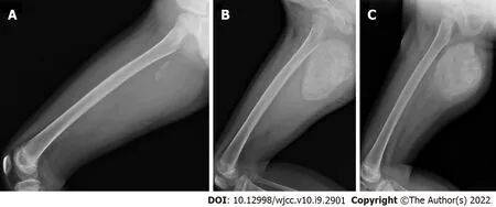
Imaging examinations
Informed written consent was obtained from the patient for publication of this report and any accompanying images.
FURTHER DlAGNOSTlC WORK-UP
Performing the relevant examination and limiting the movement of the limb,the CT examination on March 22,2019,showed that the periosteal reaction had increased compared with the previous period,and the possibility of malignant transformation was not ruled out.An incision biopsy was performed under anesthesia on March 25,2019.The postoperative pathology found no evidence of malignant a tumor,and nontraumatic myositis ossificans was suggested.
FlNAL DlAGNOSlS
Nontraumatic myositis ossificans of the right thigh.
After the graduation from the university in Xi an in 2001, I had been an English tour guide for over two years in Guilin, which is enjoying the reputation of having the most beautiful mountains and rivers in the world
TREATMENT
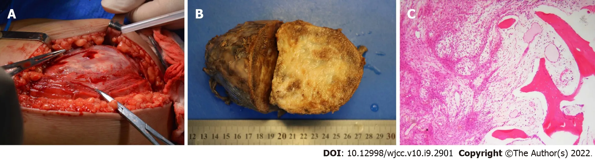
After 9 mo of conservative treatment,the child was treated with nontraumatic myositis ossificans resection of the right thigh on October 14,2019,because the mass still persisted and affected the child's sitting posture.The right hip joint was placed in flexion and abduction positions,a longitudinal incision was made along the skin medial to the surface of the mass,and the surrounding muscle tissues were separated.Exploration showed that the mass was located between the adductor magnus and the vastus medialis,partly adhered to the surrounding muscle tissues,and reached the lesser trochanter (Figure 4A).The mass was completely excised,and the wound was completely hemostatic.The excised mass was 14 cm × 8.7 cm × 8.2 cm in size and had a complete capsule(Figure 4B).Postoperative pathology showed a zonation phenomenon(Figure 4C).She recovered and was discharged one week postoperatively.

OUTCOME AND FOLLOW-UP
The authors declare that they have no conflict of interest.
DlSCUSSlON
Nontraumatic myositis ossificans,which is rare,has no large case report article[4,5],and such a huge case of nontraumatic ossifying myositis in children is very rare.Nontraumatic myositis ossificans is a benign,self-limiting condition in which calcified masses occur within skeletal muscle,and the most common sites are located in the muscle groups of the extremities[6].At present,the pathogenesis of nontraumatic myositis ossificans is incompletely understood.The release of inflammatory factors leads to the formation of nontraumatic myositis ossificans due to some conditions that lead to tissue ischemia[7,8].Kan[9]demonstrated that,when skeletal muscle injury induces a local inflammatory cascade,resulting in the release of cytokines(bone morphogenetic protein-2 and transforming growth factor),it causes local stem cell dysfunction and thus heterotopic bone formation.When vascular endothelial cells of skeletal muscle are exposed to an inflammation-rich environment,these endothelial-derived mesenchymal stem cells may differentiate into chondrocytes or osteoblasts,which in turn form chondroosteoblasts in the extraosseous tissue[10].However,the specific mechanisms of these local inflammatory cascades are still different.At present,the clinical staging of nontraumatic myositis ossificans is defined similarly to the stages of traumatic myositis ossificans,which are early,intermediate,and mature.
A tumor was found in the upper right thigh of this child,and the disease progressed rapidly.This phenomenon needed to be differentiated from malignant tumors.For example,extraosseous osteosarcoma is more aggressive on imaging,and calcifications are usually distributed inside soft tissue masses[18].There are atypical tumor cells in pathologically.Bone tissue is the most obvious in the center of the tumor,with denser surrounding cells,which is contrary to the pathology of nontraumatic myositis ossificans;nontraumatic myositis ossificans are characterized by zonaled hyperplasia of fibroblasts and osteoblastic osteoblasts,which continue to progress with the course of the disease[2,3].However,nontraumatic myositis ossificans can also become cancerous or become combined with cancer.When the diagnosis of the disease is difficult,a biopsy can be performed to assist in the diagnosis,but early biopsy is not currently recommended[19].If other diseases can be excluded well and ultrasound and imaging are in line with the typical showing of nontraumatic myositis ossificans,biopsy may not be rushed,and close observation and follow-up can be performed.The patient received a biopsy to assist in the diagnosis at approximately 9 wk after the onset of disease.To reduce the test error,we adopted an open biopsy.To obtain sufficient pathological specimens,samples were taken from the periphery to the core of the lesion.To prevent the spread of the lesion,we also combined puncture needles and other tools to assist in obtaining specimens.
THERE was once a sculptor1, named Alfred, who having won thelarge gold medal and obtained a travelling scholarship, went to Italy,and then came back to his native land. He was young at that time-indeed, he is young still, although he is ten years older than hewas then. On his return, he went to visit one of the little towns inthe island of Zealand. The whole town knew who the stranger was; and one of the richest men in the place gave a party in his honor, and all who were of any consequence, or who possessed2 some property, were invited. It was quite an event, and all the town knew of it, so thatit was not necessary to announce it by beat of drum.
The child had fever and redness on the skin surface of the mass in the early stage.Blood tests showed that the inflammation index was increased.The infection should be differentially diagnosed.The child had no rash and no recent history of upper respiratory tract infection,and the fever symptoms were short-lived and low-grade.The mass was not fluctuant on palpation.Images showed irregularities in the dense calcification of the tumor.These are different from soft tissue infections.The fever symptoms of soft tissue infections in children are more obvious and easier to repeat,but the patient is not.If the infected site was relatively superficial,it easily felt fluctuation and obvious tenderness,but the mass was hard to palpate.Images and ultrasound showed that the lesions were edema and necrotic liquefaction,and calcification was relatively rare and occurred later,but the patient had calcification early[12-14].
There was a giant calcification-like tumor in the thigh muscle tissue,and it needed to be distinguished from calcified myonecrosis disease.Calcified myonecrosis disease is more common in trauma,such as fractures complicated by ischemic muscle necrosis,calcification,and other types of masses that gradually appear.The common site is the lower leg,and compartment syndrome is a potential cause[15].Radiographs show prismatic masses with calcifications of plaques distributed longitudinally along the compartment.Calcifications are manifested around the muscles.The central area is mostly liquefied tissue,which can cause bone destruction and periosteal reaction.When the mass increases gradually and the pressure increases,it can produce layered changes in cortical bone[16,17].This is similar to the patient's magnetic resonance imaging results,but the patient had never received fracture surgery in the past,there was no history of trauma,and the patient was only 8 years old.
26. If a man yields once he s done for, and so, because he had given in the first time, he was forced to do so the second: The Grimms are preaching their own philosophy concerning a man s role in his home. Once again, their patriarchal view is emphasized in the story. This patriarchal element is thought to be one reason why the tale was so popular after its publication. Return to place in story.
The patient was an 8-year-old female child who was previously healthy.There was no history of trauma,and the early presentation was primarily pain and restriction of movement in the right lower limb.Early blood tests indicated that some inflammation indices were too high,and the images displayed that the lesions showed signs of gradual calcification.Because nontraumatic myositis ossificans has different presentations in the early stage,such as pain,fever,and limited mobility,and the disease progresses rapidly,diagnosis is difficult[11].The mass can appear as the disease progresses.Blood tests indicated that some inflammation indicators were high.Images indicated that there was a tumor in the soft tissue,some of which appeared to be calcified.At this time,the possibility of nontraumatic myositis ossificans should be considered,but other diseases should also be considered and differentiated.
At present,nontraumatic myositis ossificans is still treated conservatively,mainly by close observation and symptomatic treatment.There are reports suggesting that drugs such as ibuprofen can be used for treatment[20,21].The child was treated with ibuprofen for anti-inflammatory and analgesic treatment,which had a certain effect.This child was treated with antibiotics because it could not be differentiated from the infectious disease in the early stage.However,reviewing the evolution of the disease,if the disease can be diagnosed early,such as by obtaining blood culture test results,unnecessary antibiotic use can be avoided.For whether to undergo surgical resection,the indications are the same as in previous studies,in which those afflictions that have affected the appearance or function of the child and that cannot resolve spontaneously,or if malignant transformation is suspected,surgical treatment should be considered[1].The patient underwent surgical resection approximately 9 mo after the onset of the disease.To reduce the possibility of recurrence,hemostasis was completely achieved during the operation and postoperative immobilization(2 wk)was mandated.The child had no recurrence and is now recovering as before.
CONCLUSlON
Although the medical history of nontraumatic myositis ossificans is not typical,the early symptoms can present differently,and the images shown in different stages are different,which increases the difficulty of diagnosis,especially in the early stage,through close follow-up and periodic review of imaging examination,if necessary combined with biopsy as early as possible.Treatment is still based on conservative treatment and surgical resection if necessary.
No family history of inherited diseases and malignant tumor diseases was recorded.
ACKNOWLEDGEMENTS
We would like to thank Dr.Han JM for guidance on the treatment of the child.
FOOTNOTES
Wang JS was responsible for designed and reviewed the papers;Xia AN was mainly responsible for collected data and manuscript drafting.
Some inflammatory indicators were abnormally high,and blood calcium was slightly low at the time of admission.Blood analysis revealed white blood cells of 11.3 × 10/L,platelets of 858 × 10/L,C-reactive protein of 74.1 mg/L,erythrocyte sedimentation rate of 98 mm/h,and blood calcium of 2.04 nmol/L.
Radiographs and computed tomography(CT)scans showed a large soft tissue mass on the upper right posterior thigh with progressive calcification inside the mass(Figure 2 and Figure 3).
Following-up after more than 1 year,the tumor did not recur,and the patient's lower limbs moved freely.The child was satisfied.
Ring was quite deceived by her, and never guessed that she was not Princess Signy at all, but a strong, gigantic, wicked witch bent10 on deceiving him under a beautiful shape
The authors have read the CARE Checklist(2016),and the manuscript was prepared and revised according to the CARE Checklist(2016).
This article is an open-access article that was selected by an in-house editor and fully peer-reviewed by external reviewers.It is distributed in accordance with the Creative Commons Attribution NonCommercial(CC BYNC 4.0)license,which permits others to distribute,remix,adapt,build upon this work non-commercially,and license their derivative works on different terms,provided the original work is properly cited and the use is noncommercial.See: http://creativecommons.org/Licenses/by-nc/4.0/
China
Then he started off in pursuit, making his way through bushes and briars, and never stopped all day; but in the evening the stag entirely disappeared, and when golden lad came to look about him he found himself just opposite a hut in which lived a witch
An-Ning Xia 0000-0003-2598-8185;Jiang-Sheng Wang 0000-0003-3672-5962.
Xing YX
A
Xing YX
杂志排行

World Journal of Clinical Cases的其它文章
- Malignant struma ovarii with papillary carcinoma combined with retroperitoneal lymph node metastasis:A case report
- Upper gastrointestinal bleeding from a Mallory-Weiss tear associated with transesophageal echocardiography during successful cardiopulmonary resuscitation:A case report
- lpsilateral hemifacial microsomia with dextrocardia and pulmonary hypoplasia:A case report
- Esophageal myoepithelial carcinoma:Four case reports
- Turner syndrome with primary myelofibrosis,cirrhosis and ovarian cystic mass:A case report
- Acute coronary artery stent thrombosis caused by a spasm:A case report