Application of electron microscopy in gastroenterology
2022-06-09MasayaIwamuroHaruoUrataTakehiroTanakaHiroyukiOkada
Masaya Iwamuro, Haruo Urata, Takehiro Tanaka, Hiroyuki Okada
Masaya Iwamuro, Hiroyuki Okada, Department of Gastroenterology and Hepatology, Okayama University Graduate School of Medicine, Dentistry, and Pharmaceutical Sciences, Okayama 700-8558, Japan
Haruo Urata, Central Research Laboratory, Okayama University Medical School, Okayama 700-8558, Japan
Takehiro Tanaka, Department of Pathology, Okayama University Hospital, Okayama 700-8558,Japan
Abstract Electron microscopy has long been used in research in the fields of life sciences and materials sciences. Transmission and scanning electron microscopy and energy-dispersive X-ray spectroscopy (EDX) analyses have also been performed in the field of gastroenterology. Electron microscopy and EDX enable (1)Observation of ultrastructural differences in esophageal epithelial cells in patients with gastroesophageal reflux and eosinophilic esophagitis; (2) Detection of lanthanum deposition in the stomach and duodenum; (3) Ultrastructural and elemental analyses of enteroliths and bezoars; (4) Detection and characterization of microorganisms in the gastrointestinal tract; (5) Diagnosis of gastrointestinal tumors with neuroendocrine differentiation; and (6) Analysis of gold nanoparticles potentially used in endoscopic photodynamic therapy. This review aims to foster a better understanding of electron microscopy applications by reviewing relevant clinical studies, basic research findings, and the state of current research carried out in gastroenterology science.
Key Words: Transmission electron microscopy; Scanning electron microscopy; Energydispersive X-ray spectrometry; Gastrointestinal disease, gastroesophageal reflux disease;Pathogens
INTRODUCTION
In light microscopy, visible light is used to obtain magnified views of the object. As the resolution is related to the wavelength of light used to image a specimen, the resolution of an optical microscope is theoretically limited to approximately 200 nm. Thus, nanostructures cannot be observed using light microscopy. In contrast, electron beams are used in electron microscopy. As the wavelength of an electron beam is shorter than that of visible light, electron microscopy has extremely high resolution and provides sharp, finely detailed images of the surface or interior of biological and nonbiological specimens. In addition, energy-dispersive X-ray spectroscopy (EDX), which is a chemical microanalysis technique used in conjunction with electron microscopy, enables the analysis of elements or chemical characterization of a sample. Since the development of the first prototype in 1931, electron microscopes have been widely used in various fields, such as physics, chemistry, engineering, biology, and medicine[1]. Based on its versatility, electron microscopy analysis has been used in several studies covering various aspects of clinical samples obtained from patients with gastrointestinal diseases. This paper briefly discusses the fundamentals of electron microscopy and reviews the literature concerning the application of electron microscopy in gastroenterology science.
ANALYTICAL METHODS IN ELECTRON MICROSCOPY
Analytical methods in electron microscopy can broadly be categorized into three types: Transmission electron microscopy, scanning electron microscopy (SEM), and EDX. The different types of electron microscopes used in these methods are related and often applied concurrently in the field of biology.
A transmission electron microscope irradiates a specimen with an electron beam. The object must be cut into very thin cross-sections because it is visualized through the spatial distribution of the transmitted electron beam. Although the use of transmission electron microscopy is limited to engineering science at the outset, it has been extensively used in the field of biology since the 1950s largely due to improvement of the microtome for ultrathin slice preparation using a diamond knife and the development of staining techniques based on heavy metals, such as osmium.
A scanning electron microscope produces an image using electrons reflected or generated from the surface of the specimen. The specimen is placed in a high vacuum state, and the surface is scanned with an electron beam focused by an electric or magnetic field. SEM produces a characteristic threedimensional appearance that is useful for understanding the surface ultrastructure of a sample.
EDX is an X-ray system used to identify the elemental composition of a material. It has a semiconductor detector to detect the fluorescent X-rays generated when the primary X-ray beam illuminates the sample. The fluorescent X-rays emitted from the material have a spectrum of wavelengths characteristic of the types of atoms present in the specimen. EDX enables both qualitative and semiquantitative analyses of the elements based on the energy and number of generated electron-hole pairs. EDX is more suited for analyses of inorganic materials than organic materials.
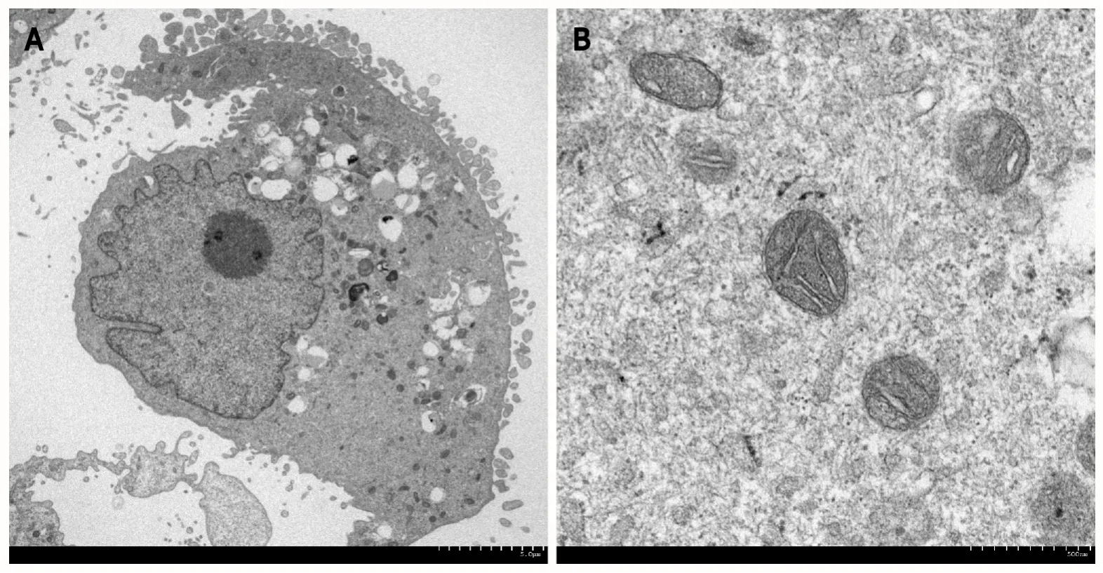
In the field of gastroenterology, transmission and SEM and EDX analyses have been used to visualize cells (Figure 1) and pathogens, including parasites, bacteria, viruses, biofilms, and elements deposited in the gastrointestinal mucosa. Nonbiological materials, such as stents, powders, and bezoars, have also been analyzed at subnanometer resolution. In the following sections, we review examples of electron microscopy analyses in association with the pathophysiology of gastrointestinal disorders.

Figure 1 Transmission electron microscopy image.

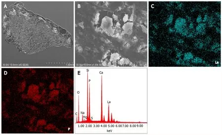
Figure 2 Transmission electron microscopy images and spectra obtained by energy-dispersive X-ray spectrometry.
EXAMPLES OF ELECTRON MICROSCOPY ANALYSES
Intercellular spaces of the esophageal epithelium
The most typical example of electron microscopy analysis in gastroenterology is evaluation of the intracellular spaces of esophageal epithelial cells. Notably, some of the articles on this topic have been published in high-impact journals. Intercellular spaces in the esophageal epithelium are known to be dilated in patients with nonerosive reflux disease and in patients with esophagitis. Following several animal studies, endoscopic esophageal biopsy specimens taken from patients with (n= 11) and without(n= 13) recurrent heartburn were investigated in 1996 using transmission electron microscopy[2]. A dilated intercellular space diameter was observed in 8 of the 11 patients with heartburn, while none of the asymptomatic individuals exhibited this feature. Dilated intercellular space was also present in the normal-appearing, nonerosive mucosa of patients with symptomatic reflux disease. Other authors have provided further evidence that detached interepithelial cell junctions, which are observed as dilated intercellular spaces assessed by electron microscopy[3-5], correspond to early esophageal damage induced by acid reflux[6-8]. Dilatation of intercellular spaces in the esophageal epithelium is not observed in patients with functional heartburn, suggesting that this microscopic feature is specific to acid reflux[9]. Proton pump inhibitor therapy resulted in complete recovery of dilated intercellular spaces in > 90% of cases with nonerosive reflux disease and erosive esophagitis, indicating that the electron microscopy features are reversible[10,11].
Dilated intracellular spaces arise along the distal and proximal esophagus of patients with nonerosive reflux disease, suggesting that they may be an underlying mechanism accounting for the enhanced perception of proximal acid reflux[12]. Duodenal gastroesophageal reflux has also been reported to cause dilatation of intercellular spaces in the esophageal epithelium[3,13]. Similarly, in patients with laryngopharyngeal reflux and sore throat, this feature appears at the squamous basal and suprabasal levels in oropharyngeal biopsy specimens[14,15]. An investigation of patients with bronchial asthma[11,16] and children with reflux-related cough[17] revealed that the intracellular spaces in the esophageal epithelium are significantly dilated compared with those in control patients, suggesting a pathophysiological correlation between gastroesophageal reflux and the development of these respiratory tract symptoms.
Although the width of the intracellular spaces can be measured using light microscopy[18], the sensitivity of light microscopy was 79.3%, and the specificity was 75.0%[19]. Owing to the inferior specificity of light microscopy analysis, electron microscopy seems to be more suitable for measuring intercellular spaces in the esophageal epithelium. Chuet al[20] reported the possible utility of in vivo confocal laser endomicroscopy to examine microalterations of the esophagus in patients with nonerosive reflux disease[20].
Eosinophilic esophagitis
Eosinophilic esophagitis is a chronic, allergic inflammatory condition of the esophagus. Dilated intracellular spaces are evident in the esophageal epithelium of patients with eosinophilic esophagitis,which are significantly reduced after treatment[21]. Transmission electron microscopy revealed a significant decrease in the number of desmosomes[22] and an increased autophagic vesicle content[23]in active eosinophilic esophagitis compared with observations in normal individuals and inactive eosinophilic esophagitis patients. Thus, electron microscopy may be useful for investigating the pathophysiology of eosinophilic esophagitis.
Lanthanum deposition
Lanthanum carbonate is a phosphate binder taken orally and is commonly used in patients with chronic kidney disease. Although its tolerability and safety profile have been reported in hemodialysis patients,lanthanum deposition in the gastric and duodenal mucosa of these patients, in the form of lanthanum phosphate, has been reported in the literature[24-28]. On light microscopy examination of hematoxylin and eosin-stained specimens, deposited lanthanum is visible as a fine, amorphous, eosinophilic material. SEM revealed bright areas in the deposited lanthanum (Figure 2A). Images at higher magnification showed deposition as the accumulation of minute particles (Figure 2B). EDX analysis provided evidence directly related to the presence of lanthanum and phosphate (Figure 2C). Elemental mapping by EDX revealed that lanthanum (Figure 2D) and phosphate (Figure 2E) showed an identical location to that of the bright areas on SEM. Although lanthanum deposition in the gastrointestinal tract can be clinically diagnosed with conventional light microscopy observation of the fine, amorphous,eosinophilic material and medication information from the patient’s current or past use of lanthanum carbonate, SEM has advantages in the detection of deposited lanthanum, as it is easily identified as a bright area.
Enteroliths and bezoar s
Enteroliths are calculi that occur in the intestines and include two types: “True” and “false” enteroliths.True enteroliths, for example, cholic acid and calcium stones, are generated from the sediments of substances found in enteric contents. False enteroliths, such as bezoars, gallstones, and foreign objects,are formed from indigestible substances stuck in the alimentary tract. Infrared spectroscopy is generally used to identify the chemical substances constituting enteroliths removed from patients. Electron microscopy and EDX have the advantages of imaging the microstructure and analyzing elements,allowing clarification of the nature of enteroliths.
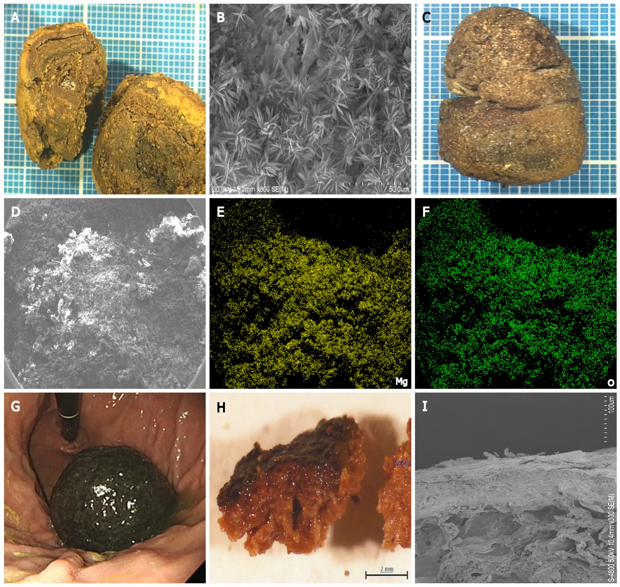
Figure 3 shows examples of enteroliths and bezoars that we previously investigated. One patient had an enterolith in the stomach composed of bilirubin calcium, calcium carbonate, and fatty acid calcium[29] (Figure 3A and B). Another patient had a rare pharmacobezoar in the stomach, which was composed of magnesium oxide (Figure 3C–F)[30]. We also investigated the ultrastructure of the persimmon phytobezoar in the stomach (Figure 3G–I)[31]. Thus, electron microscopy and EDX analyses offer insights into the microstructure and elemental composition of enteroliths.

Figure 3 Images of enteroliths.
Pathogens including bacteria, parasites, and viruses in the gastrointestinal tract
Electron microscopy has been widely used in microbiology to elucidate the number, distribution, and adherence of microorganisms in clinical samples. One of the typical applications of electron microscopy for pathogens in gastroenterology is the detection ofHelicobacterspecies, such asHelicobacter pylori[32-36] andHelicobacter heilmannii[37]. These bacteria have a spiral form, which is a distinct difference from other bacteria. Another example isTropheryma whipplei[38-41], which causes the rare systemic infectious disorder Whipple's disease. Electron microscopy revealed thatTropheryma whippleishows a characteristic trilamellar plasma membrane. Other rare pathogens identified by electron microscopy include anisakiasis[42], amoebiasis[43], intestinal spirochetosis[44],Sutterella wadsworthensis[45],Giardia intestinalis[46], andBrachyspira aalborgi[47].
A biofilm is a thick layer formed by microorganisms attached to the surface of a solid material or liquid. SEM has been used to visualize the shape and localization of biofilms and the steps of the biofilm formation process. For instance, several authors have investigated the efficiency of the cleaning,disinfection, and sterilization processes of biofilm-contaminated endoscopes[48,49].
Gastrointestinal tumors with neuroendocrine differentiation
Neuroendocrine and mixed neuroendocrine neoplasms can arise in most of the epithelial organs of the body and are not rare in the gastrointestinal tract. Transmission electron microscopy revealed that neuroendocrine tumor cells in the gastrointestinal tract contained numerous dense-core secretory granules of variable sizes and shapes in the cytoplasm. Because these neurosecretory granules are characteristic of neuroendocrine tumors, electron microscopy analysis has been used to support its diagnosis. For instance, neuroendocrine differentiation was assessed using electron microscopy images in cases of malignant peripheral nerve sheath tumors of the esophagus[50], gangliocytic paraganglioma in the duodenum[51], mixed acinar-endocrine carcinoma arising in the ampulla of Vater[52], combined adenocarcinoma and neuroendocrine tumors in the stomach[53], neuroendocrine carcinoma in the stomach[54], mixed acinar-endocrine neoplasm in the stomach[55], and large cell neuroendocrine carcinoma in the esophagogastric junction[56].
Gold nanoparticles potentially used in endoscopic photodynamic therapy
Based on the properties of absorption and scattering of electromagnetic radiation, gold nanoparticles are emerging as promising agents and are of particular interest for applications in photothermal therapy, in addition to efficient drug carriers and diagnostic agents. For instance, endoscopic fluorescence-guided near-infrared photothermal therapy using gold nanoparticles is in development for the treatment of gastrointestinal tumors[57]. The size, morphology, and composition of synthesized gold nanoparticles and their location within tissue can be assessed using transmission electron microscopy and EDX analysis[58].
CONCLUSION
Electron microscopy enables (1) Observation of ultrastructural differences in esophageal epithelial cells in patients with gastroesophageal reflux and eosinophilic esophagitis; (2) Detection of lanthanum deposition in the stomach and duodenum; (3) Ultrastructural and elemental analyses of enteroliths and bezoars; (4) Detection and characterization of microorganisms in the gastrointestinal tract; (5) Diagnosis of gastrointestinal tumors with neuroendocrine differentiation; and (6) Analysis of gold nanoparticles potentially used in endoscopic photodynamic therapy. Therefore, electron microscopy has had a profound impact on our knowledge and understanding of various digestive tract diseases. We hope that this article will help gastroenterologists widely utilize electron microscopy analysis for clinical diagnosis and basic research.
FOOTNOTES
Author contributions:Iwamuro M designed the research study and wrote the paper; Urata H and Tanaka T critically reviewed the manuscript for important intellectual content; Okada H approved the manuscript.
Conflict-of-interest statement:There are no conflicts of interest associated with any of the senior authors or other coauthors who contributed their efforts in this manuscript.
Open-Access:This article is an open-access article that was selected by an in-house editor and fully peer-reviewed by external reviewers. It is distributed in accordance with the Creative Commons Attribution NonCommercial (CC BYNC 4.0) license, which permits others to distribute, remix, adapt, build upon this work noncommercially, and license their derivative works on different terms, provided the original work is properly cited and the use is noncommercial.See: http://creativecommons.org/Licenses/by-nc/4.0/
Country/Territory of origin:Japan
ORCID number:Masaya Iwamuro 0000-0002-1757-5754; Haruo Urata 0000-0002-0268-6187; Takehiro Tanaka 0000-0002-1509-5706; Hiroyuki Okada 0000-0003-2814-7146.
S-Editor:Fan JR
L-Editor:A
P-Editor:Fan JR
