Neuroprotective effect of Phyllanthus urinaria extract on ischemic brain injury in rats
2022-06-08XiaoLongWenJunPengLiXinXinLiuZongHuiLiYunYunXiongDaoRuiYu
Xiao-Long Wen, Jun-Peng Li, Xin-Xin Liu, Zong-Hui Li, Yun-Yun Xiong, Dao-Rui Yu
1. Department of Pharmacy, the Second Affiliated Hospital of Hainan Medical University, Haikou 570311, China
2. School of Clinical Medicine, Hainan Medical University, Haikou 571199, China
3. School of Basic and Life Science, Hainan Medical University, Haikou 571199, China
ABSTRACT Objective: To investigate the neuroprotective effect of Phyllanthus urinaria on ischemic stroke and its primary mechanism. Methods: Adult SD rats were selected as the research object, and the right middle cerebral artery infarction rat model was established by the modified suture method(MCAO).Observe the neurological deficit score at 24h, 48h and 72h after the model is successfully prepared,then TTC staining method to detect the area of cerebral infarction,the content of superoxide dismutase (SOD), nitric oxide (NO) and endothelial NOS (eNOS)in brain tissue;Immunofluorescence method was used to detect the expression of Caspase-3 positive cells in brain tissue; Western blot method was used to detect the expression of PI3K and AKT protein in brain tissue.72 experimental animals were randomly divided into 4 groups, sham operation group, model group (MCAO), extract of Phyllanthus urinaria low dose group(PuL5 g/kg), high dose group (PuL10 g/kg), and make MCAO model after 7 days of continuous administration, and continue to infuse the medicine once/d until the material is obtained. Results: No neurological deficits in the sham operation group, The 24h, 48h and 72h of the modelling showed that the neurological impairment of the two doses of extract of Phyllanthus urinaria and the MCAO group was more severe than that of the sham operation group (P<0.01),however, with the prolongation of the modeling time, the neurological function scores of the two doses of extract of Phyllanthus urinaria were lower than those of the MCAO group, the most significant at 72h (P<0.01);The infarct size of the rats in the two dose groups of extract of Phyllanthus urinaria was lower than that of the MCAO group (P<0.01), and there was no dose dependence between the two groups,the content of SOD in the MCAO group was reduced, and the content of NO and eNOS was increased than the sham operation group(P<0.05),Compared with the MCAO group, the two administration groups significantly increased the content of SOD, decreased the content of NO and eNOS (P<0.05);Although the expression of Caspase-3 positive cells in the two administration groups was higher than that in the sham operation group, it was significantly lower than that in the MCAO group (Plow<0.05,Phigh<0.01);The expression of PI3K and AKT protein in the brain tissue of the MCAO group was significantly lower than that of the sham operation group (P<0.05),but the expression of PI3K and AKT protein in brain tissue of extract of Phyllanthus urinaria in high and low dose groups was significantly higher than that in MCAO group (P<0.05). Conclusion: Extract of Phyllanthus urinaria can improve neurological damage and cerebral infarction area in rats,and reduce the expression of Caspase-3 positive cells in MCAO rats, and then increase the expression levels of PI3K and AKT proteins to protect ischemic brain injury.
Keywords:Extract of Phyllanthus urinaria Rats MCAO Nerve function
1. Introduction
Stroke is a common cardiovascular and cerebrovascular disease.It is a disease that causes brain damage due to abnormal blood circulation in the brain tissue. It is clinically divided into hemorrhagic and ischemic strokes. The proportion of ischemic strokes is 80%[1].Throughout the world’s cerebrovascular diseases, acute ischemic brain injury caused by cerebral artery occlusion is a key factor. It is more common in elderly people at the age of onset, and its morbidity and mortality are higher[2].According to a report from the Institute of Health Economics of the Ministry of Health of my country, the annual expenditure of more than tens of billions of yuan is due to cerebrovascular diseases, which has caused an increasing social and economic burden[3].Once cerebral ischemic injury occurs, there will be a series of changes that cause swelling, degeneration, and death of neurons and other brain tissues, such as oxidative stress, release of inflammatory factors, release of excitatory amino acids, etc[4].At present, there are few effective drugs for the treatment of the disease,and patients benefit little from it, and are prone to risk of bleeding and reperfusion injury. Therefore, it is urgent to explore effective neuroprotective drugs in depth, and it is particularly important to develop work in the field of neuroscience.
Phyllanthus is a plant of the Euphorbiaceae Phyllanthus genus,also known as N. vulgaris, which grows widely in tropical and subtropical regions. my country mainly grows in Guangdong,Guangxi and Hainan[5].As a traditional Chinese medicine, its taste is slightly bitter, and its coolness has the effects of clearing heat and detoxification, hydrating and reducing swelling, etc. It is used in the treatment of dysentery, diarrhea and unknown swelling and pain caused by enteritis[6].Modern pharmacology shows that Phyllanthus urinaria has anti-tumor, anti-oxidant and anti-thrombotic effects[7].It has low toxic and side effects and has good clinical application potential. And Phyllanthus has no in-depth reports on the treatment of acute cerebral ischemia.Therefore, in this study, SD rats were used to establish the right middle cerebral artery occlusion (MCAO)model of rats through the modified suture method.Investigate the protective effect of Phyllanthus urinaria extract on acute cerebral ischemia injury and its mechanism of action, and provide theoretical support for the development of new drugs for the treatment of the disease.
2. Materials and Methods
2.1 Main reagents and instruments
SOD test kit: Solarbio, production batch number: 20181117;NO test kit: Nanjing Jiancheng Biotechnology Co., Ltd., production batch number: 20180609;eNOS test kit: Biyuntian Biotechnology Company, production batch number: S1817;Rabbit antirat Caspase-3 polyclonal antibody: Abcam, batch number:ab18137;PI3K polyclonal antibody: Abcam, production lot number:ab191607;Akt polyclonal antibody: Abcam, production lot number:ab18785;Chromogenic agent DAB: Invitrogen, production batch number: 191013;Laser Doppler blood flow measuring instrument:British company moor;Fluorescence microscope: Olympus company szx16 type;Professional image processing and analysis system Image Pro Plus v6.0: American Media Cybernetics.
2.2 Preparation and administration of Phyllanthus urinaria extract
The Phyllanthus urinaria was collected by the Variety Resources Research Institute of the Chinese Academy of Tropical Agricultural Sciences, and the Phyllanthus urinaria extract was prepared by the extraction and preparation process provided by the Pharmacy Entrance Examination Office of Hainan Medical University.Weigh an appropriate amount of Phyllanthus urinaria, soak in distilled water for 30 minutes and then heat it with fire. After boiling, adjust to simmer and cook for 1 hour, then stir and filter.The dregs are repeated twice in the same way, and the filtrate is combined, and finally concentrated with a rotary evaporator, and diluted with water according to the required concentration, and stored in a refrigerator at 4℃.Before MCAO modeling, intragastric administration is given once per day,Rats in Phyllanthus urinaria extract group were given 10% of the maximum lethal dose to determine the dose, and the dose was continuously administered at two doses of 5 g/kg and 10 g/kg respectively,Rats in sham operation group and MCAO group were given the same volume of normal saline,After 7 days of continuous administration, a rat experimental cerebral ischemic injury model(MCAO) model was made, and the medicinal solution was continuously perfused once a day until the material was obtained.
2.3 Experimental animal grouping and MCAO model preparation
Healthy adult SD rats: Changsha Tianqin Biotechnology Co., Ltd.,72 in number, clean grade, body mass: 280-350 g. Animal certificate number: SCXK (Xiang) 2018-001.Raised in separate cages at room temperature, and after adapting to the laboratory environment for a week, they were randomly divided into sham operation groups with 18 animals in each group,Phyllanthus urinaria extract low and high dose group (PuL5 g/kg, PuL10 g/kg).The experimental animals used in this study are based on the Hainan Medical College ethics committee standards based on the principle of minimizing the number of animals. At the same time, the clinical use of anesthesia is simulated during the operation, and the administration is strictly regulated to reduce discomfort. At the end of the experiment, the undead animals are dislocated. The euthanasia method is used for treatment, and it is temporarily stored in the school’s laboratory animal refrigerator for unified processing by the school.Start modeling on the 7th day 1 hour after the administration,Prepare rat experimental ischemic model (MCAO model) with reference to the modified Longa suture method[8].The specific operation is as follows: Rats are anesthetized by intraperitoneal injection of 1.5%sodium pentobarbital according to their body weight. Next, they are fixed on the rat board in the direction of exposing the neck, cut the neck skin, and bluntly separate the muscle tissue with hemostatic forceps. , Until the carotid triangle is exposed.Separate the common carotid artery, use a surgical thread to pull the common carotid artery, and then carefully separate the internal carotid artery and external carotid artery at the bifurcation.Block the distal end of the external carotid artery with No. 4 black silk thread, and block the common carotid artery and internal carotid artery with arterial clips.Use ophthalmic scissors to cut a small opening at the position of the common carotid artery near the bifurcation at an angle of 45 degrees., The pre-prepared fishing line is inserted from the common carotid artery to the internal carotid artery to a depth of about 18 mm,blocking the start of the middle cerebral artery to make it ischemic.After two hours, the fishing line is pulled out for a short period,and the model is completed. The sham operation group was only separated without intubation.
2.4 Main testing indicators and testing methods
2.4.1 Neurological score
After the last administration of rats in each group, MCAO rat models were prepared. The rat neurological function score was performed at 24, 48, and 72 hours. Operate according to Longa neural scoring standard.A score of 0 to 5 is used as a sign of the successful preparation of the MCAO model, and will be used as a follow-up experimental observation to evaluate whether the test drug has the effect of protecting nerves.
2.4.2 Brain tissue infarct area detection
TTC staining experiment was performed at 72 hours after MCAO model was successfully established. 6 rats from each group were randomly taken out and then anesthetized with 1.5% sodium pentobarbital. The whole brain tissue was immediately decapitated and the blood stains on the surface were washed with ice normal saline. Then put it in a suitable rat brain mold slot. Place it in a refrigerator at -20℃ until it hardens and then take it out. Use a scalpel blade to cut into coronal slices with an interval of about 2 mm between each slice.Place the sliced brain slice in the prepared 1% TTC solution for staining. It can be seen that the infarct area is not stained, while the rest of the area turns red. Finally, take pictures to measure the whole brain area and infarct area. This step is completed with a professional image analysis system (Image Pro Plus v6.0). Next, calculate the ratio of the infarct area to reflect the size of the infarct. The formula is: infarct area / Whole brain area ×100%.
2.4.3 Detection of SOD, NO, eNOS content in brain tissue
After scoring the neurological function of the rats at 72h, 6 rats from each group were randomly selected to be anesthetized with 1.5% sodium pentobarbital. Separate the heart at 3000 rpm in a centrifuge for 15 minutes. Take the supernatant and place it in the refrigerator for later use. Take out the prepared supernatant during the test. Operate strictly according to the instructions in the SOD,NO, eNOS kit, and test each group of rats. The content of SOD, NO,eNOS in brain tissue.
2.4.4 Caspase-3 immunofluorescence staining detection
The remaining rats in each group were anesthetized after 72 hours of neurological function scores, and were perfused with 4℃PBS buffer and 4% paraformaldehyde. After perfusion and fixation, the brains were taken, and the coronal was cut into two on average,and one brain tissue was placed at -80℃ Stored in the refrigerator,another brain tissue was embedded in paraffin and sliced. This operation prepares several brain tissue slices. Randomly select a brain tissue slice from each group after deparaffinization and water leakage, and then antigen restoration, circle with a marker The brain tissue on the slice, add Caspase-3 antibody dropwise to the brain tissue in the circle, and place it in the refrigerator at 4℃. The next day, take out and add the fluorescent secondary antibody dropwise.Leave it at room temperature for 1 hour, rinse with PBS buffer, and iodize Pyridine was counterstained, glycerin was fixed, sealed and dried. Observe the positive expression of caspase-3. In this step, use a fluorescence microscope to collect pictures, and then analyze its expression with a professional image analysis system (Image Pro Plus v6.0). Each specimen was counted and stained in 5 different fields to determine the average number of positive cells.
2.4.5 Detection of PI3K and AKT protein expression levels in brain tissue
Take out another brain tissue from the refrigerator at -80℃, add it to the lysis buffer and homogenize it, and bathe it on ice for 10 min.The total protein of the brain tissue of each group of rats is extracted for use. According to the literature [10] "Qiao Huimin et al. The procedure of the Western blot method in "Exploring the protective effects and mechanism of cerebral ischemia in rats" is carried out.Finally, the ECL luminescent liquid is added dropwise to develop color, and the gel image processing system is used to expose and take photos. GAPDH was used as a control to analyze the expression of PI3K and AKT protein in the brain tissue with professional image software.
2.5 Statistical analysis
All data were analyzed by statistical software SPSS 26.0, and two independent samples were analyzed by t-test. The measurement data was expressed by ±s, and the difference was statistically significant with P<0.05.
3. Result
3.1 The effect of Phyllanthus urinaria extract on the score of neurological function in rats
After the MCAO model was successfully established in each group of rats, the neurological function scores of the rats in each group were significantly higher than those in the sham operation group during the three time periods (P<0.01);After the MCAO model was successfully established in each group of rats, the neurological function scores of the rats in each group were significantly higher than those in the sham operation group during the three time periods(P<0.01),[(2.67±0.41)、(2.65±0.39)vs(2.92±0.33);FLow=0.543,FHigh=0.672;tLow=2.045, tLow=1.759;Paverage>0.05];With the prolongation of cerebral ischemia time, the score of MCAO group increased after 48h, but the low and high dose of Phyllanthus urinaria extract decreased significantly compared with the highdose groups,[(2.34±0.34)、(2.30±0.34)vs(3.09±0.34);FLow=0.013,FHigh=0.155;tLow=5.449, tHigh=5.314;Paverage<0.05];By 72h, there was a downward trend as a whole, especially the neurological function scores of the low and high doses of Phyllanthus urinaria extract were the most obvious, and the difference was statistically significant,[(1.59±0.29)、(1.31±0.35)vs(2.67±0.31);FLow=0.104,FHigh=0.002;tLow=7.111, tHigh=7.789;Paverage<0.01].It is suggested that Phyllanthus urinaria can effectively protect rats from cerebral ischemia injury, and the dose-dependence is not significant. As shown in Figure 1.

Figure 1 Phyllanthus urinaria extract on neurological function score of rats(n = 18)
3.2 Effect of Phyllanthus urinaria Extract on Cerebral Infarct Area in Rats
After the surgical model was established, the percentage of cerebral infarction area of the rats in the MCAO group and the administration group increased significantly, indicating that the model was successful.After 72 hours of cerebral ischemia in rats, the percentage of cerebral infarction area in the low and high dose of Phyllanthus urinaria extract group was significantly lower than that of the MCAO group,[(21.21±2.78)、(20.72±2.75)vs(34.73±3.19);FLow=0.04,FHigh=0.06;tLow=25.875, tHigh=12.663;Paverage<0.01].The results suggest that Phyllanthus urinaria can effectively reduce the cerebral infarction area of the experimental cerebral ischemia rat model to protect the cerebral nervous system. Although the percentage of cerebral infarction in rats with high dose of Phyllanthus urinaria extract was slightly lower than that of low dose group, there was no significant difference between the two groups. See Figure 2.

Figure 2 The effect of Phyllanthus urinaria extract on cerebral infarct area in rats (n = 6)
3.3 The effect of Phyllanthus urinaria extract on SOD, NO,eNOS content in rat brain tissue
After 72 hours of cerebral ischemia, the levels of SOD, NO and eNOS in the brain tissue of rats in the sham operation group were(219.10±9.766), (40.41±3.44), (18.21±3.16), The levels of SOD,NO and eNOS in the brain tissues of rats in the model group were(142.09±4.36), (84.61±4.14), (58.80±3.15),Compared with the sham operation group, the difference between the model group and the sham operation group is statistically significant,(FSOD=3.45、FNO=0.628、FeNOS=0.066;tSOD=24.83、tNO=-42.44、teNOS=-28.41,Paverage<0.05), was prompt the success of cerebral ischemia model.Compared with the model group, the level of SOD content in low and high doses of PuL was significantly increased,[(173±3.99)、(180.31±5.36)vs(142.09±4.36);FLow=0.173, FHigh=0.309;tLow=-13.368, tHigh=-30.605;Paverage<0.05] ;The levels of NO at low and high doses of PuL decreased to a certain extent, but there was no statistical significance; the levels of eNOS at low and high doses of PuL decreased significantly,[(29.91±3.48)、(26.93±4.77)vs(58.80±3.15);FLow=0.102, FHigh=1.453;tLow=17.449,tHigh=18.121;Paverage<0.05];Looking at the levels of SOD, NO,and eNOS in the two dose groups of PuL, the content of the highdose group was slightly better than that of the low-dose group, but there was no significant difference between the two groups. Tip:Phyllanthus urinaria can resist oxidative stress to protect brain cell damage. See Figure 3.
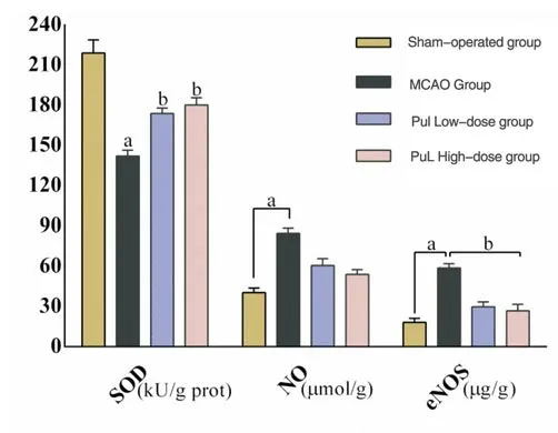
Figure 3 The levels of SOD, NO and eNOS in the brain tissues of rats in each group (n=6)
3.4 Phyllanthus urinaria extract on the count of activated Caspase-3 positive cells in rat brain tissue
After 72 hours of cerebral ischemia in each group, rats in each group were stained by immunofluorescence, and fluorescein isothiocyanatespecific markers were used to emit light. Caspase-3 positive cells expressed green fluorescence. It can be seen under the microscope that Figure 4A shows the sham operation group. Scattered Caspase-3 positive cells can be seen in mouse brain tissue(3.03±0.59);Figure 4B is the MCAO group, Figure 4C is the PuL low-dose, and Figure 4D is the high-dose group. The Caspase-3 positive cell counts in the rat brain tissue are (40.30±4.42), (26.82±4.75),(21.11±4.17), significantly higher than the sham operation group(FMCAO=42.435、FLow=9.208、FHigh=33.373;tMCAO=-26.408、tLow=-15.683、tHigh=-13.553, Paverage<0.01);Compared with the MCAO group, the Caspase-3 positive cell counts in the brain tissue of the PuL low-dose and high-dose groups were significantly reduced, and it was statistically significant,[(26.82±4.75)、(21.11±4.17)vs(40.30±4.42);FLow=0.174、FHigh=0.247;tLow=6.573、tHigh=9.982, PLow<0.05、Paverage<0.01].Tip: Phyllanthus urinaria can inhibit Caspase-3 from executing apoptotic protein to prevent the proliferation of nerve cells. See Figure 4.
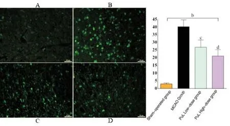
Figure 4 Count of activated Caspase-3 positive cells in the brain tissue of rats in each group (immunofluorescence staining ×200, n=6)
3.5 The effect of Phyllanthus urinaria extract on the expression of PI3K and AKT protein in rat brain tissue
After 72 hours of cerebral ischemia in each group, Figure 5B shows the expressions of PI3K and AKT protein in the brain tissue of the MCAO group were (0.64±0.17) and (0.58±0.13), Compared with Figure 5A, the expression of PI3K and AKT protein (1.19±0.15)and (1.61±0.24) in the brain tissue of rats in the sham operation group were significantly lower, and the difference was statistically significant,(FPI3K=0.016、FAKT=1.594;tPI3K=6.269、tAKT=11.351,Paverage<0.05).Figure 5C and Figure 5D are PuL low and high dose groups, and the expressions of PI3K and AKT protein in rat brain tissue are (0.81±0.18), (0.95±0.19); (1.27±0.17), (1.05±0.14),respectively Significantly higher than the MCAO group,among them, the expression of PI3K protein in the brain tissue of the PuL high-dose group was significantly different from that of the MCAO group,(F=0.133, t=-3.192, P<0.05);The expression of AKT protein in the brain tissue of the PuL low-dose group was significantly different from that of the MCAO group,(F=0.435, t=-8.835, P<0.05).The results are shown in Figure 5. It is suggested that Phyllanthus urinaria can inhibit the apoptosis of brain neurons and make neuronal cells survive through anti-apoptosis.

Figure 5 The expression of PI3K and AKT protein in the brain tissue of rats in each group (n=6)
4. Discussion
Cerebrovascular diseases are increasing year by year. Ischemic brain injury is one of the common diseases. After the onset, the blood supply arteries often function abnormally, resulting in insufficient blood supply to the brain tissue[11].At present, there are many reports on the application of traditional herbal medicine to reduce ischemic brain injury, which shows that traditional herbal medicine has certain advantages in this field. By improving cerebral circulation,protecting neurons, clearing freely, and reducing secondary reactions to cerebral ischemia[12].In this study, Phyllanthus urinaria extract was used to interfere with cerebral ischemia rats and found that the neurological damage in rats was lower than the MCAO group at the three time periods of cerebral ischemia at 24h, 48h and 72h,suggesting that the body after brain injury progressed with time It may cause self-clearing damage to restore neurological function,but the results show that Phyllanthus urinaria extract has stronger clearance ability than MCAO group rats at each time point after intervention, and the percentage of cerebral infarction area is also significantly reduced, indicating that the sublobium Beads may help the body's self-cleaning function to protect brain cells after ischemic stroke in rats, accelerate the recovery of nerve function, and reduce the area of cerebral infarction.
After cerebral ischemic injury, many pathological cascades occur, such as damage caused by inflammation, damage caused by oxidative stress, and the beginning of cell apoptosis. These factors will influence and interact with each other, eventually forming a vicious circle of induction loop, leading to cell necrosis or apoptosis. It is also the main difficulty in the clinical treatment process.eNOS is the main subtype and main source of NO in blood vessels, and its deficiency can lead to vascular diseases and various pathophysiological diseases[13].High levels of nitric oxide can induce neuronal apoptosis. Studies have found that the increase in apoptosis after cerebral ischemia may be related to the production of reactive oxygen species, which is manifested as a decrease in the level of antioxidant enzyme SOD activity[14].The results of this study found that after giving Phyllanthus urinaria extract to intervene in MCAO model rats, the contents of NO, eNOS and SOD in the rat brain tissue were improved to a certain extent, suggesting that Phyllanthus urinaria extract may play a certain role in oxidative stress damage.It can improve the oxidative stress damage after cerebral ischemic injury in rats, thereby inhibiting the apoptosis of nerve cells to protect the cerebral ischemic injury.
Cerebral ischemic damage forms the central ischemic zone, the brain tissue in the ischemic zone may suffer programmed death and increase the degree of brain damage. Caspase-3 is also an important protease. If in the acute phase of cerebral ischemic injury, it can promote cell apoptosis and can lead to neuronal death; while in the recovery phase of cerebral ischemic injury, it can inhibit The growth of nerve cells and prevents the regeneration of brain function after injury[15].It is also reported that the expression of caspase-3 in the brain tissue of humans or rats will be up-regulated when cerebral ischemic injury occurs[16].Therefore, Caspase-3 has been identified as an important mediator of cell death. When caspase-3 is activated,brain tissue cells will involuntarily undergo apoptosis.It has also been found that in the rat model of cerebral infarction by blocking or inhibiting the effect of drugs that block or inhibit its high expression of caspase-3, it has exerted a significant neuroprotective effect[17].In this study, immunofluorescence was used to observe whether there was apoptosis in the expression of caspase-3 in rat brain tissue.The results showed that after 72 hours of cerebral ischemia, the caspase-3 positive cell counts in the other three groups except the sham operation group All were significantly increased, but after the intervention of Phyllanthus urinaria extract, the caspase-3 positive cell count was much lower than that of MCAO group rats, indicating that Phyllanthus urinaria may inhibit the expression of caspase-3 to prevent neuronal apoptosis and protect brain deficiency Injury after blood.
Akt is a threonine kinase that acts downstream of phosphatidylinositol-3 kinase (PI3K). It relies on the PI3K pathway to play a role in brain cell survival after brain injury. The PI3KAkt signaling pathway can prevent nerve cell apoptosis and prevent bleeding and deficiency. Bloody stroke has a neuroprotective effect. Therefore, PI3K and Akt have the functions of accelerating cell proliferation and anti-apoptosis. Studies have shown that this signaling pathway plays a key role in ischemic damage to the brain,heart and kidney[18].In addition, PI3K and Akt play a major role in protecting brain tissue and nerve cell death in cerebral ischemia and hypoxic brain injury[19].The results of this study showed that after cerebral ischemic injury, except for sham operation, the expression of AKT and PI3K proteins in the other groups decreased to varying degrees, especially in the MCAO group, and the rat brain after the action of Phyllanthus The expression of AKT and PI3K proteins in the tissues was greatly improved, which was significantly higher than that in the MCAO group. It is suggested that Phyllanthus urinaria protects against cerebral ischemia by reducing cell apoptosis and possibly by stimulating PI3K-Akt signaling pathway.
In summary, the protection of traditional medicine on the nervous system is gradually becoming critical.This study started from the aspect of anti-oxidation. The results showed that although Phyllanthus urinaria has a certain regulatory effect on several key indicators of cerebral ischemia injury, the scope is too wide, and the deeper and more detailed mechanisms such as gene regulation have not been clarified. Therefore, it is a shortcoming of this research. I hope that we can further dig deeper and continuously verify the key aspects of its dominance, and expand the content of this field. It also provides basic experimental basis for the future clinical application of Phyllanthus urinaria.
杂志排行
Journal of Hainan Medical College的其它文章
- Analysis on medication rule of traditional Chinese medicine treating chemotherapy-induced diarrhea based on traditional Chinese medicine(TCM) inheritance computing platform system
- Study on medication rules of traditional Chinese medicine for Meniere's disease based on data analysis
- Efficacy and safety of traditional Chinese medicine in the treatment of coronary heart disease complicated with anxiety and/or depression after PCI: A systematic review and meta-analysis
- Effect of Xifeng Capsule on blood stasis in patients with rheumatoid arthritis by regulating miR-126-VEGF/PI3K/AKT signaling pathway
- Design and characterization of a bifunctional bybrid antibacterial peptide LLH for bactericidal/endotoxin neutralization effects
- Box-Behnken response surface method combined with fingerprint to optimize the extraction process of total anthraquinone from Cassia seeds