Neuroretinal dysfunction in patients affected by neurofibromatosis type 1
2022-05-15AntoniettaMoramarcoLucaLucchinoFabianaMalloneMichelaMarcelliLudovicoAlisiVincenzoRobertiSandraGiustiniAlessandroLambiaseMarcellaNebbioso
INTRODUCTION
Neurofibromatosis type 1 (NF1), also known as Von Recklinghausen disease, is a rare genetic disorder that is transmitted in an autosomal dominant fashion, with complete penetrance and variable expressivity. It is caused by a mutation in the
gene located on chromosome 17q11.2 which encodes for neurofibromin, a tumor suppressive protein involved in RAS signaling pathways
. The disease is 50%sporadic or inherited, and it occurs with an estimated frequency of approximately 1:2500-1:3500, without any known gender or ethnic predilections. Individuals with NF1 are prone to the development of both malignant and benign nervous system tumors, skeletal dysplasia, and skin abnormalities
.
医学研究生在文献信息检索及应用方面主要存在以下几方面的问题:文献信息意识淡薄;文献信息检索知识短缺;文献信息使用能力薄弱。目前研究生大多使用网络搜索引擎来查找专业资料,并且大部分学生并不知道有很多专业数据库可提供所需的专业文献资源,而在文献类型的利用上,对会议论文、专利文献、标准文献和科技报告的利用率不高[3]。同时,我国高等教育机构的文献信息知识教育体系不够完善,大部分高校的文献检索课程是选修课,教学大纲、教材 、课时 、考核等各校没有统一的标准,不利于研究生对文献信息知识的系统掌握。
①刘志彪:《为高质量发展而竞争:地方政府竞争问题的新解析》,《河海大学学报》(哲学社会科学版)2018年第2期;何艳玲、李妮:《为创新而竞争:一种新的地方政府竞争机制》,《武汉大学学报》(哲学社会科学版)2017年第1期。
阿尔及利亚盖尔达耶是一座矗立在撒哈拉北部已有千年历史的神秘古城,它曾给柯布西耶、原广司、山本理显、隈研吾和西泽立卫等建筑师带来启示,默默地影响着世界建筑的走向。盖尔达耶在不断的发展与更新中衍生出独特的聚落特征,对抗着沙漠中的光与热,也以其独特的格局庇护着守卫它的生灵。
根据《意见》要求,各高校对创新创业教育理论和实践都进行了有益的探索,如开发就业创业课程体系、制定学分转换政策、搭建创新创业实践平台、建立创客空间等,为大学生进行个性化指导和持续性帮扶,并取得了一定的成绩。在校创业学生可以享受到专业导师指导、固定场地保障、浓厚创业氛围等有利条件。可一旦毕业,这些学生即将面临优厚待遇“失效”的窘境。这也使学生的创业面临更多困难,高校不能充分发挥“扶上马,送一程”的责任。这时就需要社区创客空间发挥其服务终身学习、致力创新创业继续教育的优势。
However, electrophysiological abnormalities were also reported in the absence of optic gliomas in NF1 patients.Specifically, abnormal VEPs were described in NF1 regardless of the presence of gliomas of the optic pathways or of the brain. These findings were ascribed to a primary abnormality of visual processing in NF1
. Similarly, our group demonstrated subclinical impairment in the conduction of visual stimuli in patients with NF1 and absence of any condition affecting the optical pathways, as assessed on VEPs and frequency-doubling technology (FDT) campimetry
.
Unlike the optical pathways, electrophysiological evaluation of the neuroretina as an earlier indicator of the damage to the axons forming the optic nerve in NF1 has scarcely been characterized. Experimental studies on murine models of NF1 and OPGs showed progressive loss by apoptosis of retinal ganglion cells (RGCs) occurring in early phases of OPG development
. In accordance, inner retinal dysfunction was reported in a subgroup of patients with NF1 and OPGs on electroretinogram (ERG) examination
.
However, neuroretinal function in NF1 in the absence of OPGs is a relatively unknown topic.
Each subject underwent detailed clinical examination including: Snellen measurement of the BCVA, biomicroscopic examination of the anterior segment, Goldmann applanation tonometry, mydriatic indirect fundus biomicroscopy, crosssectional spectral domain-optical coherence tomography(SD-OCT) and SD-OCT in near-infrared reflectance (NIR)modality, mfERG exam. Additionally, all NF-1 patients underwent 1.5-Tesla magnetic resonance imaging (MRI) scan of the brain to assess the presence of OPGs.
The mfERG is a technique that allows local ERG responses to be recorded simultaneously from many regions of the retina
. Specifically, through the simultaneous stimulation of multiple retinal areas and recording of each response independently, mfERG provides a topographic measure of retinal electrophysiological activity in the central 25 degrees retina.
Therefore, the aim of this study was to examine retinal function by using the mfERG test in NF1 patients without OPGs and any other disorder of the visual pathways.
SUBJECTS AND METHODS
This observational, cross-sectional study was conducted at the University of Rome ‘Sapienza’, Umberto I Hospital, Italy, from June 2019 to February 2020. The study was prospectively reviewed by the Ethics Committee of the Sapienza University of Rome. The research followed the tenets of the Declaration of Helsinki, and informed consent was obtained from all subjects of the study.
传统游戏很有趣,但是面对快速发展的社会,幼儿更加愿意接受新事物。幼儿教师本来在年龄上与幼儿就有很大的距离,为了近距离和幼儿的心灵接触,就要在保持一颗童心的基础上与时俱进,这样才会更容易被幼儿所接受和信任。教学中我们要根据教学大纲开发具有时代特征的游戏。
如何正确运用单因素方差分析——药物研究中的统计学(二)……………………… 《药学与临床研究》编辑部(2·159)
SD-OCT scans were obtained with the Spectralis OCT(Spectralis Family Acquisition Module, V 5.1.6.0; Heidelberg Engineering, Heidelberg, Germany), following a standardized protocol.
Inclusion criteria were: best corrected visual acuity (BCVA)not less than 20/20 in each eye, refractive defects less than±4 D (spherical equivalent), absence of OPGs and any other disorder of the visual pathways, absence of ocular, systemic and/or neuroretinal pathologies that could affect retinal function, absence of NF1-related manifestations at fundus oculi examination.
Exclusion criteria included: poor collaboration which prevented the correct execution of diagnostic exams, excessive signal-to-noise ratio, artifacts and non-uniform waveforms at mfERG.
Healthy subjects were recruited from outpatients of the eye clinic of the University of Rome ‘Sapienza’.
In the present study, we assessed neuroretinal function by using the multifocal electroretinography (mfERG).
2018年3月初,顺丰收购新邦71%股份,成立新公司“顺心”。新邦原有资产和业务将转移到新公司,而顺丰则通过此次收购的方式,快速布局零担市场。
We included 35 consecutive patients (35 eyes; 21 females and 14 males) between 18 and 55 years of age (mean age:31±10.1y) with a diagnosis of NF1 based on the National Institutes of Health (NIH) criteria
and 30 healthy subjects(HC group; 30 eyes; 17 females, 13 males) between 18 and 60 years of age (mean age 33.30±6.0y) for control group.
The mfERG exam was performed after administration of 1%tropicamide topical solution in both eyes, followed by an adaptation to daylight for about 30min. The mfERG recording was performed by using an ERG-Jet corneal contact lens active electrode under topical anesthetic solution of 0.5% benoxinate topical solution in the subject eye. The reference electrode was attached next to the corresponding outer canthus. The neutral electrode was applied with conduction gel on the patient’s earlobe. The examination was performed individually for each eye for a duration of approximately 5-7min, with application of a bandage on the fellow eye. Each patient was placed on the chin-guard of the visual stimulator at 33 cm from the display and was corrected with a temple lens for near vision, where required, due to the pharmacologically induced accommodative block. The stimulus was represented by a pseudorandomized sequence of alternating light and dark hexagonal flashes, with any given flash having a 50% possibility to change in each single frame
. Electrodes were connected with a junctional box from which the amplified signals were delivered to a digital recording system for graphic transformation into a path of negative and positive waves. The mfERG signals were analyzed on the computerized Optoelectronic Stimulator Vision Monitor MonPack 120 Metrovision (Perenchies,France) with reference to the International Society for Clinical
Electrophysiology of Vision (ISCEV) guidelines
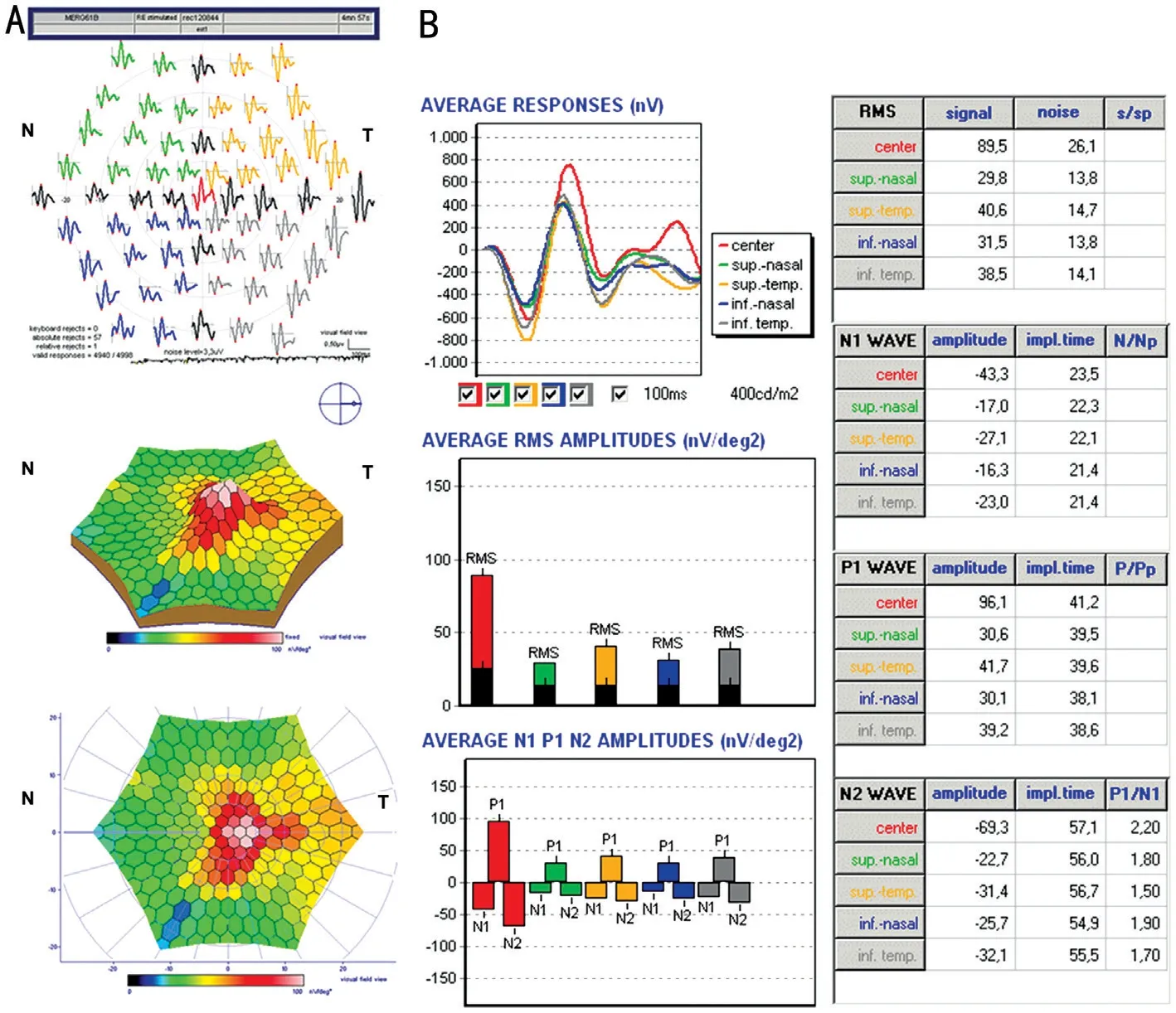
. The first order Kernel mfERG component was used to evaluate amplitude and implicit times of N1, P1, and N2 wave peaks.The areas were analyzed in quadrants from 2 to 25 degrees of eccentricity relative to fixation and the analysis generated a histogram for each of the extended zones. The analysis was carried out on 6 zones: the 2 central degrees, the 4 quadrants from 2 to 25 degrees of eccentricity, and the overall average of the 4 quadrants, for an array of 61 hexagons scaled with eccentricity by means of the 61 program. The analysis of mfERG data was performed in two steps: analysis of the trace arrays, evaluating the shape of the wave; and group averages,evaluating the differences of the absolute values of amplitude and implicit time of P1 wave between NF1 and HC groups.
The right eye was selected for data analysis in each study subject, and assessors were masked to whether or not the patients and controls had NF1.
The normal distribution of data was assessed using the D’Agostino-Pearson test. Statistical significance was determined by Fisher’s exact test for qualitative variables.Comparisons between groups were performed by Student
-test and Mann-Whitney
test, respectively for normally and not normally distributed data. Comparisons between more than 2 groups were performed by repeated measures ANOVA test and Friedman test, respectively for normally and not normally distributed data. A value of
≤0.05 was considered statistically significant. Statistical analysis was performed using Graph Pad vers. 8.0.2 and IBM
SPSS
Statistics version 24.0 (IBM Corp., Armonk, NY, USA) on the Windows 10 Home edition platform. v22 (IBM SPSS Statistics, IBM
, IL, USA).
中国城乡二元结构体系的基本特征将公民分为两类,对城镇居民和农民实行不同的政策。然而长期实行这种体制的后果就是农村和城市的发展不均衡,城乡差距越来越大,对农民的生产积极性和生产力造成了严重束缚。打破城乡二元结构体系,可以从户籍制度改革和土地集体所有制入手。政府应采取相关政策,解除户籍制度的限制,实现农民与城镇居民户口的平等;并改革土地所有制,对土地制度进行一定的调整,制定相关详细的法规并加以规范,充分保障农民的土地财产权。只有实行户籍制度和土地制度改革,才能打破城乡二元结构体系,保障农民的土地产权。
RESULTS
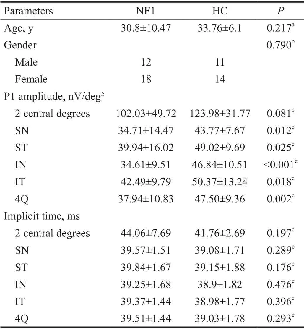
Five eyes in NF1 group and 5 eyes in the HC group were excluded due to excessive signal-to-noise ratio on mfERG.Therefore, the final samples consisted of 30 patients (18 females, 12 males), mean age 30.8±10.47y, for a total of 30 eyes examined; whereas HC group consisted of 25 subjects(14 females, 11 males), mean age 33.76±6.1y, for a total of 25 eyes examined. There were no significant differences between groups in terms of age and gender (Table 1).
The BCVA was 20/20 in all patients, as per inclusion criteria.Lisch nodules were detected in 25 eyes (83.3%) of NF1 patients and none of the HC group. Intraocular pressure was within normal limits in the totality of patients. No pathologic alterations were identifiable at mydriatic indirect fundus biomicroscopy exam in both the NF1 and HC group.


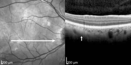
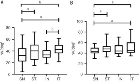
At cross-sectional SD-OCT and NIR-OCT evaluation, 28 eyes out of 30 (93.3%) in the NF1 group showed the presence of choroidal nodules variously distributed to the posterior pole,whereas no choroidal abnormalities were recognizable in the HC group (Figure 1). At MRI evaluation, no patient had optic nerve gliomas or other lesions involving the optic pathways.The analysis of the trace arrays showed no differences in the uniformity of the waveforms between NF1 patients and HC subjects. NF1 patients had significantly lower values of the P1-wave amplitudes in all of the 4 quadrants when compared to HC (Table 1, Figures 2 and 3), whereas, there were no differences of the P1-wave amplitude in the 2 central degrees between the groups. In addition, a statistically significant difference was observed among the P1 wave amplitudes as recorded in the 4 quadrants within the NF1 group. Specifically,lower amplitudes were recorded in the nasal quadrants(Table 2, Figure 4). Similar results were obtained for the HCs as summarized in Table 2. Table 2 shows the comparison analyses among the P1-wave amplitudes as evaluated in the 4 quadrants in NF1 patients and HC subjects. No statistically significant differences were observed in the absolute values of implicit time between NF1 patients and HCs (Table 1).Moreover, no differences were observed in the implicit times as recorded in the 4 quadrants within the NF1 group and among HCs.


DISCUSSION
The purpose of our study was to examine neuroretinal function in NF1 patients without OPGs compared to a group of HCs by the use of mfERG. The mfERG is a validated methodology for clinical evaluation of several conditions,including retinitis pigmentosa, hydroxychloroquine toxicity,glaucoma, ocular vascular occlusive disorders
. Revealing subclinical abnormalities of retinal function, mfERG can identify patients at risk of retinal damage progression allowing for early therapeutic interventions
. Currently, there are no available studies in the literature investigating the value of mfERG in NF1 patients. Limited studies demonstrated retinal electrofunctional anomalies in NF1 regardless of the presence of OPGs, although the clinical meaning of these findings is still unknown
. Lubiński
performed electro-oculogram(EOG) and full-field flash ERG evaluations in patients diagnosed with NF1 and variable ocular manifestations including optic nerve gliomas (<5%), and compared results to normal HC. These authors reported a significative increase in the Arden indexes of the EOG test in NF1 patients, whereas there were no recorded abnormalities in the flash ERG examination. The reported EOG changes were attributed to calcium level variations caused by melanin abnormalities related to reduced expression of neurofibromin
.
To our knowledge, this is the first study evaluating neuroretinal function in NF1 patients in the absence of OPGs.

Moreover, the NF1 group presented a high percentage of chorioretinal alterations consisting of choroidal nodules when compared to HCs.
The exact origin of the P1 wave is still debated. There is evidence that the same cellular elements that contribute to the full field ERG b-wave formation could be the source of the P1 wave. The major contribution may derive from the depolarization of bipolar cells activated by a light source,therefore, it is believed that the b-wave is generated by cells in the inner retina
.
There are studies showing that NF1 RGCs exhibit shortened neurite length and reduced growth cone areas, with decreased survival in response to different types of injury compared to wild-type counterparts. These defective neuronal phenotypes have been suggested to reflect an abnormal neurofibrominmediated cyclic adenosine monophosphate (cAMP) generation
.CAMP is a derivative of adenosine triphosphate (ATP) and serves as second messenger for intracellular signal transduction.Recently, it has been found that changes in cAMP levels have regulatory influences on the phototransduction cascade.This action exerts expanding the adaptation contingent of photoreceptors to illumination conditions, decreasing its sensitivity in bright light and increasing its sensitivity during the dark part of the day
.
Neurofibromin is a positive regulator of cAMP levels in various cell types including neurons, and its deficit leads to a reduction in cAMP basal levels
. Therefore, we hypothesize that the lower levels of mfERG P1 wave amplitude in NF1 patients found in our study may be attributable to an altered intracellular signal transduction due to abnormal neurofibromin-mediated cAMP generation.
The eye and ocular adnexa are frequently involved in NF1.Some ocular manifestations of NF1 including optic pathway gliomas (OPGs), iris Lisch nodules, orbital and eyelid neurofibromas, eyelid café-au-lait spots, are diagnostic of the disease, whereas additional, recently described ocular features and are not currently diagnostic for NF1 and include choroidal nodules, retinal microvascular abnormalities, and hyperpigmented spots of the fundus oculi
.The presence of electrophysiological changes in NF1 was previously investigated on visual evoked potentials (VEPs) in patients with related OPGs. Notably, abnormal visual evoked responses allowed for early detection of optic gliomas in NF1 and earlier intervention prior to significant visual loss
.
This hypothesis provides a possible biological explanation of the electrofunctional results obtained.
In our study, we identified electro functional disorders in NF1 patients consisting of P1 wave alterations. Specifically, NF1 patients showed a statistically significant reduction in the mfERG P1 wave amplitude in the 4 quadrants when compared to HC, with no recorded differences of the P1 wave amplitude in the 2 central degrees between the groups. These alterations were subclinical as the recruited patients all presented with normal or corrected to normal visual acuity and no underlying disease that could affect retinal function. Moreover, the absence of ERG alterations in the 2 central retinal degrees,corresponding to the fovea centralis, reflected the absence of visual impairment in our patients.
Variable in number and morphology, mostly located at the posterior pole, choroidal nodules are a frequent manifestation of ocular involvement in NF1 patients
. These lesions represent amounts of proliferating Schwann cells, melanocytes and ganglion cells around axons of the ciliary nerves innervating the choroid
. They appear as hyperreflectivewhitish lesions at SD-OCT in NIR modality, with variable features from well-defined to dull, confluent margins,according to previous evidence
. A few authors described a thinning of the overlying retinal tissue in correspondence of choroidal nodules in NF1, expression of sub-atrophy of retinal layers
. More recently, low flow areas overlying choroidal nodules were demonstrated at the level of choriocapillaris on OCT-angiography in a single-case report, showing topographical matching with areas of reduced chorioretinal thickness
. However, we are currently unable to establish any correlation between the impairment of retinal function and the presence of choroidal nodules. Further investigation aimed at evaluating choroidal nodules-related low flow to the retina and corresponding retinal functional alterations detected by the use of mfERG is encouraged.
3.2 无菌操作 操作中必须树立牢固的无菌观念,严格执行无菌操作技术。头部毛发较多,毛囊内容易滋生细菌,因此穿刺前必须仔细备皮,认真清洁皮肤,严格消毒,保证足够面积的无菌区,至少为10 cm×10 cm。剃头发时动作轻柔,避免剃破皮肤,引起感染。
Additional findings from our study consisted in significative differences in the mfERG P1 wave amplitudes in the 4 quadrants within the NF1 group, with lower amplitudes detected in the nasal quadrants.
In HCs, we observed similar differences regarding amplitude values with lower values registered in the supero-nasal quadrant if compared to temporal quadrants (SN
ST; SN
IT; Table 2).
These results are in agreement with previous evidence
,showing that lower values of amplitudes in the nasal quadrants appear to be physiological in multifocal evaluation.
This may explain why, although significantly reduced amplitudes are detectable in each quadrant of the mfERG evaluation in NF1 patients compared to HCs, the amplitudes in the nasal quadrants appear to be the most affected in both groups.
The clinical significance of the recorded electro-functional abnormalities in NF1 patients remains unclear. Prospective studies are needed to evaluate long-term responses in patients with NF1 and potential correlation with progressive visual impairment. In summary, mfERG evaluation in patients affected by NF1 showed a decreased amplitude of the P1 wave between 2 and 25 central retinal degrees attributable to retinal function impairment. This abnormality is subclinical as all patients did not have a reduced visual acuity nor had any underlying disease that could have affected the outcome of the research. These observations suggest a possible use of mfERG as subclinical retinal damage indicator with a potential utility in clinical practice for the follow-up of NF1 patients.
项目名称:海南市三亚老干部休养所建筑设计:美国丹尼尔连设计事务所建筑施工:上海韩进建筑有限公司项目所在地:海南
Study conception and design:Moramarco A, Nebbioso M. Acquisition of data: Moramarco A,Lucchino L, Nebbioso M. Analysis and interpretation of data:Moramarco A, Nebbioso M, Lucchino L, Roberti V. Drafting of manuscript: Lucchino L, Mallone F, Marcelli M, Nebbioso M,Alisi L. Critical revision: Moramarco A, Giustini S, Nebbioso M,Lambiase A.
None;
None;
None;
None;
None;
None;
None;
None;
None.
1 Cimino PJ, Gutmann DH. Neurofibromatosis type 1.
2018;148:799-811.
2 Wilson BN, John AM, Handler MZ, Schwartz RA. Neurofibromatosis type 1: new developments in genetics and treatment.
2021;84(6):1667-1676.
3 Ly KI, Blakeley JO. The diagnosis and management of neurofibromatosis type 1.
2019;103(6):1035-1054.
4 Kinori M, Hodgson N, Zeid JL. Ophthalmic manifestations in neurofibromatosis type 1.
2018;63(4):518-533.
5 Moramarco A, Mallone F, Sacchetti M, Lucchino L, Miraglia E,Roberti V, Lambiase A, Giustini S. Hyperpigmented spots at fundus examination: a new ocular sign in neurofibromatosis type 1.
2021;16(1):147.
6 Moramarco A, Sacchetti M, Franzone F, Segatto M, Cecchetti D, Miraglia E, Roberti V, Iacovino C, Giustini S. Ocular surface involvement in patients with neurofibromatosis type 1 syndrome.
2020;258(8):1757-1762.
7 North K, Cochineas C, Tang E, Fagan E. Optic gliomas in neurofibromatosis type 1: role of visual evoked potentials.
1994;10(2):117-123.
8 Vagge A, Camicione P, Pellegrini M, Gatti G, Capris P, Severino M, di Maita M, Panarello S, Traverso CE. Role of visual evoked potentials and optical coherence tomography in the screening for optic pathway gliomas in patients with neurofibromatosis type I.
2021;31(2):698-703.
9 Iannaccone A, McCluney RA, Brewer VR, Spiegel PH, Taylor JS,Kerr NC, Pivnick EK. Visual evoked potentials in children with neurofibromatosis type 1.
2002;105(1):63-81.
10 Nebbioso M, Moramarco A, Lambiase A, Giustini S, Marenco M,Miraglia E, Fino P, Iacovino C, Alisi L. Neurofibromatosis type 1:ocular electrophysiological and perimetric anomalies.
2020;12:119-127.
11 Hegedus B, Hughes FW, Garbow JR, Gianino S, Banerjee D, Kim K,Ellisman MH, Brantley MA, Gutmann DH. Optic nerve dysfunction in a mouse model of neurofibromatosis-1 optic glioma.
2009;68(5):542-551.
12 Abed E, Piccardi M, Rizzo D, Chiaretti A, Ambrosio L, Petroni S,Parrilla R, Dickmann A, Riccardi R, Falsini B. Functional loss of the inner retina in childhood optic gliomas detected by photopic negative response.
2015;56(4):2469-2474.
13 Hoffmann MB, Bach M, Kondo M, Li SY, Walker S, Holopigian K,Viswanathan S, Robson AG. ISCEV standard for clinical multifocal electroretinography (mfERG) (2021 update).
2021;142(1):5-16.
14 National Institutes of Health Consensus Development Conference Statement: neurofibromatosis. Bethesda, Md., USA, July 13-15, 1987.
1988;1(3):172-178.
15 Asanad S, Karanjia R. Multifocal electroretinogram. 2021 Dec 12. In:
. Treasure Island (FL): StatPearls Publishing; 2022 Jan-.
16 Tsang AC, Ahmadi S, Hamilton J, Gao J, Virgili G, Coupland SG,Gottlieb CC. The diagnostic utility of multifocal electroretinography in detecting chloroquine and hydroxychloroquine retinal toxicity.
2019;206:132-139.
17 Tanaka H, Ishida K, Ozawa K, Ishihara T, Sawada A, Mochizuki K,Yamamoto T. Relationship between structural and functional changes in glaucomatous eyes: a multifocal electroretinogram study.
2021;21(1):305.
18 Nebbioso M, Livani ML, Steigerwalt RD, Panetta V, Rispoli E.Retina in rheumatic diseases: standard full field and multifocal electroretinography in hydroxychloroquine retinal dysfunction.
2011;94(3):276-283.
19 Özmert E, Arslan U. Management of retinitis pigmentosa by Wharton’s jelly derived mesenchymal stem cells: preliminary clinical results.
2020;11:25.
20 Nebbioso M, Grenga R, Karavitis P. Early detection of macular changes with multifocal ERG in patients on antimalarial drug therapy.
2009;25(3):249-258.
21 Cetınkaya E, Inan S, Yıgıt K, Sabaner MC, Inan ÜÜ. Spectral-domain optical coherence tomography and multifocal electroretinography results in the long-term follow-up of glaucoma patients.
2020:22-32.
22 Sahay P, Kumawat D, Gupta S, Tripathy K, Vohra R, Chandra M,Venkatesh P. Detection and monitoring of subclinical ocular siderosis using multifocal electroretinogram.
(
) 2019;33(10):1547-1555.
23 Lubiński W, Zajaczek S, Sych Z, Penkala K, Palacz O, Lubiński J.Electro-oculogram in patients with neurofibromatosis type 1.
2001;103(2):91-103.
24 Lubiński W, Zajaczek S, Sych Z, Penkala K, Palacz O, Lubiński J.Supernormal electro-oculograms in patients with neurofibromatosis type 1.
2004;2(4):193-196.
25 Creel DJ. Electroretinograms.
2019;160:481-493.
26 Brown JA, Gianino SM, Gutmann DH. Defective cAMP generation underlies the sensitivity of CNS neurons to neurofibromatosis-1 heterozygosity.
2010;30(16):5579-5589.
27 Astakhova LA, Kapitskii SV, Govardovskii VI, Firsov ML. Cyclic AMP as a regulator of the phototransduction cascade. Vol. 44,
. Springer New York LLC,2014:664-671.
28 Deraredj Nadim W, Chaumont-Dubel S, Madouri F, Cobret L, De Tauzia ML, Zajdel P, Bénédetti H, Marin P, Morisset-Lopez S.Physical interaction between neurofibromin and serotonin 5-HT6 receptor promotes receptor constitutive activity.
2016;113(43):12310-12315.
29 Viola F, Villani E, Natacci F, Selicorni A, Melloni G, Vezzola D, Barteselli G, Mapelli C, Pirondini C, Ratiglia R. Choroidal abnormalities detected by near-infrared reflectance imaging as a new diagnostic criterion for neurofibromatosis 1.
2012;119(2):369-375.
30 Kurosawa A, Kurosawa H. Ovoid bodies in choroidal neurofibromatosis.
1982;100(12):1939-1941.
31 di Nicola M, Viola F. Ocular manifestations in neurofibromatosis type 1.
. Cham:Springer International Publishing, 2020:71-84.
32 Chilibeck C, Shah S, Russell H, Vincent A. The presence and progression of choroidal neurofibromas in a predominantly pediatric population with neurofibromatosis type-1.
2021;42(5):1-7.
33 Ayata A, Unal M, Ersanli D, Tatlipinar S. Near infrared fluorescence and OCT features of choroidal abnormalities in type 1 neurofibromatosis.
2008;36(4):390-392.
34 Abdolrahimzadeh S, Plateroti AM, Recupero SM, Lambiase A.An update on the ophthalmologic features in the phakomatoses.
2016;2016:3043026.
35 Kumar V, Singh S. Multimodal imaging of choroidal nodules in neurofibromatosis type-1.
2018;66(4):586-588.
36 Hood DC. Assessing retinal function with the multifocal technique.
2000;19(5):607-646.
猜你喜欢
杂志排行
International Journal of Ophthalmology的其它文章
- Multimodal imaging in immunogammopathy maculopathy secondary to Waldenstrom’s macroglobulinemia: a case report
- Periorbital necrotizing fasciitis accompanied by sinusitis and intracranial epidural abscess in an immunocompetent patient
- Multimodal imaging in Purtscher-like retinopathy associated with sarcoidosis: a case report
- Can a sneeze after phacoemulsification cause endophthalmitis? A case report
- Persistent macular oedema following Best vitelliform macular dystrophy undergoing anti-VEGF treatment
- Genetic, environmental and other risk factors for progression of retinitis pigmentosa
