Prophylactic juxtapapillary laser photocoagulation in pediatric morning glory syndrome
2022-05-15YiHuaZouKaiQinSheJiaNingRenTingYiLiangPingFeiYuXuJingLiXiangZhangJiePengPeiQuanZhao
INTRODUCTION
Morning glory syndrome (MGS) is a rare congenital cavitary anomaly of the optic disc, that was first named by Kindler
in 1970 because of its resemblance to the morning glory flower. It is characterized by an enlarged and excavated optic disc with juxtapapillary chorioretinal pigment disturbance, radial retinal blood vessels, and a central white glial tuft. The prevalence of this condition is 3.6/100 000 in children, and the exact pathogenesis of MGS remains unknown
.
Juxtapapillary laser treatment alone is generally used as a supplement to vitrectomy in RD of MGS. The mechanism of laser treatment is to produce adhesion, through the thermal effect, between the retinal pigment epithelium (RPE),neuroepithelium and choroid to create a barrier to prevent subretinal fluid migration
. Prophylactic juxtapapillary laser treatment is controversial in the literature for the prevention of RD in MGS, as the likely risk of visual loss is attributed to the papillomacular bundle (PMB) injury. However, more animal and human studies have demonstrated that lasers in the region of the PMB do not cause loss of visual function
. Hence,this study aimed to report the preliminary anatomic and visual outcomes of prophylactic juxtapapillary laser treatment alone in a cohort of pediatric MGS patients from a single institution.
It should be mentioned that echography is an important method of examination for MGS. B-scan echography of all MGS eyes in this study showed excavation of the posterior pole, and no RD band was found during the entire followup period. Cennamo
reported that spectral-domain optical coherence tomography could sometimes show RD when echography could not, suggesting that echography may miss the diagnosis of RD sometimes compared to optical coherence tomography. In this study, it was indefinite whether mild subretinal fluid or intraretinal fluid existed in some cases.Despite the good RPE response of the laser in all patients, the progression of serous RD may be observed in a longer time,and the efficacy of laser adhesion as a barrier may be reduced gradually, which needs to be followed up for a longer time.
In the literature, the treatment of advanced RD in MGS is challenging. Some cases are treated when total RD occurs,which requires silicone oil or long-acting gas tamponade
. In addition, cases that have recurrent RD always need multiple interventions
. Complications of these operations are severe in the long term, such as silicone oil emulsification,keratopathy and photophobia
. Despite the reports of cases with successful retinal attachment after treatment
, the follow-up periods in these studies were not long enough,and long-term complications were ignored. In addition, the visual acuity prognosis is very poor in these conditions since RD has occurred for a long time. Considering these findings,early prophylactic treatment is actually needed to improve the anatomical and visual prognosis.
MGS could be complicated with other ocular diseases, such as persistent fetal vasculature (PFV), cataracts, microphthalmia and retinal detachment (RD). It has been reported that approximately 1/3 of those with MGS can develop RD, and a small number of cases have spontaneous resolution
. The development process of RD may result from various factors,mainly abnormal communications between the subarachnoid space and subretinal space or retinal breaks, which contribute to the migration of cerebrospinal fluid or vitreous humor to the subretinal space and the traction of preretinal glial tissues
.
气化炉高压氧气切断阀主要用于氧气输送管路以及氧气吹扫管路快速关闭或者开启。由于介质是氧气,具有助燃性能,阀门一旦发生热量累积、泄漏等,容易引发燃烧甚至爆炸,从而会严重影响装置的平稳运行,因而气化炉高压氧气切断阀对阀门的密封性能、材料选择、使用寿命等性能有非常严格的要求。
SUBJECTS AND METHODS
会宁县是西北教育名县,自恢复高考以来,全县累计向全国大中专业院校输送优秀毕业生5.1万名,其中获得博士学位者500多人,硕士学位者近2000多人,学士学位者近2万人。“领导苦抓,教师苦教,学生苦学,家长苦供,亲友苦帮”的“五苦”精神已经是我们这一区域的特有的教育文化。我们在班级文化建设中深入挖掘教育中可歌可泣的感人的事例,培育学生的核心素养。
Consecutive patients with a diagnosis of MGS from April 2015 to February 2021 were enrolled. The patients underwent comprehensive ophthalmic examinations before and after treatment. All patients were examined with binocular indirect ophthalmoscopy with a +20 D lens or slitlamp microscope by the same clinician (Zhao PQ). Widefield fundus images were taken with RetCam III (Clarity,Pleasanton, California, USA) or Optos Optomap 200Tx (Optos,PLC, Dunfermline, Scotland, UK). A/B-scan echography(CineScan, BVI Co., France) examinations were performed to measure the axial length, maximal depth and width of excavation and to evaluate the status of the whole globe. The frequencies of the probe of the A-scan and B-scan echography were 11 MHz and 10 MHz, respectively. Intraocular pressure(IOP) was measured using a non-contact tonometer (Topcon CT-90, Tokyo, Japan) or rebound tonometer (iCare, Tiolat Oy,Helsinki, Finland) every time.
The criterion for laser treatment was diagnosis of MGS without obvious RD in those aged 0-15y. Patients with refractive media opacity, PFV, tractional or rhegmatogenous retinal detachment (RRD) or without intact data were excluded. All the patients were under general anesthesia monitored by an experienced pediatric anesthesiologist. Fundus photography and fluorescein angiography (FA) were performed according to the reported literature
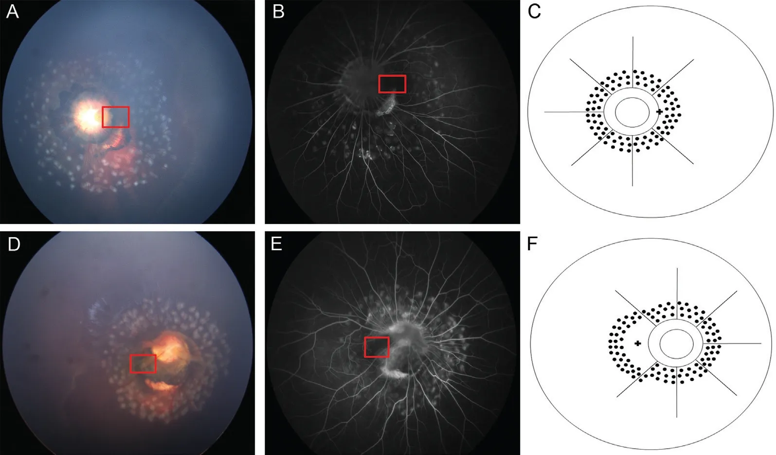
. All patients underwent prophylactic laser treatment alone by two experienced clinicians (Xu Y and Peng J) after fundus photos were taken. This treatment was three to five rows of confluent gray-white laser spots at 360° of the retina around the edge of the optic disc through a 520 nmwavelength, a power of 180-250 mW and a 300ms-duration laser with an indirect ophthalmoscope (Novus, Lumenis, USA).The laser spots were grid, and the intensity was moderate(Figure 1). The laser spots were given around the border of these areas, including atrophy of RPE and small folds around the optic disc (Figure 2A). The visible macular area was spared with laser carefully to protect the PMB (Figure 1).
Baseline data for age, gender, laterality of MGS and symptoms at presentation were collected.Ophthalmic evaluations, including visual acuity, axial length,IOP and ocular complications, were noted one-month, six months and after every year postoperatively.
Cranial magnetic resonance imaging (MRI) was also suggested for all patients.The main outcomes were the status of the retina and best corrected visual acuity (BCVA), which were evaluated during follow-ups.
Statistical analyses were performed using IBM SPSS Statistics, version 22.0 software (Armonk, NY:IBM Corp). Paired
-tests and Mann-Whitney
tests were used to compare the differences in several parameters between two subgroups of patients who were younger than 12 months old at the time of treatment and the others. A
value of less than 0.05 was considered statistically significant.
RESULTS
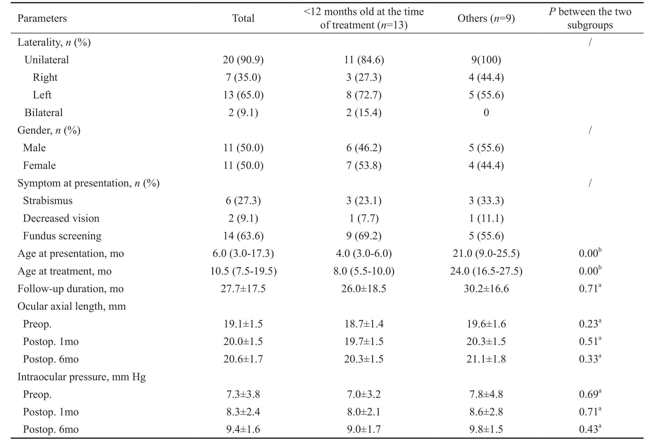
A total of 24 eyes from 22 patients were included in this study. Two (9.1%) patients underwent bilateral treatments.At the time of treatment, 13 (59.1%) patients were younger than 12 months old. The baseline data of the patients are listed in Table 1. The median age at treatment was 10.5mo(interquartile range: 7.5-19.5mo), and the mean follow-up duration was 27.7±17.5mo. Symptoms at presentation included strabismus in 6 (27.3%), decreased vision in 2 (9.1%) and routine fundus screening in 14 (63.6%) patients (Table 1). At the preoperative, postoperative one-month and postoperative six-month follow-ups, the mean ocular axial length and IOP are listed in Table 1. There was an increasing trend of the axial length with age, but no significantly shortened axial length or elevated IOP was found. Fifteen (68.2%) patients underwent cranial MRI examinations. Three of the 15 (20.0%)patients had various abnormal findings in the central nervous system, including one moyamoya, one encephalocele, and one intracranial arteriostenosis.


On fundus photography and B-scan echography at the preoperative and postoperative one-month, postoperative sixmonth and following follow-ups, the anatomic outcomes of all eyes remained stable (Figures 2 and 3). None of them developed obvious RD during the entire follow-up course.Preoperative BCVA acquired from 2 (9.1%) patients ranged from light perception to 20/200. Postoperative acuity acquired from 11 (50.0%) patients ranged from light perception to 20/125 (Table 2). Patient noncompliance limited this examination for others. No visual losses were reported from any patients or their parents.
2)通常情况下,GM(1,1)预测模型要求所采用的数据序列必须是等时间间隔的,而在实际工作中,观测的原始数据往往是非等时间间隔的数据序列,所以,针对此种情况,需要进行数据序列的转换,即把非等间隔序列变换成等间隔序列[3]。


No obvious ocular or systemic complications were reported during the entire follow-up course. No hemorrhage, retinal holes, cataracts or keratitis were found in any patients. No evidence of the spontaneous resolution of RD, such as new subretinal proliferation, was noted. One patient had transient conjunctivitis after treatment and recovered by using topical antibiotics. Due to the poor visual acuity and young age of most patients, visual field examinations were not performed.
This study adhered to the tenets of the Declaration of Helsinki (2008) and was approved by the Ethics Committee of Xinhua Hospital affiliated with the Shanghai Jiao Tong University School of Medicine. Written information consent was acquired from the parents of all patients.
DISCUSSION
MGS is a rare congenital cavitary anomaly of the optic disc.There was a unilateral tendency and no significant difference in occurrence between genders
, which is in agreement with the data in this study. Abnormal findings in the nervous system suggest the importance of cranial examinations for MGS patients in this study, which has also been reported in previous studies
. The median age at presentation was six months old (range: 3.0-17.3mo) in this cohort, suggesting that early fundus screening is worthwhile for early diagnosis and management.
Considering these findings, early prophylactic treatment is truly needed to improve the anatomical and visual prognosis.In the above studies, laser photocoagulation around the optic disc is generally used as a supplement to vitrectomy. However,there remains some ambiguity about the safety of lasers in the region of the PMB. Early animal and human studies suggested that there was no obvious detriment to applying a laser in the region of the PMB
. Recently, more human studies have demonstrated that laser-associated retinal damage is limited to the outer retinal layers. The immediate morphological changes of the inner retina suggested that the RPE prevents damage to the inner retina by absorbing most of the energy of the laser
. More recently, Bloch and da Cruz
reported the safety of juxtapapillary laser photocoagulation in the region of the PMB for optic disc pit maculopathy. No anatomical or perimetric findings consistent with nerve fiber layer damage in the region of laser treatment were reported in any of the patients, suggesting that lasers in the region of the PMB would not cause loss of visual function. On the other hand,it is controversial to treat the fovea directly with the laser for patients with good vision. Fovea-sparing laser treatment has been applied for diabetic macular edema
. As the macula in MGS often develops abnormally or is displaced toward the edge of the excavation, it is important to distinguish the probable site of the fovea, which truly requires some effort.Normal macula is usually yellowish on fundus photography,with a clear foveal reflection and a clear foveal avascular zone(FAZ) on fundus FA
. Therefore, despite the limitation of exactly anchoring the fovea in pediatric MGS patients, the probable whole area of the macula was dignified and spared to protect the fovea and the PMB. Pre- and postoperative visual acuities of two patients who were observed to be unaffected also supported the safety of scrupulous juxtapapillary laser photocoagulation in these patients.
It has been reported that approximately 80% of MGS could be complicated with other ocular diseases, such as PFV, cataracts,microphthalmia and RD
. As one of the most severe complications of MGS, RD has been reported to occur in 30%-38% of MGS patients
. Haik
reported that the natural course of MGS with RD occurred in 11 of 32 eyes during a mean follow-up of 10.3y. Of the 11 eyes, four had spontaneous reattachment, and two of them exhibited redetachment.Evidence of the spontaneous resolution of RD in MGS, such as new subretinal proliferation, was not observed during the follow-up period, which suggested that RD did not occur in all the patients in this study.
In the literature, the treatment of advanced RD in MGS is challenging. Harris
reported an MGS case with near total RD and a tiny retinal break treated with one vitrectomy and two fluid-gas exchanges. The retina remained attached for 14mo after the final surgery. Jo
reported an MGS case with a retinal hole and RD treated with vitrectomy and long-acting gas tamponade. The retina was redetached one month later, and silicone oil tamponade was then used. The retina retained attached for five years after the removal of the silicone oil. Recurrent RD and multiple interventions, such as in these cases, are intolerant for some patients. Sakamoto
reported two MGS cases with total RD and contractile movements treated with vitrectomy, and the retina failed to be reattached in both cases, suggesting the considerable difficulty of the treatment of these conditions. Sen
reported nine MGS cases with RD treated with vitrectomy. Silicone oil tamponade was used in 7/9 eyes, and the rate of retinal reattachment was 66%. However, the long-term complications of the surgeries were unclear. Visual improvement is quite limited in these patients.
存储在机构知识库中的数字信息资源是它的核心所在。如何高效准确地获取信息资源,并且保持数据资源持续更新,是制约机构库发展的关键因素,也是机构知识库建设的最重要的工作。机构库信息资源获取困难制约和阻碍了机构库建设和发展。根据美国网络信息联盟发布的调查报告可以知道,制约高校机构知识库发展的关键因素是高校的教职工和科研人员无法及时向机构库提交信息,困难重重[7]。
For the rationale of laser treatment, retinal laser photocoagulation results in permanent chorioretinal scars as a barrier for preventing subretinal fluid migration
. The scars could be well achieved before an accumulation of intraretinal fluid.Retinal laser photocoagulation has been utilized widely in most retinal vascular diseases, particularly diabetic macular edema and retinal vein occlusions
. Although the pathogenesis of these diseases is different from RD in MGS, lasers in both conditions might decrease retinal edema by stimulating RPE cell to reabsorb subretinal fluid
. In addition, retinal laser photocoagulation has also been extensively used in the treatment of retinal breaks, which can reduce progression to RD. For asymptomatic retinal breaks, prophylactic laser retinopexy is needed in some cases to prevent RD
. In addition, prophylactic laser treatment proved useful in the prevention of RD associated with congenital chorioretinal coloboma
. Uhumwangho and Jalali
showed that RRD developed in 2.9% of laser-treated eyes in contrast to 24.1%of untreated eyes, suggesting that prophylactic laser treatment around the coloboma had a protective effect for the prevention of RRD. For RD in MGS, prophylactic laser treatment around the optic disc may play a role by impeding the migration of subretinal fluid, which may originate from the retinal holes or abnormal communications among various compartments within the excavation to prevent the progression of RD. The data of this study provided the preliminary anatomical and visual outcomes of prophylactic juxtapapillary laser treatment for a cohort of pediatric MGS patients. The results showed that the retinas of all the patients remained flat during a mean follow-up duration of 27.2mo.
《网络安全法》第21条明确规定国家实行网络安全等级保护制度[3],各级教育主管部门也高求高校要落实等级保护制度工作,开展等级保护制度有助于查明单位信息系统与国家的标准是否存在差距,明确系统存在的安全隐患,通过整改之后,提高系统的安全防护能力,降低安全风险,是信息安全工作的头等大事。
Several limitations in this study should be considered. First,the number of patients was small and had unavoidable selection bias. Second, this was a retrospective study, and some examinations, such as optical coherence tomography,visual field examinations and electroretinograms (ERGs), were unavailable due to patient noncompliance. Third, this study lacked a control group without laser treatment. Despite these limitations, this study provides the preliminary anatomic and visual outcomes of prophylactic juxtapapillary laser treatment alone for pediatric MGS patients in a short-term followup. Further long-term clinical observation will be needed to confirm its safety.
2.1.1 3组小鼠每分钟静息通气量比较 3组小鼠在6、18、36 h后的每分钟静息通气量比较,差异均无统计学意义(P>0.05)。见图1a。
In conclusion, this study showed the preliminary anatomic and visual outcomes of prophylactic juxtapapillary laser treatment alone in pediatric MGS patients, which were relatively stable in a short-term follow-up. No severe ocular or systemic complications were reported in any patients. Further long-term clinical observation will be needed to confirm its safety.
Supported by the Shanghai Sailing Program(No.20YF1429700); the Clinical Research Plan of SHDC (No.SHDC2020CR5014-002).
世界卫生组织公布的数据显示,全球癌症发病率和死亡率仍呈迅速上升趋势。全球每年约有880万人死于癌症,占全球每年死亡总人数近1/6,死者大多数在中低收入国家[13-14]。每年有1 400多万新发癌症病例,预计到2030年将增加到2 100多万。癌症带来的经济影响较大且在不断加剧,2010年由癌症导致的年度经济总费用约为1.16万亿美元[15]。目前30%~50%的癌症可通过避免危险因素和落实现有循证预防策略进行预防,通过癌症早期发现和癌症患者管理可有效降低癌症负担[16-17]。如果做到早期诊断和充分治疗,许多癌症会有很高的治愈率[18-19]。
None;
None;
None;
None;
None;
None;
None;
None;
None;
None.
1 Kindler P. Morning glory syndrome: unusual congenital optic disk anomaly.
1970;69(3):376-384.
2 Ceynowa DJ, Wickström R, Olsson M, Ek U, Eriksson U, Wiberg MK, Fahnehjelm KT. Morning glory disc anomaly in childhood - a population-based study.
2015;93(7):626-634.
3 Haik BG, Greenstein SH, Smith ME, Abramson DH, Ellsworth RM.Retinal detachment in the morning glory anomaly.
1984;91(12):1638-1647.
4 Inoue M. Retinal complications associated with congenital optic disc anomalies determined by swept source optical coherence tomography.
2016;6(1):8-14.
5 Lytvynchuk LM, Glittenberg CG, Ansari-Shahrezaei S, Binder S.Intraoperative optical coherence tomography assisted analysis of pars plana vitrectomy for retinal detachment in morning glory syndrome: a case report.
2017;17(1):134.
6 Sen P, Maitra P, Vaidya H, Bhende P, Das K. Outcomes of vitreoretinal surgery in retinal detachment associated with morning glory disc anomaly.
2021;69(8):2116-2121.
7 Harris MJ, de Bustros S, Michels RG, Joondeph HC. Treatment of combined traction-rhegmatogenous retinal detachment in the morning glory syndrome.
1984;4(4):249-252.
8 Toklu Y, Cakmak HB, Ergun ŞB, Yorgun MA, Simsek Ş. Time course of silicone oil emulsification.
2012;32(10):2039-2044.
9 Bartz-Schmidt KU. Communication between the subretinal space and the vitreous cavity in the morning glory syndrome.
1996;234(6):409.
10 Jo YJ, Iwase T, Oveson BC, Tanaka N. Retinal detachment in morning glory syndrome with large hole in the excavated disc.
2011;21(6):841-844.
11 Saab MG, Cordahi GP, Rezende FA. Fibrin sealant in the treatment of retinal detachment in morning glory syndrome.
2011;5(4):326-329.
12 Jain N, Johnson MW. Pathogenesis and treatment of maculopathy associated with cavitary optic disc anomalies.
2014;158(3):423-435.
13 Garoon RB, Smiddy WE, Flynn HW Jr. Treated retinal breaks:clinical course and outcomes.
2018;256(6):1053-1057.
14 Bloch E, da Cruz L. Dense laser at the papillomacular bundle does not cause loss of visual function.
2021;31(4):2160-2164.
15 Kriechbaum K, Bolz M, Deak GG, Prager S, Scholda C, Schmidt-Erfurth U. High-resolution imaging of the human retina
after scatter photocoagulation treatment using a semiautomated laser system.
2010;117(3):545-551.
16 Laatikainen L. The fluorescein angiography revolution: a breakthrough with sustained impact.
2004;82(4):381-392.
17 Wang YY, Zhou KY, Ye Y, Song F, Yu J, Chen JC, Yao K. Moyamoya disease associated with morning glory disc anomaly and other ophthalmic findings: a mini-review.
2020;11:338.
18 Poillon G, Henry A, Bergès O, Bourdeaut F, Chouklati K, Kuchcinski G,Caputo G, Lecler A; Morning Glory Disc Anomaly Study Group. Optic pathways enlargement on magnetic resonance imaging in patients with morning glory disc anomaly.
2021;128(1):172-174.
19 Fei P, Zhang Q, Li J, Zhao PQ. Clinical characteristics and treatment of 22 eyes of morning glory syndrome associated with persistent hyperplastic primary vitreous.
2013;97(10):1262-1267.
20 Sakamoto M, Kuniyoshi K, Hayashi S, Yamashita H, Kusaka S.Total retinal detachment and contractile movement of the disc in eyes with morning glory syndrome.
2020;20:100964.
21 Paulus YM, Jain A, Gariano RF, Stanzel BV, Marmor M, Blumenkranz MS, Palanker D. Healing of retinal photocoagulation lesions.
2008;49(12):5540-5545.
22 Blair CJ, Gass JD. Photocoagulation of the macula and papillomacular bundle in the human.
1972;88(2):167-171.
23 Saxena S, Mishra N, Ruia S, Akduman L.
early retinal structural alterations following laser photocoagulation using threedimensional spectral domain optical coherence tomography.
2016;2016:bcr2016215743.
24 Bolz M, Kriechbaum K, Simader C, Deak G, Lammer J, Treu C,Scholda C, Prünte C, Schmidt-Erfurth U, Vienna DRRG.
retinal morphology after grid laser treatment in diabetic macular edema.
2010;117(3):538-544.
25 Filloy A, Chong V, Solé E. Subthreshold yellow laser for foveainvolving diabetic macular edema in a series of patients with good vision: effectiveness and safety of a fovea-sparing technique.
2020;20(1):266.
26 Everett LA, Paulus YM. Laser therapy in the treatment of diabetic retinopathy and diabetic macular edema.
2021;21(9):35.
27 Ogata N, Ando A, Uyama M, Matsumura M. Expression of cytokines and transcription factors in photocoagulated human retinal pigment epithelial cells.
2001;239(2):87-95.
28 Flaxel CJ, Adelman RA, Bailey ST, Fawzi A, Lim JI, Vemulakonda GA, Ying GS. Posterior vitreous detachment, retinal breaks, and lattice degeneration preferred practice pattern
.
2020;127(1):P146-P181.
29 Uhumwangho OM, Jalali S. Chorioretinal coloboma in a paediatric population.
(
) 2014;28(6):728-733.
30 Tripathy K, Chawla R, Sharma YR, Venkatesh P, Sagar P, Vohra R, Singh HI, Kumawat B, Bypareddy R. Prophylactic laser photocoagulation of fundal coloboma: does it really help?
2016;94(8):e809-e810.
31 Cennamo G, de Crecchio G, Iaccarino G, Forte R, Cennamo G.Evaluation of morning glory syndrome with spectral optical coherence tomography and echography.
2010;117(6):1269-1273.
猜你喜欢
杂志排行
International Journal of Ophthalmology的其它文章
- Multimodal imaging in immunogammopathy maculopathy secondary to Waldenstrom’s macroglobulinemia: a case report
- Periorbital necrotizing fasciitis accompanied by sinusitis and intracranial epidural abscess in an immunocompetent patient
- Multimodal imaging in Purtscher-like retinopathy associated with sarcoidosis: a case report
- Can a sneeze after phacoemulsification cause endophthalmitis? A case report
- Persistent macular oedema following Best vitelliform macular dystrophy undergoing anti-VEGF treatment
- Genetic, environmental and other risk factors for progression of retinitis pigmentosa
