Non-alcoholic fatty liver disease and hepatocellular carcinoma:Clinical challenges of an intriguing link
2022-02-18LamprosChrysavgisIliasGiannakodimosPanagiotaDiamantopoulouEvangelosCholongitas
Lampros Chrysavgis, Ilias Giannakodimos, Panagiota Diamantopoulou, Evangelos Cholongitas
Abstract Non-alcoholic fatty liver disease (NAFLD) has emerged as the most common liver disorder worldwide mainly attributed to the epidemic spread of obesity and type 2 diabetes mellitus. Although it is considered a benign disease, NAFLD can progress to non-alcoholic steatohepatitis, liver cirrhosis and hepatocellular carcinoma (HCC). Most data regarding the epidemiology of NAFLD-related HCC are derived from cohort and population studies and show that its incidence is increasing as well as it is likely to emerge as the leading indication for liver transplantation, especially in the Western World. Although cirrhosis constitutes the main risk factor for HCC development, in patients with NAFLD, HCC can arise in the absence of cirrhosis, indicating specific carcinogenic molecular pathways. Since NAFLD as an underlying liver disease for HCC is often underdiagnosed due to lack of sufficient surveillance in this population, NAFLDHCC patients are at advanced HCC stage at the time of diagnosis making the management of those patients clinically challenging and affecting their prognostic outcomes. In this current review, we summarize the latest literature on the epidemiology, other than liver cirrhosis-pathogenesis, risk factors and prognosis of NAFLD-HCC patients. Finally, we emphasize the prevention of the development of NAFLD-associated HCC and we provide some insight into the open questions and issues regarding the appropriate surveillance policies for those patients.
Key Words: Non-alcoholic fatty liver disease; Hepatocellular carcinoma; Epidemiology; Risk factors; Surveillance; Risk stratification
INTRODUCTION
Non-alcoholic fatty liver disease (NAFLD) is defined as the presence of triglycerides ≥ 5% into the hepatic tissue (i.e.steatosis), in the absence of excessive alcohol consumption and other competing liver disorders such as chronic viral hepatitis or administration of steatogenic drugs[1]. The disease may progress to non-alcoholic steatohepatitis (NASH) which is characterized by steatosis and liver inflammation, with or without fibrosis. NAFLD is considered as the global epidemic of the 21stcentury in the field of liver diseases and is strongly associated with the increased prevalence of obesity[1,2]. In 2016, the World Health Organization estimated the number of overweight or obese adults to be more than 1.9 billion worldwide. NAFLD is also correlated with other metabolic comorbidities, besides obesity, namely type 2 diabetes mellitus (T2DM), hyperlipidemia, arterial hypertension and it is considered the hepatic manifestation of the metabolic syndrome (MetS)[3]. Concerning epidemiology of NAFLD, a recent large meta-analysis, including 45 studies reported an estimated global prevalence of NAFLD as high as 25.24% with highest prevalence in the South America (30.45%) and Middle East (31.8%) and lowest in Africa (13.5%)[4]. Of note, during the past decade, a consistent rise of NAFLD prevalence was observed, increasing from 15% in 2005 to 25% in 2010[4].
Concerning the advanced form of the disease, the pooled NASH prevalence among NAFLD patients with an indication for liver biopsy was 63.45% for Asian region, 69.25% for Europe and 60.64% for North America[4]. Oppositely, NASH prevalence among NAFLD patients without an indication for biopsy was 6.67% for Asia and 29.85% for North America, while no corresponding data were available for European territory[4]. Moreover, approximately 41% of NASH patients experienced fibrosis progression with an average annual progression rate of 0.09%[4]. A smaller, but significant proportion of NAFLD and mainly NASH patients will ultimately develop cirrhosis or even hepatocellular carcinoma (HCC), thus facing life-threatening liverassociated complications[5]. To our point of view, since prevalence of NAFLD is increasing rapidly globally, HCC in patients with NAFLD will become a major public health issue and will emerge as a leading cause for liver transplantation (LT) in the near future. The vast underestimation of the true burden of NAFLD, especially in the developing countries, leads to reduced and delayed access of patients to specialized medical centers for appropriate surveillance and treatment of HCC and its complications, since early diagnosis constitutes a fundamental factor for effective therapy.
In this current review, we discuss the latest data concerning epidemiology, risk factors and prognosis of NAFLD-related HCC as well as we emphasize the prevention, and the appropriate surveillance polices which shall be conducted to improve patients’ attentiveness and care.
LITERATURE SEARCH
We reviewed the current literature from the inception of this current review until March 2021. For our scope, we used “PubMed” database and we included only studies written in English. We used the following search terms: “Non-alcoholic fatty liver disease-related hepatocellular carcinoma”, “NAFLD-related HCC”, “NAFLD-HCC”, “Non-alcoholic steatohepatitis-related hepatocellular carcinoma”, “NASH-related HCC”, “NASH-HCC” and we retrieved the results of our search for the epidemiology, pathogenesis, risk factors, prognosis/outcomes, surveillance, and prevention of NAFLD/NASH-related HCC. Also, the references of the research articles were scrutinized for relevant studies.
EPIDEMIOLOGY OF NAFLD-RELATED HCC
HCC, as an entity, is the fifth most frequently diagnosed cancer and the second leading etiology of cancer-related mortality worldwide[6]. It is the predominant histological type of primary liver cancer accounting for 70%-85% of all liver cancer cases[6] and is estimated to be the fastest growing cause of cancer-related mortality among United States male population[7]. Although most HCC cases occur in the setting of chronic viral hepatitis or alcoholic (ALD) cirrhosis, a significant proportion of patients with NAFLD/NASH may develop HCC. According to the aforementioned recent meta-analysis, the annual incident rate of HCC in NASH patients was 5.29per1000 person-years (PY), whereas for NAFLD patients that percentage dropped to 0.44per1000 PY[4]. Noteworthy, another meta-analysis which included only studies of Asian populations reported that the incidence rate of HCC was 1.8 casesper1000 PY on NAFLD patients, while the corresponding data for NASH patients were unavailable due to the design of that meta-analysis[8]. The prevalence of NAFLDrelated HCC is rising worldwide, and data derived from studies conducted in the past decade estimated that 4%-22% of all HCC were attributed to NAFLD in Western countries with the corresponding percentage to be 1%-2% in Asian region, where viral hepatitis remains endemic[9]. However, these studies underestimate the true NAFLDrelated HCC prevalence, as they ignore the impact of “cryptogenic” cirrhosis. It is widely suspected that half of the cryptogenic cirrhosis-related HCC, which accounts for 15% to 30% of all HCC cases, arise from NAFLD[10].
Regarding the prevalence of HCC among NASH patients (cirrhotic or not), as compared to other liver diseases, a recent meta-analysis demonstrated that it was 22.5% for all NASH and 13.6% among all other non-NASH patients[11]. More recently, a large health-care database study in the United States identified NAFLD or NASH as the most predominant underlying risk factor for HCC, being present in 59% of cases[12]. In addition, NAFLD accounted for 34.8% of HCC events in England, while a yearby-year increase of HCC attributed to NAFLD between 2000 and 2010 was identified[12,13]. In a recent analysis of the Scientific Registry of Transplant Recipients including data from 158347 candidates for LT in the United States between 2002 and 2016, the proportion of NAFLD/NASH in HCC was increased 7.7-fold (from 2.1% to 16.2%), while the corresponding proportion for other-etiologies HCC remained relatively stable[14]. Of note, during this period the prevalence of HCC in LT candidates with NASH increased 11.8-fold[14]. Consistent with that, during a 20-year period, among a French cohort of histologically confirmed HCC patients who underwent surgical resection, the prevalence of NAFLD-related HCC increased from 2.6% in the period of 1995-1999 to 19.5% in 2010-2014, while the corresponding hepatitis C virus (HCV)-related fraction was decreased from 43.5% to 19.5%[15]. Along this line, Hesteret al[16] in their recent cross-sectional study including 13648 HCC patients, identified NAFLD as the predominant cause of HCC in both inpatient and outpatient population, accounting for 32.07% and 20.22% of all cases respectively, followed by HCV infection[16].
Of note, a major cause for concern is the incidence of HCC among T2DM patients considering both the high prevalence of T2DM globally and the fact that > 70% of T2DM patients have NAFLD[17]. A large observational study revealed that HCC was the most incident malignancy among 457473 T2DM patients (Hazard Ratio: 3.31), while a large population-control study further confirmed that T2DM was independently associated with 2-3-fold increase of HCC risk regardless of other wellestablished risk factors for HCC[18]. Interestingly, Dysonet al[13] showed that the prevalence of T2DM or obesity among HCC patients was growing during a decade of follow-up (2000-2010) while, intriguingly, in one third of all HCC patients referred to this tertiary care centre, metabolic dysregulation was the only identified risk factor for HCC[13]. Of importance, numerous meta-analyses have also demonstrated similar findings[19-21]. We should emphasize that throughout the above-mentioned metaanalyses, the association between T2DM and HCC was robust across different population groups, geographic areas, and a plethora of control groups while it remained significant even after adjusting for demographic and laboratory parameters.
Of cardinal importance, a distinctive feature of NAFLD/NASH, compared to other liver diseases such as HCV or ALD, is the development of HCC even in the absence of cirrhosis[22]. In a retrospective study of 1500 patients with HCC, the incidence of HCC development without cirrhosis was higher in NAFLD patients, since 34.6% of NAFLDHCC patients were non-cirrhotic, while only 8.9% in HCV, 7.7% in hepatitis B virus (HBV) and 11.1% in ALD groups had no evidence of cirrhosis[23]. Consistently, two large independent Western studies showed that 54% and 46.2% of NAFLD and NASHrelated HCCs respectively, arose in a non-cirrhotic background[12,24], while the corresponding proportion was similar (49%) in a cross-sectional multicenter Japanese study[25]. Additionally, a recent meta-analysis confirmed those findings since in noncirrhotic NASH subjects the pooled prevalence of HCC was 38% compared to 14.2% in non-cirrhotic non-NASH[11] suggesting that the former had significantly increased odds for developing HCC[11]. Thus, NAFLD seems to be the second cause, together with HBV, where HCC can develop in non-cirrhotic liver. Studies of the last decade concerning the epidemiology of NAFLD-related HCC are summarized in Table 1[13,15,16,23,26-45], while studies including cohorts of NAFLD patients who prospectively developed HCC are summarized in Table 2[24,25,46-52].

Table 1 Epidemiology of non-alcoholic fatty liver disease-related hepatocellular carcinoma based on studies published in the last decade (2011-2020)

Table 2 Characteristics of non-alcoholic fatty liver disease-associated hepatocellular carcinoma based on studies including cohorts of non-alcoholic fatty liver disease-associated hepatocellular carcinoma patients
PATHOGENETIC PATHWAYS
Like other malignancies, NAFLD-related HCC development is a chronic process with gradual transition from the state of NAFLD to the state of cirrhosis and HCC onset. In the setting of liver cirrhosis, the pathophysiological mechanisms of HCC arising have been well-studied. Repeated cycles of hepatocyte death and subsequent liver regeneration and tissue restoring along with cellular proliferation and constant cell growth lead to tumor development[53]. Besides hepatic cirrhosis, in this current review we shed light to several other etiologies and mechanisms that have been specifically implicated in the development of NAFLD-related HCC, since a lot of noncirrhotic related HCC are associated with NAFLD[36]. The potential pathogenetic molecular pathways implicated in the NAFLD-related HCC development are illustrated in Figure 1 (Created with BioRender https://biorender.com/). Herein, we highlighted the major and well-established pathogenetic mechanisms whereas the remaining and relatively recently proposed ones are in detail described in the Supplementary material.
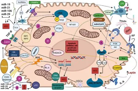
Figure 1 The molecular pathways that regulate non-alcoholic fatty liver disease-related hepatocellular carcinoma along with their interactions are represented. Activation of downstream signaling pathways is indicated by full or dot lines whereas inhibition of them is indicated by blunted lines with a circular-shaped “X”. Bidirectional arrows highlight the interplay between distinct molecular pathways. Molecules acting as mediators of signaling paths are indicated over a full, dot or blunted line in a box along with their names. MicroRNAs that promote hepatocellular carcinoma development are collectively represented in the upper left of the figure while tumor suppressor microRNAs are represented in the lower left of the schema. Cas: Caspase; DCA: Deoxycholic acid; ER: Endoplasmic reticulum; FFAs: Free fatty acids; HCC: Hepatocellular carcinoma; HDAC8: Histone deacetylase 8; HSCs: Hepatic stellate cells; IGF: Insulin growth factor; IL-6: Interleukin-6; IRec: Insulin receptor; IRes: Insulin resistance; JNK: Jun-(N)-terminal kinase; KC: Kuppfer cell; lncRNAs: Long non-coding RNAs; LPS: Lipopolysaccharides; MAPK: Mitogen-activated protein kinase; PAMPs: Pathogen-associated molecular patterns; SASP: Senescence associated secretory phenotype; STAT3: Signal transducer and activator of transcription 3; TGF-B: Transforming growth factor-B; TLR: Toll-liκe receptor.
NAFLD is closely associated with insulin resistance and subsequently increased levels of insulin and insulin-like growth factor-1 (IGF-1)[54]. Binding of these two molecules to insulin receptor and insulin-like growth factor-1 receptor (IGF1R) respectively, results in activation of PI3K/AKT and Mitogen-activated protein kinase (MAPK) molecular pathways[55]. Regarding the first, exerts its action by signaling on cyclin D1, Mdm2/p53 and mTOR and leads to inhibition of apoptosis, induction of cell proliferation and excessive cell growth respectively, while the activation of MAPK mediates the transcription of proto-oncogenes c-fos and c-jun, further affecting cell growth[56]. Moreover, MAPK pathway facilitates the activation of Wnt/β-catenin signaling cascade, which leads to liver fibrosis and promotion of hepatocarcinogenesis[57]. Moreover, insulin resistance and energy imbalance drive to excessive liver lipid accumulation, metabolic reprogramming and production of free fatty acids (FFAs)[58]. Increased mitochondrial oxidation of these FFAs induces the formation of reactive oxygen species (ROS) leading to insufficient mitochondrial respiratory chain activity as well as triggering apoptotic death pathways, such as receptor interacting protein 1 (RIP1) and RIP3- activated Jun-(N)-terminal kinase (JNK),which in turn facilitate liver inflammation and fibrosis[59,60]. In addition, the increased FFA oxidation is associated with augmented levels of endoplasmic reticulum (ER) and oxidative stress in hepatocytes. The latter promotes increased calcium release from ER that leads to mitochondrial permeabilization, disrupted ER function, liver cell injury and tumorigenesis in NASH[61]. Moreover, the crosstalk between oxidative or ER stress and ROS overproduction aggravates the progression of liver disease into NASH and HCC as the aforementioned mitochondrial dysfunction leads to further overproduction of ROS, which facilitate the activation of proapoptotic paths, mediating bycaspases 9 and 3[62,63].

NAFLD: Non-alcoholic fatty liver disease; HCC: Hepatocellular carcinoma; NASH: Non-alcoholic steatohepatitis; HCV: Hepatitis C virus; NSD: No significant difference; NA: Not applicable.
Furthermore, the products from lipid peroxidation and the elevated levels of ROS provoke the release of several pro-inflammatory and inflammatory substances such as interleukin-6 (IL-6), tumor necrosis factor-α (TNF-α) as well as affects adipokines’ secretion, namely leptin and adiponectin[64]. Increased expression of IL-6 activates the oncogenic pathway of signal transducer and activator of transcription 3 (STAT-3) which mediates cell proliferation, inhibits apoptosis and contributes to HCC development, while augmented TNF-α levels mediate the activation of pro-oncogenic paths namely nuclear factor kB (NF-kB)viaJNK and phosphorylation of inhibitor of kappa light polypeptide gene enhancer in B-cells, kinase beta (IKKβ)[65]. Consistently, leptin, as a profibrotic and proangiogenic factor exerts its action by both stimulating an intracellular signaling cascade of the inflammatory molecules namely TNF-α and IL-6 and by activating the previously mentioned JAK2/STAT-3, MAPK and PI3K pathways upon its binding to its receptor in HCC cells[66]. Noteworthy, although adiponectin is a strong anti-inflammatory mediator which modulates apoptosis under normotropic conditions, the exacerbated insulin resistance suppresses its action while ROS-induced overproduction of leptin, as an antagonistic hormone on the field of hepatic fibrogenesis with adiponectin, further inhibits its production and thus intensifies HCC development[67,68].
As the activation of the immune system considered as a prerequisite for NAFLD progression to NASH, the involvement of immunological pathways in the NAFLDrelated HCC is of much interest. Maet al[69] reported that selective intrahepatic depletion of CD4+ T cells robustly induced tumor development in a methioninecholine-deficient liver specific MYC transgenic mouse showing that CD4+ T cells mediate tumor regression[69]. Moreover, stimulation of hepatocellular lymphotoxin-β receptor (LTBR) and NF-kB signaling led to HCC onset in a MCD high fat diet mouse model whereas that same dietary pattern induced activation of natural killer T (NKT) cells and intrahepatic CD8+ T cells, which in turn facilitated NASH to HCC transition[70]. Additionally, liver damage and subsequent inflammatory response leads to activation of Kupffer cells (KCs), which are the resident macrophages of the liver and their involvement into NAFLD progression is well established in both animal models and human hepatic dysregulation[71]. Yet, these cells are also implicated in the hepatocarcinogenesis since they express the pro-inflammatory myeloid cell surface receptor TREM1 which facilitates HCC development in a diethyl nitrosamine-induced HCC mouse model[72]. KCs also express Toll-like receptor 4 and binding of Lipopolysaccharides drives the activation of the above-mentioned tumorigenic pathways of NFkB, JNK and MAPK[73]. Additionally, upon acute liver cells injury, the signaling pathway Hedgehog was triggered and reinforced the recruitment of hepatic progenitor cells at the sites of injury in order to replace the damaged hepatocytes[74]. Dysregulated signaling of this pathway leads to insufficient cell repair within the hepatic parenchyma and results in malignancy and HCC progression[75].
RISK FACTORS
Although cirrhosis constitutes the major risk factor for the development of HCC in various liver diseases, including NAFLD, HCC can also occur in non-cirrhotic NAFLD individuals[76]. Demographic, behavioral, or genetic factors contribute along with cirrhosis or even more in absence of cirrhosis to HCC. Older age, male sex and Hispanic ethnicity are strongly associated with higher risk of HCC development[5]. In a cohort study of 296707 NAFLD patients, age over 65 years comprised anindependent risk factor for HCC occurrence[77]. In the same study, the incidence of HCC was higher in males compared to females (0.22vs0.04per1000 PY respectively), in LatinovsWhite and African-American patients (0.29vs0.21 and 0.12per1000 PY, respectively) and in cirrhotic compared to non-cirrhotic patients (10.2vs0.02per1000PY, respectively)[77].
Furthermore, distinctive MetS-related features, namely obesity and T2DM have been identified as risk factors for HCC[78]. In a meta-analysis, overweight and obese patients had 17% and 89% increased relative risk for HCC respectively compared to normal-weight individuals[79]. In another study, obesity was recognized as an independent predictor for HCC development only in patients with cryptogenic [odds ratio (OR): 11.1; 95% confidence interval (CI): 1.5-87.4] and ALD-related cirrhosis (OR: 3.2; 95%CI: 1.5-6.6)[80]. Concerning the burden of T2DM, in a retrospective study including 6508 NAFLD patients, T2DM comprised an independent risk factor for the HCC (Hazard ratio: 3.21; 95%CI: 1.09-9.50)[81], while in a Mayo clinic study with 354 NASH-cirrhotic patients, T2DM along with older age and decreased serum albumin levels were identified as independent risk factors for HCC[82]. Consistently, in a casecontrol study of 185 HCC cases and 404 controls, T2DM (OR: 4.33, 95%CI: 1.89-9.86) and obesity (OR: 1.97, 95%CI: 1.03-3.79) were associated with increased HCC risk, while the combination of obesity and T2DM further exacerbated the hazard of HCC (OR: 4.75, 95%CI: 1.75-12.89)[83]. However, it should be noted that evaluating the exact independent pathogenetic burden of T2DM or obesity in the development of HCC can be really challenging, due to the strong association of these two entities[84].
Moreover, lifestyle modifiable factors, such as smoking and alcohol consumption seem to be implicated in NAFLD-related HCC[84]. In a meta-analysis of 81 studies, the risk for HCC development was higher for both current and former smokers[85], but no specific data were given regarding the relationship between smoking and the incidence of HCC in NAFLD patients[84]. Alcohol consumption is independently related to elevated risk for HCC in NAFLD patients[86], despite that some studies imply that the higher HCC risk is limited only on heavy alcohol use (e.g.> 50 g/d[87,88]), during which NAFLD is excluded by definition. Intriguingly, Sookoianet al[89] in their meta-analysis, showed that moderate alcohol consumption (< 30 g/d) is associated with decreased incidence of NAFLD occurrence[89], whereas a prospective study demonstrated a potential synergism between obesity and alcohol intake in increasing the risk of HCC[90]. However, the evidence from those studies could be hindered by potential biases, such as the observational study design which does not permit to ascertain causality, the implication of obesity as potential confounder and the overall insufficient alcohol intake assessment. Overall, the impact of moderate alcohol consumption in NAFLD-related HCC is still difficult to be clarified.
Noteworthy, genetic predisposition further aggravates the risk for NAFLD-related HCC. The possession of Patatin-like phospholipase domain-containing protein 3 (PNPLA3) rs738409C>G and Membrane Bound O-Acyltransferase Domain Containing 7rs641738 polymorphism were independently correlated with increased risk of HCC[91], with the latter being a burdened factor particularly in non-cirrhotic NAFLD patients[92]. On the contrary, a loss of function of variant rs72613567 in 17-betahydroxysteroid dehydrogenase 13 has been recently identified to protect against HCC development[93].
PROGNOSIS OF NAFLD-RELATED HCC
The prognostic outcomes of NAFLD-HCC patients as compared to their non-NAFLDHCC counterparts have been evaluated by population-based and cohort studies with controversial findings (Table 3)[15,24,30,34,40,42,94-103].

Table 3 Treatment outcomes and prognosis in patients with non-alcoholic fatty liver disease-related hepatocellular carcinoma vs other etiologies-related hepatocellular carcinoma
Based on Surveillance, Epidemiology and End Results Medicare database, Younossiet al[30] performed a large retrospective cohort study including 4979 HCC patients and showed that the one-year mortality risk was significantly higher in NAFLD-HCC compared to HCV/HBV-HCC patients[30]. On average, the former had almost 5 mo shorter survival time compared to the latter[30]. Moreover, in the multivariable model adjusted for clinical and tumour-related parameters, NAFLD was an independent factor associated with increased one-year mortality risk[30]. More recently, Golabiet al[94] based on the afore-mentioned database along with outpatients files, showed that NAFLD-HCC patients displayed markedly higher risk of 2-year mortality in comparison with patients with HCV-HCC[94]. Consistent to the abovementioned study, the magnitude of this association remained significant in the multivariable analysis[94]. The afore-mentioned findings were mainly attributed to the more advanced tumour stage at the time of diagnosis due to less intense surveillance of NAFLD patients[94]. In addition, due to the presence of increased visceral obesity, the ultrasonography, which is the current HCC screening tool, may fail to distinguish small tumours in NAFLD patients[94]. Furthermore, the increased prevalence of cardio-metabolic comorbidities along with higher age at the time of HCC diagnosis of NAFLD population may also contribute to the lower possibility of receiving LT, compared to HCC patients with viral hepatitis, and to their decreased survival time[94]. In a prospective study in Italy, among 756 HCC patients the crude 1-year and 3-year overall survival (OS) was remarkably lower in the NAFLD-HCC patients compared to HCV-HCC cohort[24]. In order to account for the potential biases of less intense surveillance and later detection of the NAFLD-related malignancy, they adjusted the whole HCC cohort of patients for the lead time who were under surveillance[24]. Intriguingly, both the mean 1-year and 3-year survival time of NAFLD-HCC patients remained significantly lower than the corresponding of HCVHCC patients[24]. Along this line, more recently, Hesteret al[96] in their multivariable analysis demonstrated that the NASH-HCC was associated with worse OS compared only to the ALD-HCC[96]. Additionally, since the magnitude of NAFLD in cryptogenic cirrhosis is major, it is of interest that Gianniniet al[97] using data form the ITALICA database, showed that patients with cryptogenic cirrhosis-related HCC displayed significantly shorter survival time compared to HCV-HCC patients during a median follow-up of 21 mo[97].
Of importance, we should note that a selection bias within clinical studies could be implicated concerning the worse prognostic outcomes of NAFLD-HCC patients in comparison to other-aetiologies-HCC. Patients who were eligible for radical surgical treatments or LT, were subsequently enrolled in the cohort studies whereas patients with major comorbidities, cardiovascular diseases or advanced age, aspects more common among NAFLD patients, were excluded.
On the contrary, numerous studies have emphasized the better prognosis of NAFLD-HCC patients, compared to other aetiologies of HCC. In a prospective study from Singapore, 844 non-NAFLD-HCC and 152 NAFLD-HCC patients who underwent total liver resection were enrolled and the latter displayed significantly increased 5-year OS as compared to the former, whereas NAFLD was independently associated with lower hazard for mortality in a multivariable model adjusted for clinical and epidemiological parameters[34]. Consistently, after a median follow-up of 50 mo, Reddyet al[42] evaluated HCC patients suffered from NASH compared to those from ALD and/or HCV who received curative treatment[42]. Although the postoperative mortality and the recurrence free survival (RFS) did not significantly differ between the two groups, NASH patients had longer OS, compared to ALD andHCV patients, independently of clinical factors and type of the curative treatment they received[42]. In another study, during a median follow-up of 17 mo, NAFLD-HCC patients displayed significantly improved OS and a trend towards increased RFS, compared to both HCV and HBV patients, in a model adjusted for demographic factors, Child-Pugh score and most definite treatment[98]. Notably, in order to assess the afore-mentioned long-term outcomes independently of the LT, authors omitted the LT recipients from all groups[98]. To this end, they showed that the NAFLD-HCC patients still had significantly improved OS rates compared to their HCV counterparts and a trend towards increased survival compared to HBV patients[98]. In 2015 Viganòet al[99], matched 96 HCC patients with MetS with 96 HCV-HCC patients who received liver resection during a 12-year study period[99]. Matching was based on age, prevalence of cirrhosis, Child-Pugh class, portal hypertension and HCC characteristics[99]. MetS-HCC patients had significantly better OS and lower recurrence rate compared to HCV-HCC cases whereas in the multivariate analysis MetS-HCC was an independent protective factor for both OS and early recurrence[99].
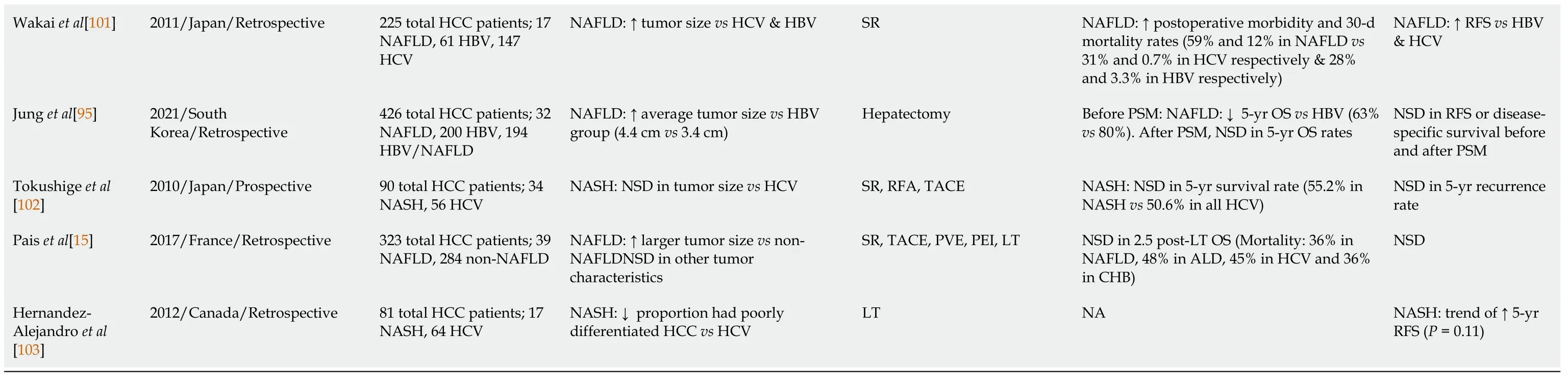
HCC: Hepatocellular carcinoma; NAFLD: Non-alcoholic fatty liver disease; AH/BC: Autoimmune hepatitis/Biliary cirrhosis; ALD: Alcohol-related liver disease; HBV: Hepatitis B virus; HCV: Hepatitis C virus; LT: Liver transplantation; OS: Overall survival; NA: Not applicable; SR: Surgical resection; TACE: Transarterial chemoembolization; BCLC: Barcelona-Clinic Liver Cancer; PEI: Percutaneous ethanol injection; BSC: Best supportive care; NSD: No significant difference; RFS: Recurrence-free survival; NASH: Non-alcoholic steatohepatitis; TARE: Transarterial radioembolization; HR: Hazard ratio; CC: Cryptogenic cirrhosis; RFA: Radiofrequency ablation; MetS: Metabolic syndrome; PVE: Portal vein embolization; OR: Odds ratio; UCSF: University of California at San Francisco.
Finally, several studies have highlighted the similar long-term outcomes regarding NAFLD and non-NAFLD-HCC patients. A Swedish retrospective study revealed that although NAFLD-HCC patients had higher age, higher prevalence of comorbidities and less HCC surveillance, they had similar survival to non-NAFLD-HCC patients, mainly attributed to the poor prognosis of HCC in general[40]. Consistently, Thanet al[100] compared the outcomes of 212 NAFLD-HCC and 275 HCV-HCC patients who were referred for LT and showed that the 3-year post-diagnosis OS was similar in the two groups[100] and this finding was confirmed in the subgroup-analysis of patients who eventually received LT[100]. Along this line, Wakaiet al[101] upon evaluating 317 HCC patients who received hepatic resection, showed that the 5-year post-resection cumulative survival rate did not differ significantly between NAFLD and non-NAFLD groups. Yet, RFS following liver resection was markedly better in the NAFLD group[101]. Additionally, Junget al[95] reviewed the outcomes of NAFLD-HCC and HBVHCC patients who underwent hepatectomy over a 10-year period. After a median follow up of 74 mo, the latter had superior 5-year OS rates, compared to the former. However, when the authors performed a propensity score matching to minimize the bias of lead time, 5-year OS was similar between the two groups[95]. In a Japanese prospective study, the OS and the RFS after curative treatment of NAFLD-HCC and HCV-HCC patients were evaluated, and both were comparable between the two cohorts[102]. Similarly, Paiset al[15] in their retrospective study evaluating a 20-year period confirmed those results[15], while a Canadian study revealed only a trend for better 5-year RFS of NASH-HCC, compared to HCV-HCC patients who underwent LT[103].
However, we should emphasize that due to less intense surveillance of HCC in general, the disease is likely to be diagnosed at advanced stages and therefore even fewer patients are eligible for radical therapy or LT. Thus, the prognostic outcomes of NAFLD-HCC patients compared to non-NAFLD-HCC ones, should be interpreted with cautiousness and accordingly to each clinical study design and type of HCCtreatment that patients received.
Moreover, another factor raising concern for defining the long-term survival outcomes of NAFLD-HCC patients is that those patients seem to have a lower MELD score waiting at LT list compared to their non-NAFLD counterparts, as they tend to have a more preserved liver function. Therefore, they are less likely to receive LT in short-term. This in turn results in longer duration in the waiting list increasing the risk for severe health-related complications and morbidity negatively affecting their survival. Indeed, when Wonget al[104] retrospectively analysing UNOS registry data concerning LT waiting list registrations in the United States, demonstrated that NAFLD patients as compared to their HCV or ALD counterparts, were markedly less likely to receive a liver transplant within 90 d and one year after their registration, ending in higher mortality while on the waiting list[104].
SURVEILLANCE
Since NAFLD patients seem to be frequently diagnosed in advanced tumor stages, their surveillance for HCC development represents quite the most challenging issue among professional societies worldwide. As for the cirrhotic-NAFLD patients, since they appear to have an expected HCC incidence of approximately 1.5%peryear, they should follow the screening guidelines for cirrhotic patients of any cause, consisted of abdominal examination with liver ultrasonography with or without alpha-fetoprotein (AFP) every 6 mo[105] (Figure 2).
Regarding non-cirrhotic NAFLD patients, there is a lack of consensus whether NAFLD patients with F3 fibrosis should undergo screening. The updated recommendation of EASL guidelines suggest that patients with F3-fibrosis might be eligible for HCC surveillance based on an individual risk stratification[106], while the clinical update of the American Gastroenterology Association also recommend the screening for patients with findings indicative of advanced fibrosis (F3), as evaluated by two or more concordant non-invasive fibrosis tests of separate categories[107]. AASLD guidelines recommend HCC surveillance only in cirrhotic (not advanced fibrosis-F3) patients[108]. Finally, the Asian guidelines do not provide specific recommendation for non-cirrhotic NAFLD patients[109] (Figure 2). However, some concerns are raised. Even if a consensus for screening of F3 patients was reached, a large proportion of HCC cases that occur in F0-F2 NAFLD patients would still be missed. Moreover, the diagnosis of F3 fibrosis based on a broad spectrum of noninvasive tests mitigates the utility of screening since HCC risk would not still be the same for all F3-patients, while screening all patients with F3 fibrosis would drastically increase the cost of the surveillance strategy. Moreover, it should be mentioned that even among cirrhotic patients, there are differences in risk for HCC and therefore they should not be aggregated into a single category. Ioannouet al[110] developed a predictive model that estimates HCC risk in cirrhotic-NAFLD patients by implicating demographic, clinical and laboratory parameters[110]. Based on this model, patients were categorized into low-risk (annual risk: < 1%), medium-risk (annual risk: 1%-3%) and high-risk (annual risk: > 3%), suggesting that individualized screening for HCC is associated with standardized benefit compared with screening of all cirrhotic-NAFLD patients[110,111]. Moreover, the identification of PNPLA3rs738409C>G or other polymorphisms might be of value for screening since it would improve prediction accuracy, but its evaluation in multi-factorial risk models would decrease the costeffectiveness of patients’ surveillance[112]. The future surveillance policies should focus on identification of prognostic factors, consisting of imaging modalities, serum biomarkers and genetic variants, testing for which would need to become cheaper, that will stratify the risk of HCC in both cirrhotic and non-cirrhotic NAFLD patients promoting the cost-effectiveness of the programmes. The afore-mentioned parameters along with well-established traditional risk factors of HCC may be incorporated in future risk-assessment models and could result in more accurate prediction of NAFLD-related HCC and optimized surveillance strategies. A proposed algorithm of NAFLD-related HCC surveillance based on future perspectives is illustrated in Figure 3.

Figure 2 Proposed algorithm for hepatocellular carcinoma surveillance in non-alcoholic fatty liver disease patients based on the latest guidelines. HCC: Hepatocellular carcinoma; NAFLD: Non-alcoholic fatty liver disease; US: Ultrasonography; AFP: Alpha-fetoprotein; CT: Computer tomography; MRI: Magnetic resonance imaging; BMI: Body mass index.

Figure 3 Proposed algorithm for hepatocellular carcinoma surveillance in non-alcoholic fatty liver disease patients based on future perspectives. HCC: Hepatocellular carcinoma; NAFLD: Non-alcoholic fatty liver disease; PNPLA3: Patatin-like phospholipase domain-containing protein 3; MiRNAs: Micro-RNAs; lnc-RNAs: Long non-coding-RNAs; ALP: Alkaline phosphatase; AFP: Alpha-fetoprotein; US: Ultrasonography; CT: Computer tomography; MRI: Magnetic resonance imaging; BMI: Body mass index.
As yet, circulating micro-RNAs (miRNAs) and long non-coding-RNAs have been shown promising results since they were associated with HCC progression in NAFLD patients and may constitute potential non-invasive tools for NAFLD-related HCC screening[113,114]. Several micro-RNAs, such as miR-29 and miR-199, mainly expressed in NASH, are associated with fibrosis progression and HCC development[113]. Furthermore, hydroxy-methylated genes are strongly related to the involvement of chromatin in the progression of HCC and form promising genetic factors for the risk classification of AFP-negative HCC patients[115]. Results derived from animal models examining the progression of HCC in NASH mice, suggested that serum osteopontin and dikkopf-1 could be possible novel biomarkers for the early detection of HCC[116]. Finally, the identification and amplification of circulating tumor-DNA can reveal critical HCC-related genetic mutations and therefore could be used for the screening of HCC patients[117]. However, the incorporation of those prognostic biomarkers in screening programmes would significantly increase the cost with ambiguous results. As until now, they comprise mostly future perspectives rather than clinical point-ofcare practice.
PREVENTION
Although weight loss is considered fundamental for the management of NAFLD, there is no current evidence to directly indicate that weight loss leads to reduction of NAFLD-related HCC. Importantly, a recent large multi-national study with 467336 individuals, demonstrated that physical exercise, defined as performing at least 2 hours of vigorous activityperweek, can reduce the risk of developing HCC independently of other risk factors of HCC[118]. Moreover, the prevention of obesity and T2DM is considered fundamental for the management of NAFLD patients, since they constitute independent risk factors for HCC development and progression[80,82,111,119]. Noteworthy, a meta-analysis of 19 studies showed that diet rich in vegetables may reduce HCC incidence, while Georgeet al[120] suggested that stricter adherence to Mediterranean diet was protective against HCC development[120]. Increased coffee consumption is also associated with decreased risk of NAFLD development and severity progression[121], with two additional coffee cupsperday to be associated with 35% lower incidence of HCC[122]. However, the exact impact of coffee consumption as a preventive measure against NAFLD and its progression to HCC needs further investigation. Although literature data are controversial regarding the role of light and moderate alcohol use in NAFLDper se[89,90], in a study of 195 cirrhotic-NASH patients, Aschaet al[86] demonstrated that patients with any alcohol consumption had higher risk of HCC incidence compared to non-drinkers[86]. Consistently, HCC occurrence was more frequent in NAFLD patients with mild alcohol intake (< 20 g/d), especially in those with advanced fibrosis, as compared to abstainers patients[123]. Concerning medication, in a prospective study of 361 NAFLD patients, daily use of aspirin was associated with significant lower odds for NASH and advanced fibrosis, while no relationship between use of non-aspirin nonsteroidal antiinflammatory drugs and risk for advanced fibrosis was outlined[124]. Furthermore, the administration of statins in cirrhotic patients provides chemo-preventive effects and is associated with the reduction of HCC occurrence, in a dose-dependent manner[125-127]. Interestingly, fluvastatin, compared with other statin interventions, exhibited the most significant effect in the reduction of HCC incidence in cirrhotic patients, while the utility of rosuvastatin against the development of NAFLD-related HCC was shown in a murine model[127,128]. As far for T2DM pharmacotherapy, dose-response anti-tumorigenic effects of metformin were observed among T2DM patients[129], while in contrast, the administration of sulfonylureas and insulin have been associated with increased risk of HCC[129].
CONCLUSION
In the 21stcentury, we are in the midst of an epidemic of obesity, T2DM and NAFLD. Consequently, the burden of NAFLD in HCC development is rapidly rising partially explaining the elevated incidence of HCC in both men and women globally. Although the exact pathogenetic mechanisms involved in NAFLD-related HCC onset are still not well-established especially regarding the non-cirrhotic hepatic parenchyma, specific risk factors for HCC concerning demographic, genetic and behavioral parameters have been already identified. Noteworthy, the surveillance of NAFLD-HCC patients is not standard in medical practice and therefore many patients do not undergo screening and that leads to diagnosis of HCC at advanced stages negatively affecting their survival and diminishing the therapeutic options. Concerning systemic treatment for HCC, the latest data[130,131] do not support the hypothesis that the therapeutic decisions should be based on the underlying HCC etiology and therefore, HCC systemic therapy was not in the field of our review. However, noteworthy, in a recent meta-analysis of 3 trials[132-134], authors suggested that immunotherapy might be less efficacious in NASH-HCC patients as compared to their viral-HCC counterparts, presumably owing to the NASH-provoked aberrant T-cells activation and subsequently flawed immune surveillance[135]. Yet, more robust evidence are needed for therapeutic decision making. Although, lifestyle modifications, such as stricter adherence to Mediterranean diet and medication namely metformin are thought to contribute to the primary prevention of NAFLD-related HCC, the appropriate strategy would be the identification of at-risk patientsviaa relatively simple score including demographic and laboratory/imaging parameters. Implementation of risk stratification programmes and high awareness of the burden of NAFLD should be the primary goals for medical clinical specialties and health authorities worldwide.
ACKNOWLEDGEMENTS
We are grateful to Dr. Panagiotis Lembessis for the language polishing of the manuscript.
杂志排行

World Journal of Gastroenterology的其它文章
- Endothelial cells and blood vessels are major targets for COVID-19-induced tissue injury and spreading to various organs
- Eliminating viral hepatitis in children after liver transplants: How to reach the goal by 2030
- Endoscopic ultrasound role in pancreatic adenocarcinoma treatment: A review focusing on technical success, safety and efficacy
- Melatonin prevents oxidative stress, inflammatory activity, and DNA damage in cirrhotic rats
- Microbiome changes in the gastric mucosa and gastric juice in different histological stages of Helicobacter pylori-negative gastric cancers
- Postoperative mortality and morbidity after D2 lymphadenectomy for gastric cancer: A retrospective cohort study