3D SERS substrate based on nanocone forests for miRNA detections
2021-10-21LIRuiruiLIYongweiXIONGJijun
LI Ruirui,LI Yongwei,XIONG Jijun
(Key Laboratory of Instrumentation Science and Dynamic Measuremen(North University of China), Ministry of Education,Taiyuan 030051,China)
Abstract:Surface-enhanced Raman scattering (SERS)is a powerful technology for obtaining vibrational information from molecules that present in different chemical or biological environments.This paper presents a 3D SERS substrate based on nanocone forests.The substrates are prepared by using plasma treatment technique,which is a simple,fast and high-throughput approach.The SERS substrate based on nanocone forests exhibits high sensitivity.In the experiment,miRNA with a concentration as low as 10-10 M can be achieved.Meanwhile,the proposed SERS substrate shows a high uniformity over a large area.These experimental results demonstrate great potential of the 3D SERS substrate in wide applications.
Key words:surface-enhanced Raman scattering (SERS)substrates;nanocone forests;three-dimensional hot spots;miRNA detection
0 Introduction
It has been assumed for several years that SERS performance can be significantly improved if 3D hot spots are formed by extending a third dimension along thez-axis.3D nanostructures,on which 3D hot spots are likely to exist,have been commonly investigated and widely used in SERS devices.To date,ZnO nanorods,CuO nanowires and polyimide nanowires with certain heights and densities have been fabricated and used as supporters for metallic nanoparticles to construct 3D hot spots[11-12].Fabrication of these nanostructures requires serial techniques such as electron beam lithography,femtosecond laser etching,vapor-liquid-solid growth,metal-assisted chemical etching and some others[13-14].These approaches either are time-consuming and labor-consuming or require complex processes,which restrict applications of the SERS devices severely.
In this paper,a 3D SERS substrate based on nanocone forests with high uniformity is proposed by using a plasma treatment technique.The preparation process only involves a plasma treatment step to form nanomasks,an anisotropic etching step to form nanocones and a metal sputtering process.The fabrication process is very simple,fast and with high-throughput.Subsequently,the substrates are applied to be the SERS devices to detect miRNA.The results show that these SERS devices based on nanocone forests exhibit a high sensitivity for miRNA.Meanwhile,the spectral intensity shows a good uniformity across large surface areas.These results demonstrate the SERS substrates have more potential as an advanced ultra-sensitive analytical tool for medical applications such as drug screening and point-of-care testing.
1 Experiments
1.1 Materials and instruments
The ordinary single-crystal silicon wafers (4-inch,single-side polished,Suzhou Research Materials Microtech Co.,Ltd.,China)were used as the substrates.Phosphate buffered solution (PB,pH 7.4)purchased from Gibco Life Technologies (AG,Switzerland)was used as the solvent.The oligonucleotides sequences were synthesized from Sangon Biological Engineering Technology &Co.Ltd.(Shanghai,China).The sequences were as follows:
Probe DNA:5’-SH-(CH2)6-CT CCA TGA TTC AGT TCT ATT TTG GAG CAGG -Rox-3’.
Placeholder DNA:5’-CCT GCT CCA AAA ATC CATT-3’.
Target miRNA-1246:5’-AAUG GAU UUU UGG AGC AGG-3’.
SEM images of nanoforests were obtained by using a Supra 55 scanning electron microscope (Carl Zeiss Inc,Germany).The SERS signal was measured by a confocal Raman microscope (Via Reflex Raman microscope,Renishaw PLC,UK).During the measurements,the excitation wavelength was 632.8 nm and the laser was guided at certain positions using a 50×objective lens.After measurements,the data were analyzed by WiRE 3.2 software.
1.2 Preparation of SERS substrates
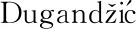
The preparation process of the SERS substrate based on nanocone forests is illustrated in Fig.1.First,a photoresist layer was spin coated on an Si substrate,then the photoresist layer was subjected to successive oxygen and argon plasma treatment.A tremendous amount of nanofibers and nanowires were synthesized.Then,these nanofibers and nanowires were acted as masks to etch Si substrate and acquire Si nanocone forests (NCFs).The residual nanofibers and nanowires were removed by wet etching using buffered hydrofluoric acid (7∶1).At last,a Au layer with a thickness of 30 nm was sputtered on the Si NCFs,thus forming the Au NCF substrate.
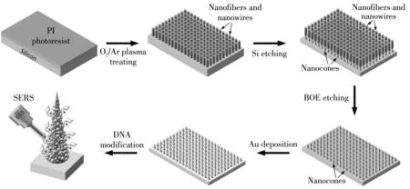
Fig.1 Schematic diagram of preparation of nanocone forest-based SERS substrate and 3D SERS substrate
1.3 Detection process of miRNA
Beforethe experiment,the Au NCF substrate was cut into 3 mm×3 mm small pieces.At first,3 μL probe DNA solution with concentration of 10 μM was mixed with 10 μM placeholder DNA solution of the same volume,and the mixture were incubated overnight at 37 ℃ for full hybridization.Then,the mixed solution was dripped onto the Au NCF substrates and incubated at 37 ℃ for 2 h to immobilize the duplex DNA on Au nanostructures.After that,these substrates were washed three times by ultrapure water to remove the excess uncombined DNA.Subsequently,3 μL miRNA solutions with different concentrations were dripped onto Au NCF substrates and incubated at 37 ℃ for 2 h to completely displace the placeholder.After the solvent was completely evaporated,these substrates were detected by confocal Raman microscope.The integration time for spectra acquisition was 5 s in each measurement and all signals were accumulated twice.
2 Results and discussion
2.1 Characterization of SERS substrates
Fig.2(a)shows SEM image of the prepared nanofibers and nanowires using oxygen and argon plasma treatment.As illustrated,nanofibers and nanowires with an average height of around 5 μm clustered together to form bundles of fiber structures.The diameter and height of the nanofibers can be controlled by changing the plasma treating time.Using these nanofibers and nanowires as masks,the Si substrate was etched to form NCFs,as shown in Fig.2(b).Due to the anisotropic etching,vertical nanostructures with certain heights on the Si substrates were formed,while a slightly lateral etching effect was also introduced.As a result,the heads of the nanostructures had smaller diameters than the bottoms,thus forming NCFs.After the residual nanofibers and nanowires on top of the NCFs were removed,the Si NCFs were obtained,as shown in Fig.2(c).The average height of the NCFs was around 1.5 μm.Fig.2(d)illustrates the sectional SEM image of NCFs.To prepare the SERS substrate,Au with a thickness of 30 nm was sputtered on the Si NCFs.SEM images of the SERS substrate based on the Au-coated nanocone forests are illustrated in Figs.2(e)-(f).After the Au sputtering step,large numbers of Au nanoparticles (NPs)with diameters of 5 nm-30 nm were distributed evenly on the Si nanocone surfaces.As illustrated in Fig.2(f),quite a lot of nanogaps between Au NPs were also formed on the nanocone surface.These NPs and nanogaps are of significant benefits for SERS enhancement owning to the plasmonic coupling effects between metallic nanoparticles.Besides,the substrate with a 3D geometric structure not only has cone tips and spacing among nanocones,but also has nanoparticles and nanogaps on the nanocone surfaces,which can greatly contribute to the SERS intensity enhancement.
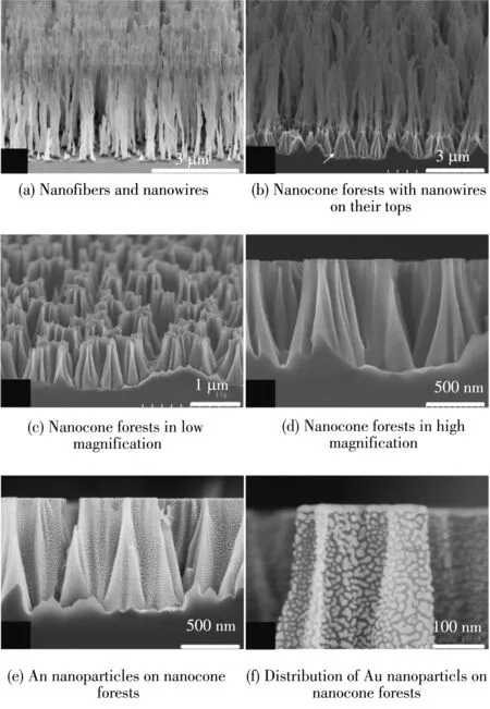
Fig.2 SEM images
2.2 Principle of miRNA detection
Fig.3 illustrates the miRNA detection principle of the Au NCF substrate.The detection of miRNA is closely related to early screening and diagnosis of cancers.In the paper,miRNA-1246 was chosen to be a target analyte to verify the SERS performance of the Au NCF substrate.After the full hybridization of probe DNA and placeholder DNA,the duplex DNA was immobilized on these Au NPs via sulfhydryl group (-SH).The Raman reporters-Rhodamine X (ROX)were far from the Au NPs,resulting in a low SERS intensity.This state was call as “OFF”state.When the target miRNA with different concentrations was added in the system,the miRNA would hybridize with the placeholder DNA in different degree due to the DNA strand-displacement.Thus,after the placeholder DNA was released,the probe DNA returned to the hairpin structure.Consequently,the Raman reporters were close to the Au NPs,resulting in a strong SERS intensity.This state was called as “ON”state.
Fig.3 Scheme of target miRNA detection by strand displacement approach
Fig.4(a)illustrates the Raman spectra of miRNA detection with “OFF”and “ON”state,respectively.Before the miRNA (10-9M)was introduced,the Raman reporters were far from the Au NPs,while the Raman reporters were very close to the Au NPs after adding miRNA,therefore,the SERS signal intensity under “OFF”state was much lower than that under “ON”state.
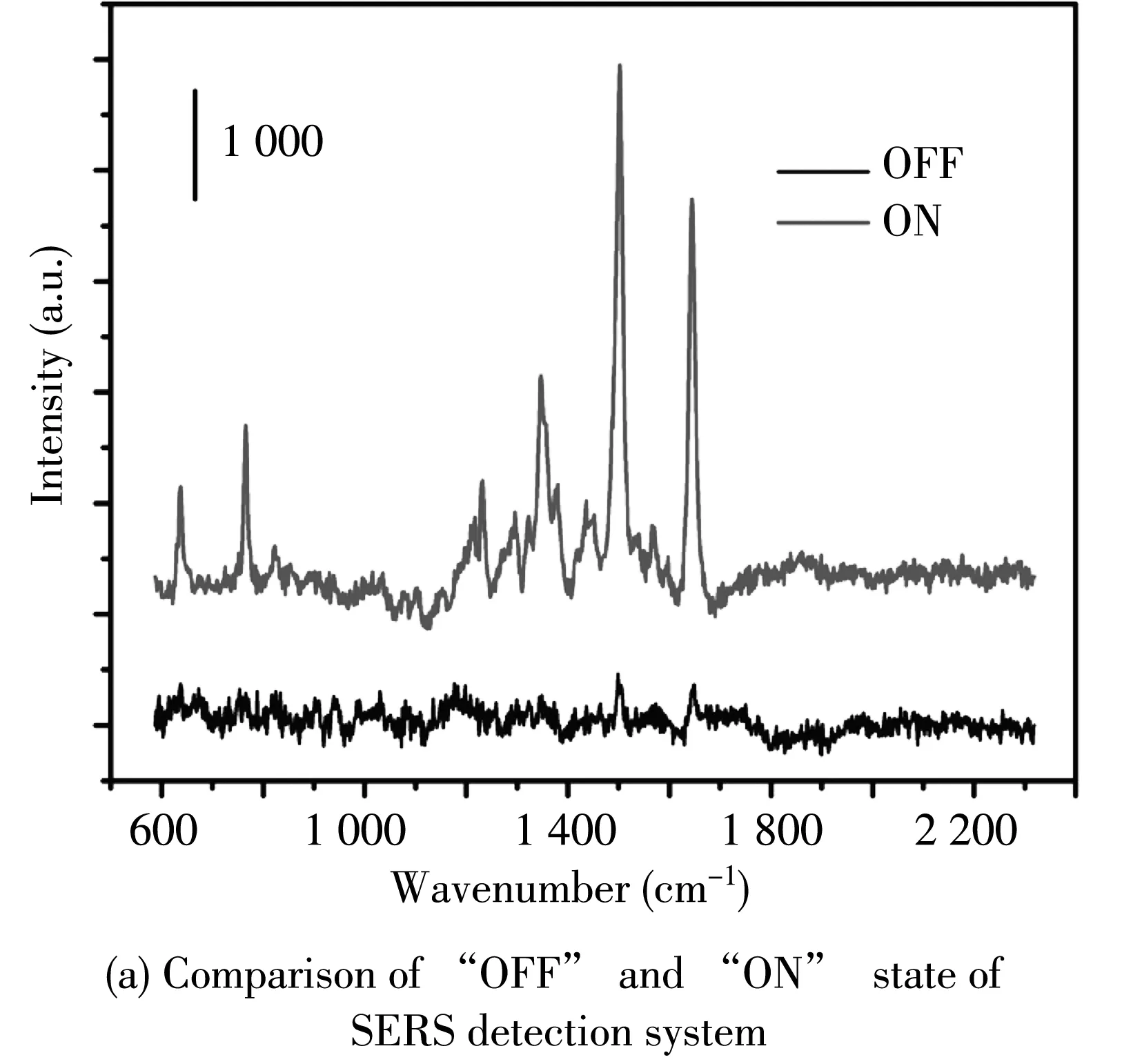
Fig.4 SERS spectra of miRNA
There were also weak signals in the “OFF”state,which ascribed to existence of the excessive and free probe DNA.To evaluate the SERS performance of the Au NCF substrates,miRNA solutions with different concentrations (10-5M,10-6M,10-7M,10-8M,10-9M and 10-10M)were added onto the substrates.After the solvent was evaporated,SERS measurements were carried out.As demonstrated in Fig.4(b),Raman spectra of miRNA with particular Raman signals of ROX were obtained by using Au NCF substrates.The characteristic peaks at 1 499 cm-1and 1 644 cm-1were completely in accord with the ROX molecule,indicating the detection ability of miRNA.
Fig.5 shows the evaluation results of SERS uniformity of Au NCF substrate.
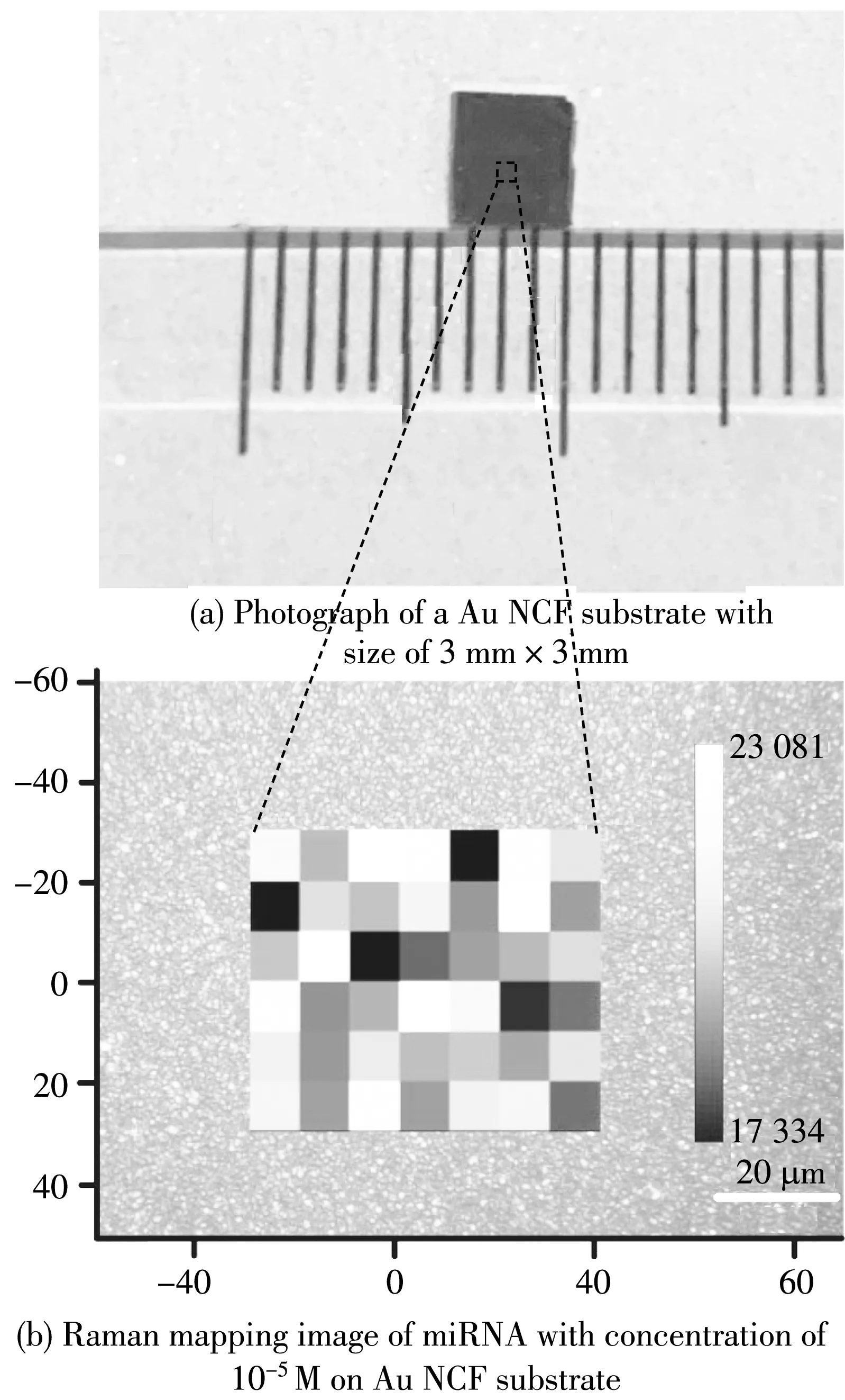
Fig.5 Evaluation of SERS uniformity of Au NCF substrate
Fig.5(a)illustrates a 3 mm×3 mm Au NCF substrate,which shows a black surface owing to the strong light absorption in the visible range caused by localized surface plasmon resonances (LSPR)effect.To evaluate the uniformity of the substrate,miRNA solution of 10-5M was added onto the substrate.After the substrate dried,the Raman signals were measured.A rectangle area of 60 μm×50 μm on the substrate was randomly selected.Fig.5(b)displays the variations of Raman signal intensity corresponding to wavenumber of 1 499 cm-1for miRNA.According to the results,the maximum SERS signal intensity variation was around 33%.However,the intensity fluctuation was found to be below 20%,and more than 80% of mapped area remained at the average level.The results suggest a high SERS uniformity of the proposed substrate,owing to the presence of ordered Si NCF with less variation in their height,and good homogeneity in size and gap of Au NPs.
3 Conclusions
In this paper,a 3D SERS substrate based on nanocone forests were successfully prepared using plasmat reating technique and Au deposition.Such a SERS substrate can provide abundant hot spots in 3D space,which are markedly advantageous to excite LSPR.As presented in this paper,the SERS substrates based on nanocone forests have high enhancement capability and good SERS uniformity over large areas.The substrate was used for miRNA detection by utilizing DNA strand-displacement process.The detection limit of miRNA can reach as low as 10-13M,which reveals that the SERS substrate has remarkable prospects for precise disease detection.
杂志排行

Journal of Measurement Science and Instrumentation的其它文章
- Tissue blood flow measurement by diffuse correlation spectroscopy based on Huber regression
- Continuous rotation north-finding algorithm based on constrained adaptive Kalman filter
- Calibration of GNSS positioning receivers
- Noise estimation and filtering method of MEMS gyroscope based on EMMAP
- Dehazing algorithm using adaptive dark channel fusion and sky compensation
- Short-time prediction for traffic flow based on wavelet de-noising and LSTM model