Role of 18F-FDG PET/CT in detection of hematogenous metastases of advanced differentiated thyroid carcinoma: a systematic review and meta-analysis
2021-07-20NataleQuartuccioDomenicoRubello
Natale Quartuccio, Domenico Rubello
1Nuclear Medicine Unit, A.R.N.A.S.Civico, Di Cristina and Benfratelli Hospitals, Palermo 90144, Italy.
2Nuclear Medicine Unit, Santa Maria della Misericordia Hospital, Rovigo 45100, Italy.
Abstract The aim of this study was to collect the evidence on the performance of 2-deoxy-2-[18F]fluoro-D-glucose(18F-FDG) Positron Emission Tomography/Computed Tomography (PET/CT) in terms of detection rate (DR) and diagnostic accuracy in identifying hematogenous metastases in patients with differentiated thyroid cancer and compare its performance with that of other imaging techniques.A comprehensive PubMed/MEDLINE database research was carried out to retrieve studies documenting the performance of 18F-FDG PET/CT in depicting hematogenous metastases in patients with differentiated thyroid cancer.Statistical analysis was performed on a per-patient and per-lesion basis.The literature search yielded 15 articles to be included in the systematic review.18F-FDG PET/CT showed a pooled DR of 85.08% on a per-patient analysis and 89.70% on a per-lesion analysis.For bone lesions, a high DR (81.78%) was found for 18F-FDG PET/CT.For the sub-group analysis of lung lesions,pooled DR was 92.77% for 18F-FDG PET/CT, 95.02% for CT, and 64.93% for magnetic resonance imaging (MRI).On a per-patient analysis, 18F-FDG PET/CT demonstrated a pooled sensitivity (SS) of 87.3% [95% confidence interval (CI): 77.3%-94%] and a pooled specificity (SP) of 95.6% (95%CI: 87.6-99.1).On a per-lesion analysis(328 metastases), PET/CT showed a pooled SS and SP of 86.3% (95%CI: 73.5%-93.5%) and 93.4% (95%CI:71.7%-98.8%) in the detection of hematogenous metastases.The presented systematic review and meta-analysis favor the use of 18F-FDG PET/CT in the detection of hematogenous metastases in patients with differentiated thyroid cancer.The limited literature warrants further studies to confirm our findings.
Keywords: PET/CT, hematogenous metastases, lung, bone, thyroid cancer, FDG, meta-analysis
INTRODUCTION
Primitive thyroid cancers can be divided into 2 main pathological entities: tumors originating from follicular cells and medullary cancer (arising from para-follicular C cells).The first group of cancers include differentiated (follicular and papillary carcinoma and their variants), poorly differentiated, and undifferentiated tumors (anaplastic carcinoma)[1].
After surgical removal of the tumor, due to the risk of disease recurrence, differentiated thyroid cancer(DTC) is followed-up by assessing serum thyroglobulin (Tg), whereas calcitonin and carcinoembryonic antigen are biochemical markers for postoperative surveillance in medullary thyroid cancer[2,3].Overall,lymph node is the most frequent metastatic site for thyroid cancer.Nevertheless, patients with thyroid cancer may present also distant metastases in other organs due to hematogenous dissemination, especially to bone and lung.Patients with extranodal metastases usually present a worse prognosis compared to patients with metastases limited to lymph nodes[4,5].It follows that it is extremely important to identify hematogenous metastases in these patients to better address clinical management.
Patients with history of DTC, who no longer respond to radioactive iodine (RAI) therapy, have developed RAI-Refractory DTC.The majority of these cases with advanced disease have a worse prognosis[6].The use of 2-deoxy-2-[18F]fluoro-D-glucose (18F-FDG) positron emission tomography/computed tomography(PET/CT) has proved beneficial in assessing recurrence of thyroid cancer in the case of a rise of Tg but no evidence of disease at131Iodium (131I) whole-body scan[7,8].This clinical scenario has been related to the so-called flip-flop phenomenon and appears to be associated with worse prognosis; thyroid cells,indeed, demonstrate an upregulation of glucose transporters (GLUT1) and a reduction of sodium-iodide symporters (NIS) when a dedifferentiation process occurs (from DTC towards a more aggressive histotype)[9].
In the literature, there are systematic reviews and meta-analyses documenting the accuracy of18F-FDG PET/CT or18F-FDG PET in assessing in patients with thyroid cancer with suspected recurrence, although these studies did not differentiate between lymph node and hematogenous metastases[10-12].
Due to the widespread availability of hybrid PET/CT scanners in current nuclear medicine facilities worldwide, we excluded from our systematic review studies involving the use of stand-alone PET scanners.Lee and coworkers recently demonstrated in a network meta-analysis the superiority of L-6-[18F]fluoro-3,4-dihydroxyphenylalnine) (18F-DOPA) over18F-FDG in medullary thyroid cancer.We, therefore, decided to focus on thyroid cancer histotypes other than medullary[13].
The primary aim of this systematic review and meta-analysis was to collect the evidence on the detection rate (DR) and accuracy (AC) of18F-FDG PET/CT in identifying hematogenous metastatic lesions in patients with advanced differentiated thyroid cancer on a per-patient and per-lesion basis.The secondary goal was the comparison of the diagnostic performance of18F-FDG PET/CT with other imaging techniques.
METHODS
The systematic review was conducted in accordance with the PRISMA guideline (Preferred Reporting Items for Systematic Reviews and Meta-Analyses)[14].
Literature search
A comprehensive PubMed/MEDLINE database research was carried out by a researcher (N.Q.) to retrieve prospective or retrospective studies, aiming the identification of distant metastases in patientswith thyroid cancer by means of18F-FDG PET/CT.The following search string was used for the literature search: (“positron emission tomography” OR “PET” OR “positron emission tomography/computer tomography” OR “PET/CT”) AND [(“differentiated” OR “papillary” OR “follicular” OR “Hurthle cell” OR“anaplastic” OR “dedifferentiated” OR “poorly differentiated”) AND (“cancer” OR “carcinoma”)] AND(“fluorodeoxyglucose” OR “FDG”) AND “thyroid”.
The literature search was updated until 22 August 2020.No date limit or language restriction was applied.All identified references were exported to a reference management software (Endnote v.X7.5, Clarivate Analytics).
Study selection
An investigator screened the titles and abstracts of the retrieved entries.Only original articles were selected.After excluding duplicates and non-original articles, the full text of the remaining articles was retrieved to verify the following inclusion criteria: (1) a study cohort or a subset of a minimum 10 patients with differentiated thyroid cancer undergoing18F-FDG PECT/CT; (2) presence of data regarding at least the detection rate of non-lymph node metastases; and (3) absence of other malignancies in the patient history.The choice of excluding articles with fewer than 10 patients and organs was made to mitigate the so-called small-study effect, for which, in brief, the observations could be due to methodological flaws, outcome reporting bias, and clinical heterogeneity.
The references of the retrieved articles were also screened for additional studies.
Data extraction
For our primary outcome (accuracy of18F-FDG PET/CT), we selected articles from which it was possible to retrieve the number of true positive (TP), false negative (FN), false positive (FP), and true negative (TN)cases on a per-patient or per-lesion basis.Lesions were grouped according to the involved organ (e.g, lung or bone).Articles providing only the number of TP or FN cases were used only for the determination of the DR of hematogenous metastases on a per-patient and per-lesion basis.
For the secondary outcome (comparison of diagnostic performance of18F-FDG PET/CT with other imaging tests), pooled DR of18F-FDG PET/CT was compared with that of other imaging tests using N - 1 Chi-squared test[15].
Methodological quality assessment
The methodological quality of the studies was assessed using version 2 of the “Quality Assessment of Diagnostic Accuracy Studies” tool (QUADAS-2)[16], which comprises four domains: patient selection, index test, reference standard, and flow and timing.The concerns about the risk of bias or applicability were described as low, high, or unclear.
Statistical analysis
Statistical analysis was carried out using MedCalc Statistical Software version 19.1.3 (MedCalc Software,Ostend, Belgium; https://www.medcalc.org; 2020), Open Meta analyst (available online at http://www.cebm.brown.edu/openmeta/downloads/open_meta_analyst_win8.zip)[17], and MetaDisc v.1.4 (available at http://www.hrc.es/investigacion/metadisc_en.htm)[18].
TheI2statistic was used to measure the degree of inconsistency across studies, withI2values of 25%, 50%,and 75% representing low, moderate, and high substantial heterogeneity.Interpretation of heterogeneity was carried out at a significance level ofP= 0.05.The choice of fixed or random effects model was made on the basis of the degree of inconsistency, selecting the random effects model in the case of moderate and high substantial heterogeneity.
The DR of18F-FDG PET/CT for hematogenous metastases was calculated for per-patient and per-lesion analyses and presented using forest plots.
Diagnostic accuracy (AC) was calculated only for the articles from which it was possible to extract the number of TP, FN, FP, and TN cases on a per-patient and/or per-lesion basis.AC was presented using summary receiver operating curve (SROC).
Significant difference between accuracy of18F-FDG PET/CT and other imaging modalities was assessed,using a significance threshold of 0.05.
RESULTS
Literature search and eligibility assessment
The comprehensive computer literature search from PubMed/MEDLINE database revealed 836 articles[Figure 1].Reviewing titles and abstracts, 725 articles were excluded.In particular, 725 were not in the field of interest of the present study or were non-original articles.Five further articles were excluded because the full-text could not be obtained despite contacting the authors.
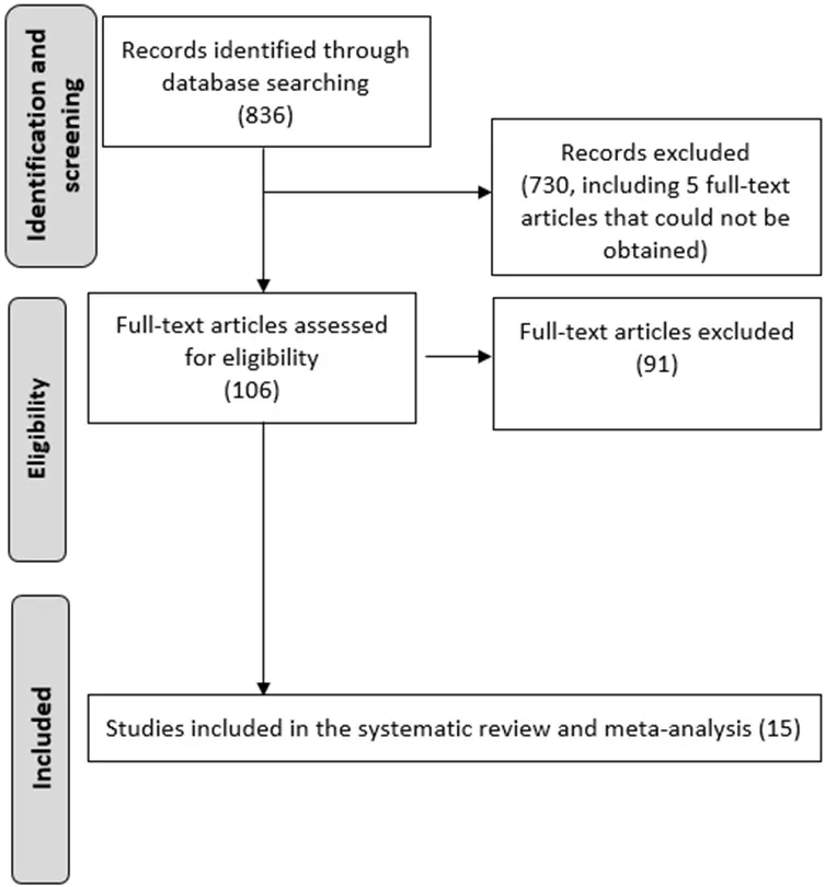
Figure 1.Flow diagram of literature search.
The full text of the remaining 106 studies was evaluated to assess the inclusion criteria.After checking the full-text, 91 articles were excluded, since at least one of the inclusion criteria was not met.No additional records were retrieved after crosschecking the references.
Ultimately, the literature search led to 15 studies suitable for the inclusion in the systematic review and meta-analysis[19-31].
Qualitative analysis (systematic review)
The characteristics of the 15 selected studies are presented in Table 1.All these articles were single-center investigations, published within the last 10 years, except one (2007), by authors from Europe (n= 8) and Asia (n= 7).Two thirds of the studies were retrospective and one third were prospective.

Table 1.Main characteristics of studies with patients with thyroid cancer and hematogenous metastases evaluated by 18F-FDG PET/CT
The patient sample was consistently relatively small across the studies ranging from 11 to 80.The histotype most represented was papillary (67%), followed by follicular (27%).Only one study recruited exclusively patients with anaplastic histotype.
In the 15 studies, patients underwent18F-FDG PET/CT scan at different moments of the diagnostic workup before or after at least one cycle of131I treatment.
The hematogenous lesions investigated were located prevalently in bone and lung.In each study, PET was compared with at least one imaging modality.In four studies,18F-FDG PET/CT was assessed in a headto-head comparison with MRI, prevalently with diffusion-weighted imaging (DWI).CT was available for comparison with18F-FDG PET/CT in three studies.Patients underwent both PET/MRI and PET/CT in three studies.The standard of reference was based on clinical imaging follow-up in most studies; histology was adopted as additional standard of reference in six studies.The risk of bias for the studies was scored as low by using the QUADAS-2 [Figure 2].
Meta-analysis: detectability of hematogenous metastatic lesions by means of 18F-FDG PET/CT
Considering the five studies (patients = 84) for which patient-based analysis was available,18F-FDG PET/CT detected hematogenous metastases in 85.08% (95% C.I.: 75.96%-91.75%) of cases [Figure 3].
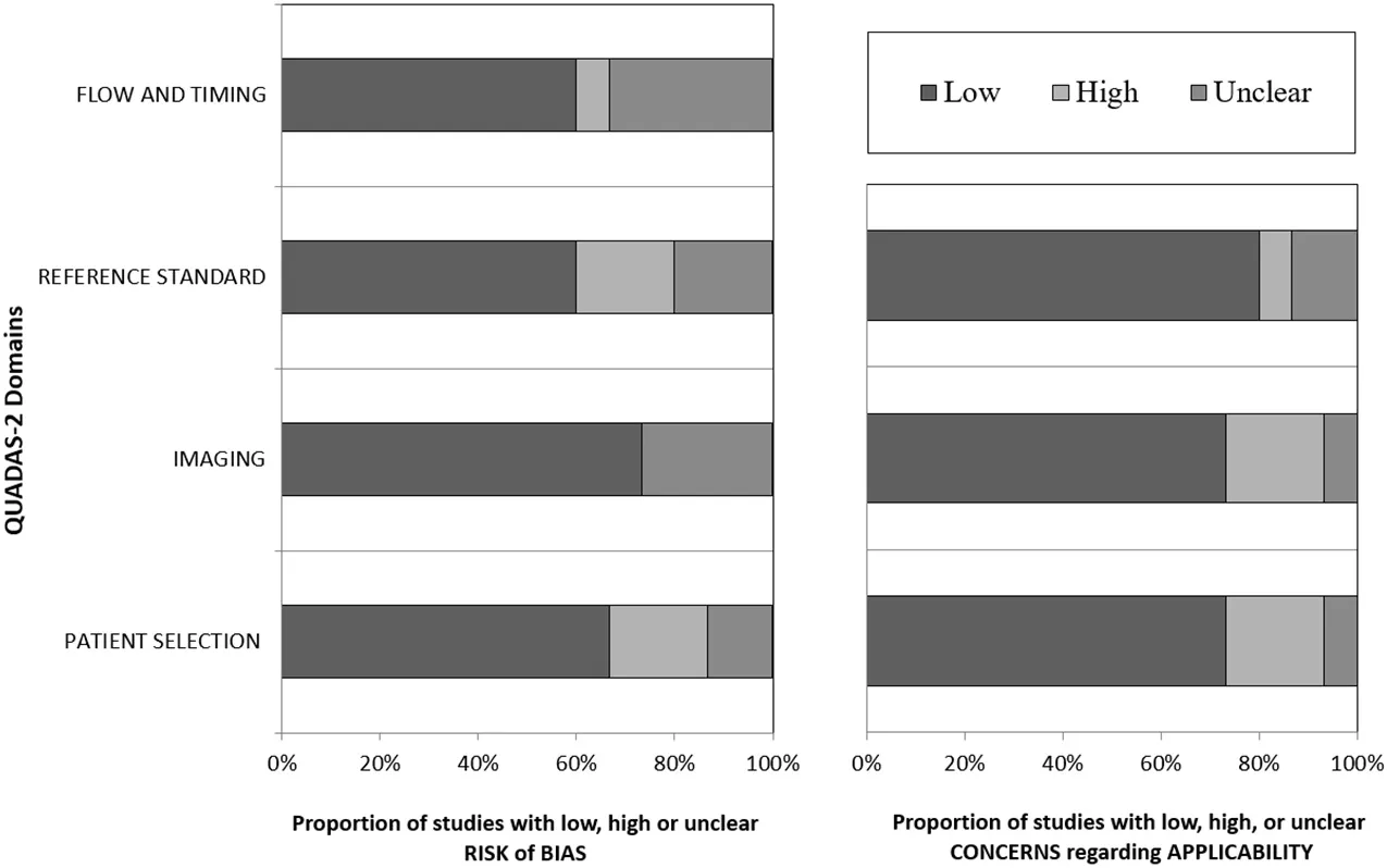
Figure 2.QUADAS-2 results.
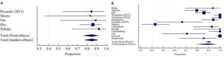
Figure 3.Forest plot of DR of 18F-FDG PET/CT for hematogenous metastases in: per-patient analysis (A); and per-lesion analysis (B).
Considering the 691 lesions (14 studies),18F-FDG PET/CT showed a pooled DR of 89.70% (95%CI: 79.61%-96.62%) with substantial heterogeneity (I2= 92.84%).
In the sub-group analysis of bone lesions (n= 229; 4 studies), a consistent (I2= 13.21%) and high DR was found [81.78%; 95%CI: 76.21%-86.52%; Figure 4].
For the sub-group analysis of lung lesions, DR for18F-FDG PET/CT, CT, and MRI were 92.77%, 95.02%,and 64.93%, respectively [Figure 5].Whereas a significant difference was found comparing the DR of18F-FDG PET/CT with that of MRI (P< 0.001), no significant difference was detected between18F-FDG PET/CT and CT (P= 0.4).
Meta-analysis: accuracy of 18F-FDG PET/CT in detecting hematogenous metastatic lesions
In the per-patient analysis (71 patients with hematogenous metastases out of 139 patients; 4 articles),18F-FDG PET/CT demonstrated a pooled sensitivity (SS) of 87.3% (95%CI: 77.3%-94%) and a pooled specificity (SP) of 95.6% [95%CI: 87.6-99.1; Figure 6], with nine FN and three FP patients.

Figure 4.Forest plot of per-lesion-based DR of 18F-FDG PET/CT for bone metastases.
In the per-lesion analysis (including 290 metastases out of 1038 suspicious lesions; 5 studies), PET/CT showed a pooled SS of 86.3% (95%CI: 73.5%-93.5%) and SP of 93.4% (95%CI: 71.7%-98.8%) in the detection of hematogenous metastases [Figure 7], with 10 FP and 66 FN findings.Substantial heterogeneity was found for both SS (I2= 72.04) and SP (I2= 82.02%).
When restricting the analysis to bone metastases (3 articles, with 170 metastatic lesions out of 614 suspicious lesions), corresponding pooled SS was 81.7% (95%CI: 75.8%-86.7%;I2= 37.9%) and pooled SP was 98.33% (95%CI: 96.5%-99.3%;I2= 88.5%) [Figure 8].Only 7 FP and 38 FN findings were found.Perlung lesion analysis was not performed due to the only 2 articles available.
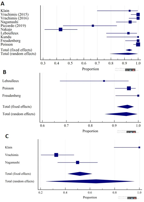
Figure 5.Forest plots of per-lesion-based DR for lung metastases of: 18F-FDG PET/CT (A); CT (B); and MRI (C).
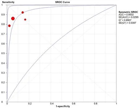
Figure 6.Summary receiver operating characteristic curve for detection of hematogenous metastases by 18F-FDG PET/CT in the patientbased analysis.
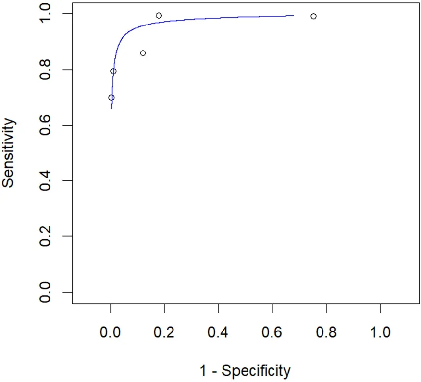
Figure 7.Summary receiver operating characteristic curve (SROC) for detection of hematogenous metastases by 18F-FDG PET/CT in the per-lesion analysis.
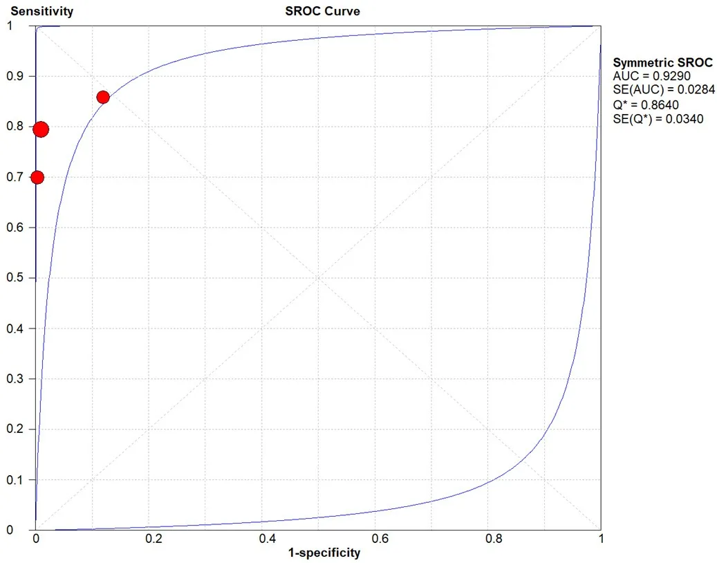
Figure 8.Summary receiver operating characteristic curve (SROC) for detection of bone metastases by 18F-FDG PET/CT in the lesionbased analysis.
DISCUSSION
Our meta-analysis demonstrated the utility of18F-FDG PECT/CT in depicting hematogenous metastases in patients with thyroid cancer.Indeed, the pooled DR for hematogenous metastases was 89.70%.Nevertheless, the substantial heterogeneity points out the small samples of the studies[32].
The location of the lesions probably also influenced the heterogeneity (I2= 92.84%) of the overall DR, as, in the subgroup analysis of bone lesions, the variability of DR across the studies was negligible (I2= 13.21%).These data may also indicate a high inter-subject repeatability of18F-FDG PECT/CT in depicting lesions suspicious for bone metastases in patients with thyroid cancer.Although MRI, with the inclusion of DWI,may assist in the differentiation of cellularity and is generally advocated in the case of vertebral metastases,Sakuraiet al.[30]demonstrated a similar DR of MRI compared to18F-FDG PECT/CT, whereas Nagamachi and colleagues documented a lower detectability of lesions by means of MRI in a patient-based analysis compared to18F-FDG PECT/CT (84.2%vs.57.6%)[23].In our meta-analysis, the three available articles[25,29,30]documented a SS of 71%, 86%, and 79% and corresponding SP of 100%%, 88%, and 99% for bone lesions for18F-FDG PECT/CT.Interestingly, Otaet al.[25]proved18F-Fluoride PET/CT as a potential optimal alternative to18F-FDG PECT/CT for the detection of bone metastases from thyroid cancer, as in their study18F-Fluoride PET/CT demonstrated higher SS and SP compared to18F-FDG PECT/CT.
For lung lesions, it was possible to compare the DR of18F-FDG PECT/CT with that of CT.As expected,CT did not perform inferiorly compared to18F-FDG PECT/CT.Nevertheless,18F-FDG PECT/CT proved superior to MRI in this setting (95.02%vs.64.93%).The sensitivity of PECT/CT in the detection of lung metastases, with corresponding TP and FN cases, was reported in only 2 studies by Vrachimis (100%, 52 lung metastases[33]; 100%, 68 lung metastases[34]).Nakajoet al.[24]reported in their study that FN lung lesions at FDG PET imaging were significantly smaller than TP lung lesions (mean size = 3.8 ± 2.1 mmvs.9.5 ± 6.2 mm;P= 0.001).
When assessing the diagnostic AC of18F-FDG PECT/CT, it appeared clear that a further advantage of18F-FDG PECT/CT in the detection of hematogenous metastases is its very high specificity (95.6% on a perpatient basis and 93.4% on a per-lesion basis).Unfortunately, due to the available literature, no comparison with the diagnostic AC of other imaging techniques was possible.In addition, for the diagnostic AC, the substantial heterogeneity of findings is attributable mainly to the small samples of the studies.
The diagnostic performance of131I-WBS (planar) was consistently reported lower compared to18F-FDG PECT/CT in the detection of hematogenous metastases.Indeed, the study of Nagamachiet al.[23]showed a lower DR for131I-WBS compared to18F-FDG PECT/CT (71.43%vs.85.71%); similarly, the performance of131I-WBS has proved substantially inferior to that of18F-FDG PECT/CT in the detection of lung metastases[22,23].Differently,131I-WBS SPECT/CT may provide a gain in the detection of bone metastases compared to planar imaging.Indeed, in the study of Qiuet al.[29], the accuracy of131I-WBS SPECT/CT was 93.92%, whereas the accuracy of18F-FDG PECT/CT was 86.49%.Unfortunately, no other study inthe literature provides a head-to-head comparison of131I-WBS SPECT/CT and18F-FDG PECT/CT in the depiction of bone metastases.
Other investigated imaging techniques that have been compared to18F-FDG PECT/CT for the detection of hematogenous metastases include18F-FDG PET/MRI and PET imaging with68Ga DOTA derivatives[20,33,34].In the study of Klainet al.[20], the use of18F-FDG PET/MRI did not result in additional benefit in the assessment of lung metastases, obtaining a lower SS (87.5%) compared to18F-FDG PET/CT (93.75%) and MRI (100%).Likewise,18F-FDG PET/MRI was outperformed by18F-FDG PET/CT in the detection of lung metastases in the study of Vrachimiset al.[34](AC: 79.01%vs.97.53%)[33].According to the limited available literature, the use of68Ga DOTA derivatives does not seem to provide noticeable advantages in terms of diagnostic accuracy for lung lesions.
Our meta-analysis presented some limitations.The selected studies provided small sample sizes.Due to the mixed population of most studies, a subgroup analysis of the performance of18F-FDG PET/CT based on the histological type was not possible.We also noticed the impossibility to extract accurate data on the diagnostic performance of18F-FDG PET/CT for subjects with unelevated Tg and/or positive131I-WBS from the included studies in the present meta-analysis; it is conceivable that the diagnostic performance of PET/CT would be inferior for this subgroup of patients compared to the general population.
A source of bias may derive from the substantially high heterogeneity of findings across the studies.Further sources of bias may arise from methodological differences across the studies, including the type of PET/CT scanner.Given the wide range of publication date (2007-2020), studies used PET scanners with different scintillating crystals and reconstruction methods.Furthermore, we noticed that no study using PET/CT scanners equipped with a solid-state detector was found in our meta-analysis.It would be interesting in the future to assess whether these PET/CT scanners may provide a better diagnostic performance in detecting metastases in patients with advanced DTC.
CONCLUSION
The present meta-analysis suggests the use of18F-FDG PECT/CT for the identification of hematogenous metastases in patients with thyroid cancer.Nevertheless, the limited number of available studies urges further research in this setting.Comparative studies are still needed to clarify the performance of18F-FDG against other radiotracers and imaging techniques.Furthermore, studies evaluating the impact of diagnostic performance of18F-FDG PET/CT on patient management are warranted.
DECLARATIONS
Authors’ contributions
Performed data search, statistical analysis and wrote the first draft of the manuscript: Quartuccio N.
Made substantial contributions to conception and design of the study and revision: Rubello D.
Provided technical and editing support: Rubello D.
Availability of data and materials
Not applicable.
Financial support and sponsorship
None.
Conflicts of interest
All authors declared that there are no conflicts of interest.
Ethical approval and consent to participate
Not applicable.
Consent for publication
Not applicable.
Copyright
© The Author(s) 2021.
杂志排行

Journal of Cancer Metastasis and Treatment的其它文章
- Diverse roles of bitter melon (Momordica charantia)in prevention of oral cancer
- Leelamine suppresses cMyc expression in prostate cancer cells in vitro and inhibits prostatecarcinogenesis in vivo
- Seaweeds’ nutraceutical and biomedical potential in cancer therapy: a concise review
- Targeting EZH2 for the treatment of soft tissue sarcomas