Correlation of testicular reproductive tumor related genes in semen of patients with testicular microlithiasis
2021-05-20ChunXuLiShaoFengChenYuanZhangLiFengChenQiangGeng
Chun-Xu Li,Shao-Feng Chen,Yuan Zhang,Li-Feng Chen,Qiang Geng
1Department of Urology,Tianjin Medical University Second Hospital,Tianjin 300211,China;
2Department of Andrology,First Teaching Hospital of Tianjin University of Traditional Chinese Medicine,Tianjin 300193,China;
3Department of Pediatrics,First Teaching Hospital of Tianjin University of Traditional Chinese Medicine,Tianjin 300193,China;
4Department of Physiology,Basic Medical College,Hebei North University,Zhangjiakou,Hebei 075000,China.
Abstract Objective:To investigate the expression of testicular tumor-related genes in the semen of patients with microlithiasis,as well as the semen concentration in patients with microlithiasis.Methods:Retrospective analysis of 2018/6-2019/6 male in our hospital clinic examination of patients with testicular color to exceed,semen specimen were collected,30 patients with testicular micro stone disease group of 15 cases,control group in 15 cases,the process of semen automatic analyzer to detect sperm concentration,by rt-pcr and Western Blot respectively detect testicular cancer related gene (KITLG KIT ligand (gene),SPRY4 (sprouty RTK signaling 4)antagonist) mRNA and protein expression.Results:The semen concentration (17.31±0.92) × 10-6/ml in the microstone group was lower than that in the control group (29.26±1.57) × 10-6/ml.The mRNA expression of semen (KITLG,SPRY4) in the microstone group was higher than that in the control group,and the difference was statistically significant.The protein expression of semen (KITLG,SPRY4) in the microstone group was higher than that in the control group,and the difference was statistically significant.Conclusion:The semen concentration of testicular microlithiasis decreased,but the expression of seminal testicular tumor genes (KITLG,SPRY4)increased.
Key words:Testicular microlithiasis,Semen concentration,Testicular malignancy related genes
Background
Testicular Microlithiasis (Testicular Microlithiasis,TM)refers to the syndrome formed by numerous calcifications with a diameter of less than 3mm that are diffusely distributed in the seminiferous tubules of the testis.Ultrasonography can detect multiple micro-calcifications [1]in the parenchyma of the testis.It is still unclear to the etiology and pathogenesis of TM.The studies have shown that TM is related to testicular tumors,and TM is a benign testicular lesion that is suspected to be precancerous and has a relatively slow progression [2].There is more and more clinical studies on the correlation between TM and testicular tumors.The Research by Mao Weilin and others [3]showed that the quality of semen decreased in patients with testicular microlithiasis.However,TM has no specific clinical manifestations,and most patients are found during routine physical examinations,pre-pregnancy examinations,or fertility disorders.In summary,TM is related to infertility and testicular tumors.Some scholars hold a view that TM is a risk factor for testicular tumors,but with controversy.This study investigates the expression of testicular tumor-related genes in the semen of patients with testicular microlithiasis,and analyze the gene expression of testicular tumors in the semen of patients with testicular microlithiasis,and the correlation between the decrease in semen quality of patients with testicular microlithias and testicular tumor genes.
Materials and methods
General information
After a reviewed analysis to the patients who had testicular color Doppler ultrasound examination in the Andrology Department of our hospital from June 2018 to June 2019 (Figure 1) and were screened,it shows that all patients had no history of mumps and normal testicular development.Among them,there were 20 patients with testicular microlithiasis.In the normal control group there were 20 patients with an average age of 35 years (20-40 years old).The average age of the microlithic group was 33 years old (20-38 years old),and the control group was 34 years old (22-40 years old.The retention time of semen samples was left after an abstinence of 2-7 days,an average of 4.5 days.
Exclusion criteria:(1) Varicocele;(2) Testis or epididymitis;(3) Male gonadal dysfunction;(4)History of mumps or testicular dysplasia;(5) Negative habits to seriously affect semen quality (Including long-term alcohol abuse,frequent sauna experience,etc.);(6) Use of drugs to impair the quality of semen;(7) Chronic prostatitis;(8) Azoospermia.
Methods
Since June 2019,we have carried out the screening to the patients in our department,collecting the semen of patients with testicular microlithiasis and normal patients reported by testicular ultrasound,which were first tested the sperm concentration before freezing.By RT-PCR,it was aimed to detect the mRNA expression in semen (KITLG,SPRY4),and by WB,it was aimed to detect the protein expression in semen (KITLG,SPRY4).
Statistical methods
The experimental data were expressed as mean ±standard deviation(±SD).All data were statistically processed by using SPSS22.0 software,which was statistically significant whenP<0.01.
Results
Effect o f t esticular microlithiasis o n se men concentration
The semen concentration of the microstone group was(17.31±0.92)×10-6/ml,and that in the control group was (29.26±1.57)×10-6/ml.The results showed that testicular microlithiasis had a bad effect on semen concentration.The concentration of semen in the TM group was lower than that in the control group (P<0.01,as shown in Table 1).
Expression of KITLG and SPRY4mRNA in semen between TM and control groups
The relative expression ratio of KITLG and SPRY4 mRNA was respectively 1.32±0.06 and 0.46±0.03,and that in the control group was 1.88±0.04 and 0.91±0.03.The expression of KITLG and SPRY4mRNA in semen of the TM group was increased in comparison with that of the control group (P<0.01,as shown in Table 2).
Expression of KITLG and SPRY4mRNA in semen between TM and control groups
The relative expression ratio of KITLG protein was 29.26±1.57,and that in the control group was 17.31±0.92.The expression of KITLG and SPRY4 protein in semen of the microlithic group was higher than that of the control group (P<0.01,as shown in Figure 2,Table 3).
Table 1 Semen concentration(±SD)
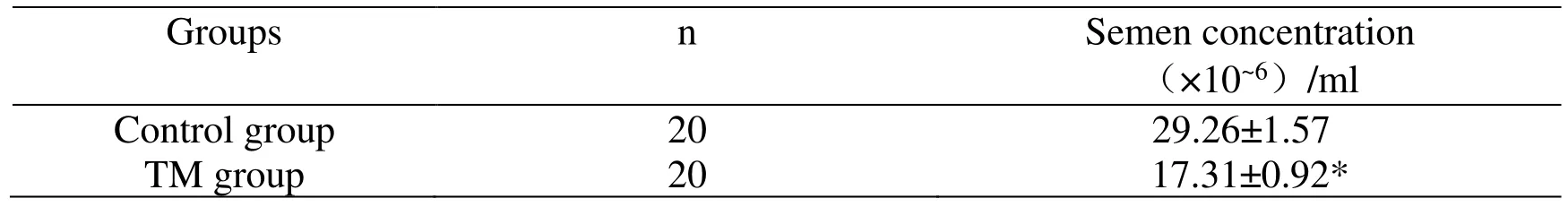
Table 1 Semen concentration(±SD)
Note:*P<0.01,compared with the control group (P<0.01)
Table 2 The expression of KITLG and SPRY4 mRNA in semen(±SD)
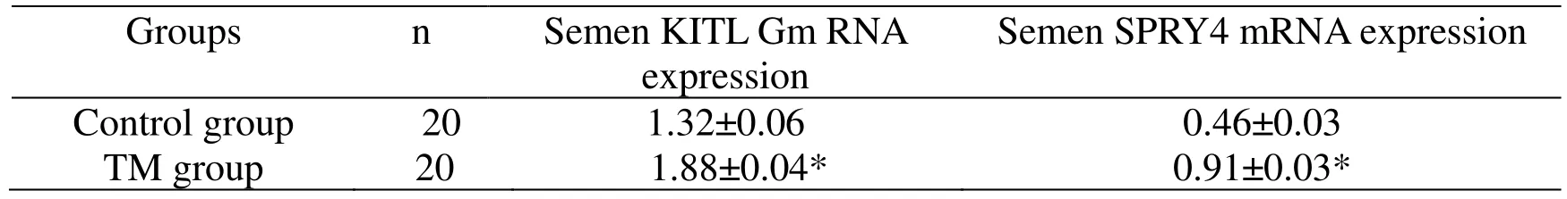
Table 2 The expression of KITLG and SPRY4 mRNA in semen(±SD)
Note:*P<0.01,compared with the control group (P<0.01)
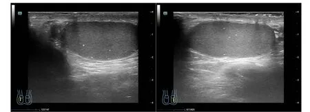
Figure 1 The appearance of testicular microstone under color ultrasound
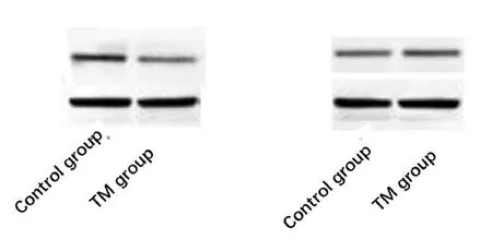
Figure 2 Western blotting of KITLG and SPRY4 proteins
Table 3 Semen KITLG and SPRY4 protein expression(±s)
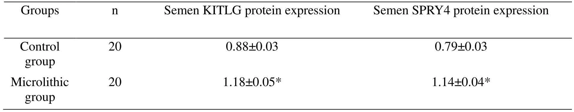
Table 3 Semen KITLG and SPRY4 protein expression(±s)
Note:*P<0.01,compared with the control group (P<0.01)
Discussion
In recent years,the diagnosis rate of testicular microlithiasis is shaped with the rising tendency,and the diagnosis is mostly found because of other diseases,with testicular microlithiasis found by the auxiliary examination.The experts believe that the formation of testicular microliths is related to obstruction. Main reason is the degeneration,necrosis and shedding of seminiferous tubule epithelial cells,and then it results in inflammation,oxidative stress,fibrosis,and increased internal pressure of the seminiferous tubules,which do harm to the blood circulation of the testis,and as a result it leads to the loss of seminiferous tubule function [4].About 30% to 60% of TM patients are found to have obstruction of the seminiferous ducts,impaired spermatogenesis,and decreased semen concentration [5].Meanwhile,sperm motility of TM patients is also characterized by decreasing,which affects sperm migration and finally it leads to infertility [6].In addition,the amount of semen and sperm motility in the patients also can be reduced by the inflammation of the seminiferous tubules and calcification in the seminiferous tubules of patients with TM,and thereby it causes infertility [4].It has a higher detection rate of TM to patients with severe oligospermia and spermatogenic dysfunction caused by reduced testicular volume [7].Therefore,it is worse to the sperm quality and testicular volume of patients with high grade TM.The existing research shows that there is a clear correlation between TM and male infertility [4].TM is partially considered to be a syndrome of testicular hypoplasia.It also has a higher detection rate in reproductive-related diseases such as low fertility,testicular atrophy and other sexual development abnormalities [8].In addition,some scholars have also reported abnormal semen quality in infertile patients with TM [9],but other scholars believe that TM has not significant relation with male infertility [10].Our research discloses that TM causes a decrease in semen concentration in comparison with infertile patients without TM.
Many studies show that testicular microliths are precancerous lesions of testicular tumors,increasing the occurrence of testicular tumors.Bach and others[11]report that about 6% to 46% of patients with TM had combined with testicular germ cell tumors,and about 27% of patients with testicular tumors had combined with TM symptom at the same time.Ringdahl and others [12]analyze a group of patients with scrotal discomfort as the main symptom,and the results of auxiliary ultrasound examination show that among 160 patients,12 is combined by TM,and 4 of 12 (33%) are testicular tumors.In the remaining 148 cases,they have not combined with TM,and 2 cases are found to have testicular tumors.The data shows that the relative risk of testicular tumors is 36.5% when TM is found in symptomatic patients,with 0.67% of the sensitivity and 0.95% of the specificity respectively.Therefore,it is speculated that it has a higher rate for the incidence of germ cell tumors combined in TM-associated tumors.Moreover,in the TM patients with symptom,the detection rate of testicular tumors is higher than that of the normal population.KITLG gene,also known as stem cell factor,is characterized of extensive biological activities and is the ligand gene for the encoded protein of the proto-oncogene e-kit.This gene plays an important role in the renewal and differentiation of spermatogonium.Studies have shown that mouse KITLG gene mutations pose a damage to the proliferation and differentiation of primordial germ cells,resulting in the decrease or loss of fertility [13].SPRY4 is located on chromosome 5q31.3.The encoded protein of this gene gets involved in the regulation process of acidic fibroblast growth factor (acid fibroblast growth factor,FGF1).FGF1 plays an important role in various physiological and pathological processes.In addition,the encoded protein of gene is also a negative regulation factor of the RAS-ERK-MAPK signaling pathway,which can be activated by the KITLG-KIT pathway.This study confirmed that the expression of KITLG,SPRY4mRNA and protein in the semen of patients with testicular microlithiasis was higher than that of the normal group,indicating that the occurrence of testicular tumors is related to the testicular tumor-related genes KITLG and SPRY4,that is,testicular microlithiasis is a risk factor for testicular tumors and is very likely that testicular microlithiasis increases the expression of testicular tumor-related genes,thereby increasing the occurrence rate of testicular tumors.In addition,this study also confirmed that the semen concentration of patients with testicular microlithiasis is lower than that of normal.Therefore,it indicates that the testicular tumor-related genes KITLG and SPRY4 may be a risk factor for male infertility.
Testicular tumor-related genes are highly expressed in the semen of patients with testicular microlithiasis,which provides experimental data for testicular microlithiasis as a risk factor for testicular tumors and increase data support.In clinical work,patients with testicular microlithiasis should be warned of timely follow-up and regular re-examinations to facilitate early detection of malignant changes,early treatment,and achieve good results.At the same time,the experiments have also confirmed that the quality of semen in patients with testicular microlithiasis is lower than that of normal patients,i.e.,testicular microlithiasis may be one of factors leading to infertility.For infertility patients with unknown reason,they can be checked with testicular color doppler,except for microlithiasis.By doing so,it can provide a new direction for the treatment of infertility.
The studies have confirmed that the expression of testicular tumor-related genes gets increased in the semen of patients with testicular microlithiasis.But the experiments still exist some deficiencies.As a small number of samples in this experiment,it may be some gaps in the results of the experiment.In the future,the number of samples will be increased so as to keep improving the related experiments.Another point is that the follow-up time of experiment is short,only about 3 months for the shortest patient,and then we will continue to pay the follow-up and observe the occurrence of testicular tumors.