Effects of Guangxi Hepu Pearl Hydrolysate on Proliferation Activity and Apoptosis of Human Hepatic Stellate Cells
2021-02-26YuePengQuanshengFengJiangLinTiejianZhaoPengLiuYanhuiCen
Yue Peng,Quansheng Feng,Jiang Lin,Tiejian Zhao,Peng Liu,Yanhui Cen
1.Guangxi University of Chinese Medicine,Nanning 530200,China;2.Chengdu University of TCM,Chengdu 611137,China
Abstract [Objective]The paper was to observe the effects of Guangxi Hepu pearl hydrolysate on proliferation and apoptosis of human hepatic stellate cells.[Method]The inhibition rate against the proliferation of human hepatic stellate cells was determined by MTT colorimetry after co-incubation with pearl hydrolysate for 48 h.The percentage of early and late apoptotic cells was determined by flow cytometry(Annexin V-FITC/PI staining).[Result]Low,medium and high concentrations of pearl hydrolysate could inhibit the proliferation of human hepatic stellate cells,and the inhibition rates at 48 h were 12.4%,27.4% and 37.8%,respectively.The low,medium and high concentrations of pearl hydrolysate could induce the apoptosis of cells.The percentages of total apoptotic cells(early apoptotic+late apoptotic)at 48 h were 8.97%,16.09% and 26.98%,respectively.[Conclusion]Pearl hydrolysate can inhibit the proliferation of human hepatic stellate cells and induce the apoptosis of these cells,which may be the mechanism of anti-hepatic fibrosis effect of pearl hydrolysate.These effects are dose-dependent.It is also found that the above cellular effects of high dose pearl hydrolysate are stronger than those of colchicine and compound Biejiaruangan tablets.
Keywords Pearls;Hepatic stellate cells;Cell proliferation;Apoptosis;Liver fibrosis
According to modern medicine,liver fibrosis refers to the process that with the increasing necrosis of liver cells after chronic continuous effect of injury factors(inflammation,physical and chemical,etc.)on liver,the liver cells are regenerating,the proliferation and degradation of extracellular matrix(various types of collagen)in the liver are out of balance,which leads to structural disorders of the liver,extensive deposition of fibrous connective tissue,resulting in a series of structural and functional abnormalities.Currently,there is a relatively unified understanding of the mechanism of the disease[1],which is due to the activation of hepatic stellate cells and significant phenotypic transformation under the regulation of various cytokines during chronic liver injury,thus obtaining the characteristics of myofibroblasts.Subsequently,the activated hepatic stellate cells migrate extensively and proliferate massively at the site of liver injury,and synthesize and secrete high levels of extracellular matrix,leading to the occurrence and development of fibrotic lesion.Hepatic stellate cells play a central role in the formation of liver fibrosis.In recent years,human hepatic stellate cells are often used as cell models in the research of hepatic fibrosis to study the pathogenesis and reversal mechanism of liver fibrosis,and even the pharmacological mechanism of anti-liver fibrosis drugs.
Liver fibrosis is the common pathological feature and destination of all types of chronic liver diseases in the middle and late development stages.The incidence of chronic liver disease is high in China,which seriously endangers the health of the people.Currently,there is no specific therapy in the clinical treatment of chronic liver disease,so the focus of treatment has gradually shifted to how to prevent the transformation of chronic liver disease into liver fibrosis and cirrhosis[2].However,efficient western drugs with low toxic and side effects are still lacked in clinical treatment of the disease,whereas traditional Chinese medicines show their characteristics and advantages in the treatment of liver fibrosis.Seawater pearls,in addition to being used as human ornaments,also have high medicinal value.Pearls can be divided into seawater pearl,freshwater pearl and western pearl according to their origin,and there is always a saying that"western pearl is inferior to freshwater pearl,and freshwater pearl is inferior to seawater pearl"[3].Pearls produced in the Beibu Gulf of China belong to seawater pearl,especially Hepu Pearl,which is the most famous and high-quality representative of seawater pearl[4].Seawater pearl is large,round,pink,tender and crystal,with thick nacre,and high medicinal value.In most cases,it is ground into pearl powder and used medically.It is cold in nature,sweet and salty in taste,which has obvious curative effect on hyperthyroidism,pharyngitis,tonsillitis,gynecological inflammation,damp-heat liver disease,and has the effect of calming the mind,clearing away heat and toxic materials,nourishing yin,and reducing phlegm and dampness[5].Compendium of Materia Medica also recorded the medical use of pearl.
In this test,Guangxi Hepu pearl hydrolysate was co-incubated with HSC-LX2,the core cell of human liver fibrosis,to observe the effect of the drug on human hepatic stellate cells,and to preliminarily explore the mechanism of action.The inhibition rate against the proliferation of cells was determined by methyl thiazolyl tetrazolium(MTT)colorimetry to infer the cytotoxicity of the drug.The percentage of early and late apoptotic cells was detected by flow cytometry (Annexin V-FITC/PI staining),and the mechanism of the drug against liver fibrosis was preliminarily investigated.
1 Materials and Methods
1.1 Materials
1.1.1 Instruments.MCO-15AC CO2constant temperature incubator was purchased from Sanyo;Eclipse Ts2 inverted microscope was purchased from Nikon;5702R low-temperature centrifuge was purchased from Eppendorf;CytoFLEX flow cytometer was purchased from Beckman.
1.1.2 Reagents.Human hepatic Stellate cell-LX2(HSC-LX2)was provided by professor Xu Liming at Institute of Liver Diseases,Shuguang Affiliated Hospital of Guangxi University of Chinese Medicine.Hepu pearl hydrolysate was provided by-Beihai Baozhulin Marine Science&Technology Co.,Ltd.Methyl thiazolyl tetrazolium(MTT)was purchased from Sigma(batch number 191M2466W);and Annexin V-FITC/PI Apoptosis Kit was purchased from ElabScience(batch number E-CK-A211).
1.2 Methods
1.2.1 Preparation of experimental drugs(pearl hydrolysate).Approximately 60 mL of concentrate pearl hydrolysate with the mass concentration of 1 g/L was added with serum-free DMEM culture solution and then diluted to a volume of 100 mL.In total,600 mg/L of experimental pearl hydrolysate was prepared following the above method,and filtered and sterilized through microporous membrane for later use.The solution was diluted into corresponding working liquid according to the concentration required by each experimental group.
1.2.2 Grouping of experimental cells.HSC-LX2 were removed from liquid nitrogen and quickly dissolved in a water bath at 37℃.After thawing,the cells were transferred to a centrifuge tube containing 5 mL of medium and centrifuged at 1 000 rpm for 5 min,and the supernatant was discarded.The cells were suspended with complete medium containing 10% fetal bovine serum,inoculated into petri dishes,blown and mixed well,then cultured in an incubator at 37℃,5% CO2and saturated humidity.When the cells were in logarithmic growth phase(good adhesion,well growth,and covering about 80% of the bottom of culture bottle),1-2 mL of 0.25% trypsin were added,and the cells were digested from the wall of culture bottle for 1-2 min.When the cells separated from each other and became round under microscope,the digestion was complete.After trypsin was discarded,complete medium was added,and the cells were blown to develop single cell suspension.After centrifugation,the supernatant was discarded,and the cells were re-suspended with fresh medium.The fluids were inoculated into 96-well or 6-well culture plates,and incubated in a CO2incubator for 48 h.After the cells were completely attached to the wall,experimental groups were designed with wells of culture plate as the units.
The cells were divided into seven groups,and the corresponding working fluids were added according to different groups.The details were as follows.(i)Group 1 was the blank control group(HSC-LX2 cells were cultured according to the standard method without any influencing factors).(ii)Group 2 was leptin activation model control group(the culture medium was added with Leptin with the final concentration of 100 μg/L for cell activation).(iii)Group 3 was the pearl hydrolysate low-concentration group(after cells were activated by Leptin,the pearl hydrolysate was added,and the final concentration was 30 mg/L).(iv)Group 4 was the pearl hydrolysate medium-concentration group (after cells were activated by Leptin,the pearl hydrolysate was added,and the final concentration was 60 mg/L).(v)Group 5 was the pearl hydrolysate highconcentration group (after the cells were activated by Leptin,the pearl hydrolysate was added,and the final concentration was 120 mg/L).(vi)Group 6 was the colchicine intervention control group (after the cells were activated by Leptin,colchicine was added,and the final concentration was 6.25 μg/mL).(vii)Group 7 was the salvianolic acid B intervention control group(after the cells were activated by Leptin,salvianolic acid B was added,and the final concentration was 10-6mmol/L).
Experiments were performed according to the above groups,and each test was repeated three times.
1.2.3 Detection of cell proliferation of HSC-LX2 by MTT colorimetry.When HSC-LX2 reached the logarithmic growth stage,it was digested with 0.25% trypsin solution and re-suspended with DMEM culture solution containing 10% fetal bovine serum,and the cell suspension (5×10⁴cells/mL)obtained was inoculated into 96-well culture plates.Each well was added with 200 μL of cell suspension.The plate was placed in an incubator at 37℃,5% CO2and saturated humidity for 48 h,and the old culture solution was discarded when the cells were in good adhesion condition.The 96-well culture plate was divided into seven groups according to the unit of wells,and each group had 12 duplicate wells.Each well was added with 200 μL of corresponding working fluid according to the requirements of experiment grouping.Subsequently,the plate was placed in an incubator at 37℃,5% CO2and saturated humidity for 48 h.MTT reagent was added into the plate,200 μL per well,and the plate was placed in the incubator for 6 h.After the old culture solution was discarded,each well was added with 200 μL of DMSO and shaken well by ultrasonic oscillator for 2 min.The OD values of cells in each group were determined by microplate reader under the wavelength of 570 nm and the reference wavelength of 450 nm.The formula for inhibition rate of cell proliferation was as follows:inhibition rate of cell proliferation=(1-OD value of drug/OD value of control)×100%.
1.2.4 Detection of cell apoptosis by flow cytometry(Annexin V-FITC/PI double staining).According to the instructions of Annexin V-FITC/PI kit,HSC-LX2 in good growth state during logarithmic growth phase was digested and blown with trypsin for cell counting.Cell suspension(5×10⁵cells/mL)was prepared with DMEM culture solution containing 10% fetal bovine serum,and the cell suspension was inoculated into 6-well culture plate at a volume of 1.5 mL per well.Afterwards,the plate was cultured in CO2incubator for 48 h.
After cells were attached to the wall perfectly,the cells were divided into seven groups and the old culture solution was discarded.According to the requirements of experimental grouping,2 mL of corresponding working liquid was added into each well,and the plate was cultured in a CO2incubator for 48 h.The adherent cells were digested and blown by trypsin and collected in a centrifuge tube.After cell counting,the tubes were centrifuged at 1 000 rpm for 5 min,and the cells were rinsed twice with PBS solution.The cells were re-suspended with 500 μL of Binding Buffer(1-fold)and mixed evenly,and then the concentration was adjusted to 1×10⁶cells/mL.The 100 μL of cell fluid was absorbed into a centrifuge tube,which was then added with 5 μL of AnnexinV-FITC and shaken,and incubated at room temperature for 5 min.Afterwards,5 μL of PI was added into each centrifuge tube and shaken,and incubated at room temperature in dark place for 15 min.The centrifuge tubes were added with 500 μL of Binding Buffer(1-fold),and the cells were mixed.The cells were detected by flow cytometry,with argon ion gas laser(wavelength 488 nm)and long-wavelength pass filter(block filter glass 530 nm).
1.2.5 Statistical treatment.The results were expressed as mean±standard deviation(mean±SD),and the data were analyzed by PEMS 3.1 statistical software.Multiple comparisons of means of multiple samples were performed by analysis of variance(Newman-Keuls test),and significance between partial data were analyzed by one-way ANOVA(F test).P<0.05 indicated statistical significance.
2 Results and Analysis
2.1 Detection results of inhibition rate of HSC-LX2 by MTT colorimetry Compared with the blank control group,the cell proliferation rate of HSC-LX2 in each administration group showed significant inhibitory effect.It could be seen that pearl hydrolysate showed significant inhibition effect on the proliferation of HSC-LX2 cells,and with the increase of administration dose,the proliferation activity of HSC-LX2 cells declined correspondingly,indicating that its inhibition effect on proliferation was dose-dependent.The differences were statistically significant(P<0.05).The inhibitory effect of high concentration of pearl hydrolysate on cell proliferation was stronger than that of colchicine and compound Biejiaruangan tablet,and the difference was statistically significant(P<0.05,Table 1).
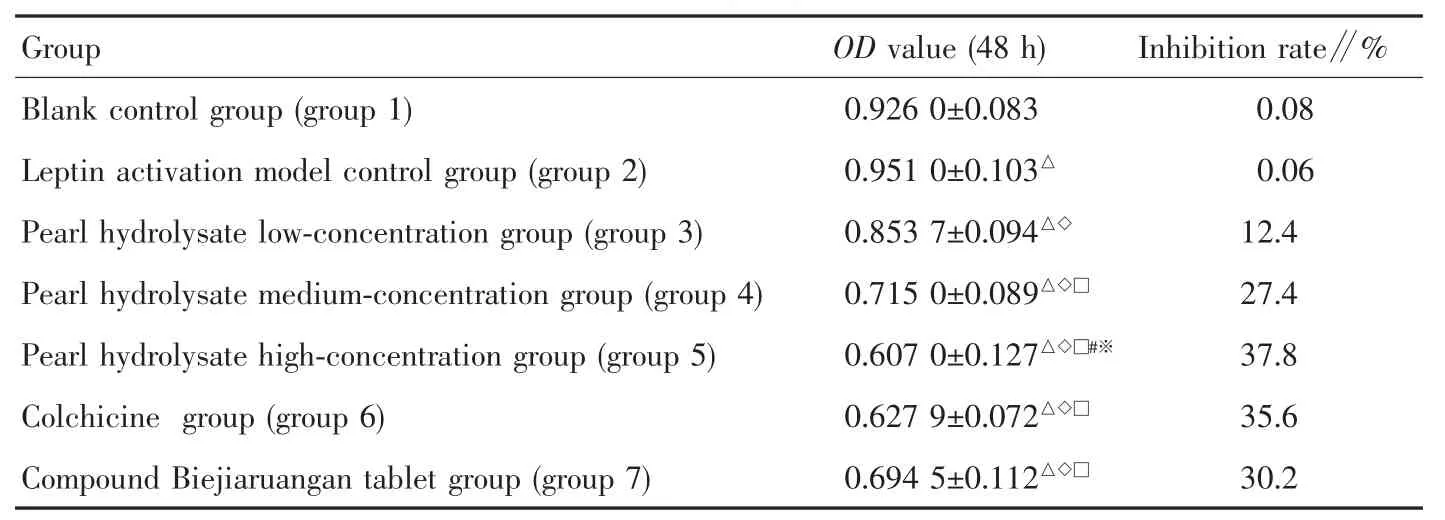
Table 1 Proliferation of HSC-LX2 cells in each group(x±s,n=3)
2.2 Cell apoptosis detected by flow cytometry Compared with group 1,the percentage of early and late apoptotic rate of HSC-LX2 cells in groups 2,3,4,5,6 and 7 were increased significantly at 48 h post administration.The percentage of early and late apoptotic rate of HSC-LX2 cells increased significantly with the increasing dose of pearl hydrolysate.The pearl hydrolysate induced apoptosis of HSC-LX2 cells in a dose-dependent manner.The differences were statistically significant(P<0.05).Compared with colchicine and compound Biejiaruangan tablet,high concentration of pearl hydrolysate had stronger ability to induce cell apoptosis(P<0.05,Table 2 and Fig.1).
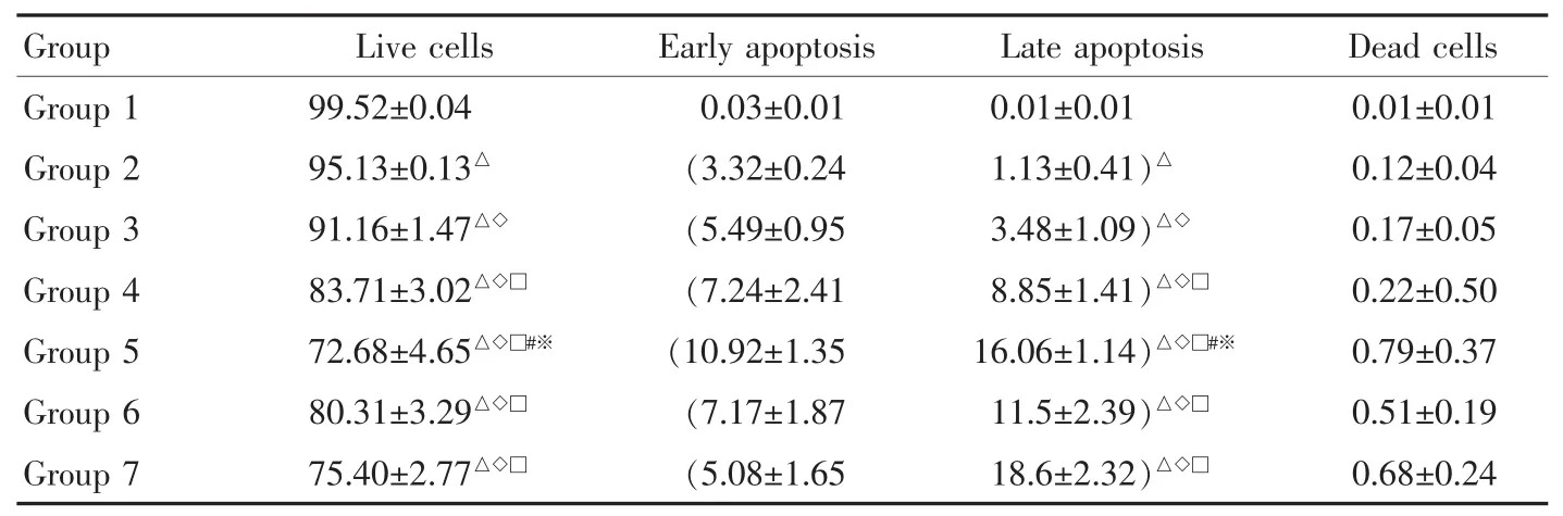
Table 2 Percentage of apoptotic rate of HSC-LX2 cells in each group(x±s,n=3) %
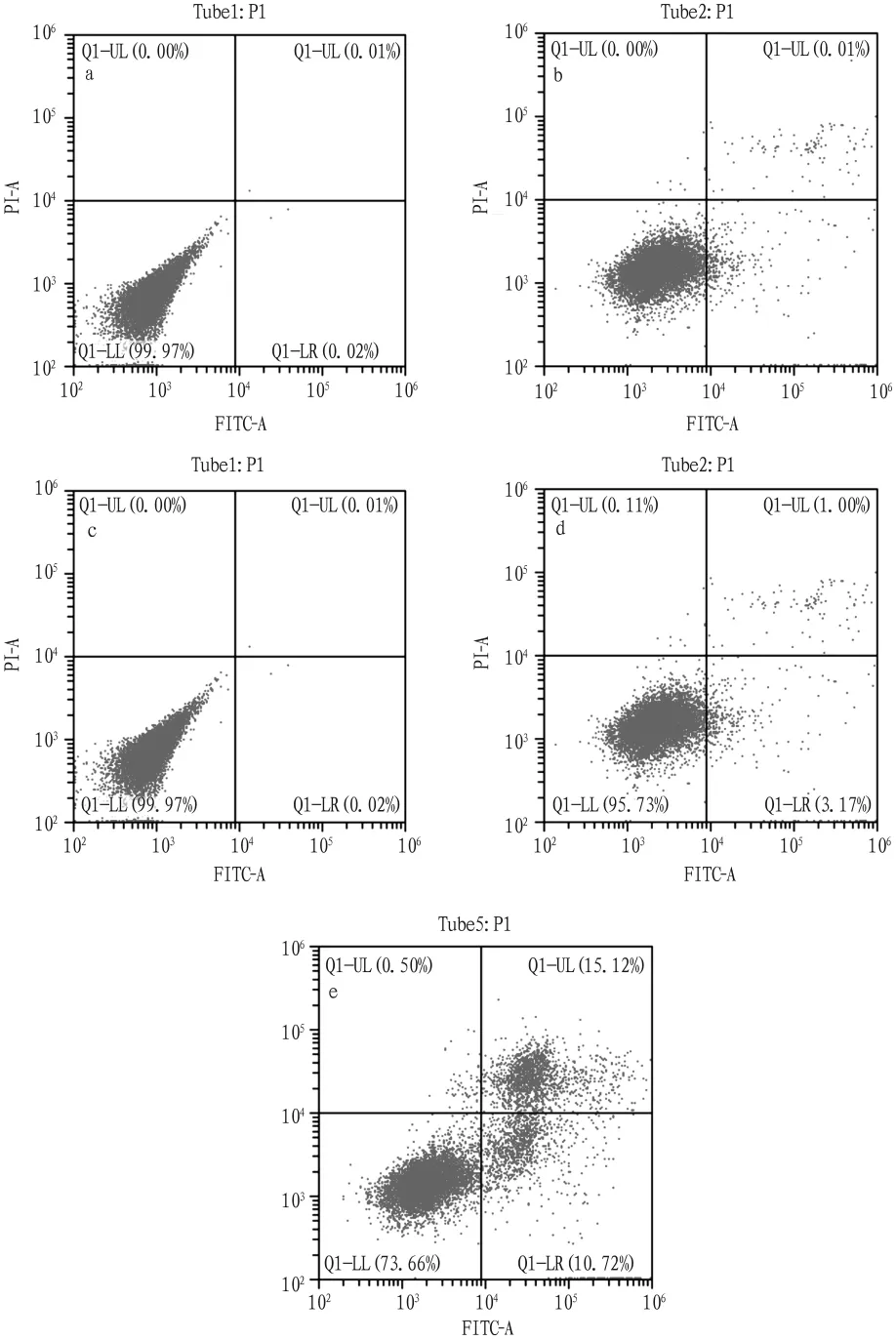
Fig.1 Four-quadrant scatter diagram of flow cytometry
3 Discussion
(i)After the death of liver cells due to continuous stimulation of pathological factors such as chronic injury or inflammation,the regeneration of liver cells and reconstruction of liver tissues continue to occur,and a large amount of extracellular matrix deposition will lead to the formation of liver fibrosis.Although many cells in the liver can synthesize extracellular matrix,hepatic stellate cells are considered to be the major sources of extracellular matrix in chronic liver disease.In the process of liver fibrosis,hepatic stellate cells undergo phenotypic transformation under the regulation of various cytokines and acquire the characteristics of myofibroblasts.The ability of hepatic stellate cells to synthesize collagen is significantly increased,which significantly enhances the contractiity of cells.Therefore,the activation and proliferation of hepatic stellate cells and the synthesis and secretion of large amount of extracellular matrix events play a key role in the formation and development of liver fibrosis[6].It can be observed that the severity of liver fibrosis is proportional to the activation degree of hepatic stellate cells in the liver tissues of liver fibrosis patients and those of animal models of experimental liver fibrosis[7].Whether a certain drug has inhibitory effect on the proliferation of hepatic stellate cells can directly reflect its reversal effect of liver fibrosis to some extent.The proliferation of HSC-LX2 cells was detected by MTT assay,and the results showed that Hepu pearl hydrolysate had obvious inhibitory effect on the proliferation of hepatic stellate cells,with significant cytotoxicity.Moreover,the cell proliferation rate was significantly reduced with the increase of the dose,and the cytotoxicity of the drug was dose-dependent.High concentration of pearl hydrolysate could inhibit the proliferation of hepatic stellate cells more efficiently than colchicine(western medicine)and compound Biejiaruangan tablet(traditional Chinese medicine).It can be concluded that the anti-fibrosis effect of Hepu pearl hydrolysate was largely related to its inhibition against proliferation activity of hepatic stellate cells.
(ii)Cell proliferation and apoptosis are two important events in cell biology.An increase in cell proliferation or a decrease in apoptosis can directly lead to excessive cell accumulation.Therefore,in addition to excessive cell activation and proliferation,the deficiency of cell apoptosis may also be a key factor in the pathological increase of hepatic stellate cells during the course of liver fibrosis.Apoptosis of hepatic stellate cells,as an internal regulation mode at the gene level,can effectively reduce the number of acti-vated hepatic stellate cells,inhibit the activation of hepatic stellate cells in another way,and also interfere with the course of liver fibrosis,or even reverse liver fibrotic lesion[8].Saile et al.[9]pointed out that the phenotype of hepatic stellate cells was difficult to be reversed once activated,and it might be an important means to reverse liver fibrosis to reduce the number of hepatic stellate cells by inducing apoptosis.Therefore,the induction of apoptosis of hepatic stellate cells has become a new hotspot in the development of anti-fibrosis drugs in recent years.In this study,the apoptotic status of HSC-LX2 cells was detected by flow cytometry(Annexin V-FITC/PI double staining),and the results showed that the early and late apoptotic rates of HSC-LX2 cells increased significantly when interfered with Hepu pearl hydrolysate for 48 h.The percentage of total apoptosis (early apoptotic rate+late apoptotic rate)was significantly increased with the increase of the dose,indicating that the apoptosis-inducing ability of the drug was dose-dependent.It was also found that high concentration of pearl hydrolysate induced higher percentage of apoptosis of hepatic stellate cells than colchicine and compound Biejiaruangan tablets.Therefore,it can be inferred that the anti-fibrosis effect of Hepu pearl hydrolysate is related to its inhibition against the activation of hepatic stellate cells,and the mechanism of the above two events is probably caused by the apoptosis of hepatic stellate cells induced by Hepu pearl hydrolysate.
EBSCO Publishing,headquartered in Ipswich,Massachusetts,is an aggregator of premium full-text content.EBSCO Publishing’s core business is providing online databases via EBSCOhost to libraries worldwide.EBSCOhost is used by libraries,schools,academic institutions,medical institutions,and corporations.The company is a subsidiary of Birmingham,Alabama-based EBSCO Industries.EBSCO Industries is located at number 196 of the top 200 privately held companies in the United States by Forbes Magazine.The company’s core business is providing online databases via its proprietary software,EBSCOhost,to libraries.EBSCO provides over 350 full-text and secondary databases.Content for these databases include full-text journals,magazines,books,monographs,reports,ebooks,business book summaries and various other publication types.It also provides databases for reference to the health and business sectors,such as DynaMed.
杂志排行

植物病虫害研究(英文版)的其它文章
- Effects of Common Mineral Trace Elements on Immune Function of Livestock and Poultry
- Comparison of the Effects of Pesticide Application on Biston suppressaria Larvae in Different Damage Stages
- Impacts of Different Weeding Methods on Weeds Control in Tobacco Fields in Anshun City
- Spectral and Photosynthetic Characteristics of Cotton under Chemical-controlled Topping Technology
- Synergistic Effect of Adjuvant Green Orange Peel Oil on Different Herbicides
- Development of"Multi-Resistance Rice"by Pyramiding of Insect(Cry1C)and Blast Resistance(Pi1 and Pi2)Genes