In-situ evaluation for activated concrete in accelerator facility with scintillation-type gamma-ray spectrometer
2021-01-28GoYoshidaHiroshiMatsumuraKoichiNishikawaAkihiroToyodaYoshiharuMiyazakiKazuyoshiMasumotoHajimeNakamuraTaichiMiura
Go Yoshida,Hiroshi Matsumura,Koichi Nishikawa,Akihiro Toyoda,Yoshiharu Miyazaki,Kazuyoshi Masumoto,Hajime Nakamura,Taichi Miura
(1.High Energy Accelerator Research Organization (KEK),1-1 Oho,Tsukuba,Ibaraki 305-0801,Japan;2.The Medical and Pharmacological Research Center Foundation,Wo-32,Inoyama-machi,Hakui,Ishikawa 925-0613,Japan)
Abstract:The assessment of activated concrete is particularly difficult during the decommissioning of an accelerator facility.Destructive analysis by core boring is the only method of investigating the activity of concrete material.To address this problem,an in-situ and nondestructive analysis method was developed to determine γ-ray-emitting nuclides and their specific activities in the concrete walls and floor by using a portable germanium semiconductor detector.In this work,we examined a substitute for Ge detector to establish a simpler and more convenient method.As candidates,we focused on some scintillation type spectrometers,and the possibility of a substitute for a Ge detector was examined by both simulation and experiment.The detection limits were roughly estimated through Monte Carlo simulation for various scintillation crystals,and it was found that 1.5-inch LaBr3,CeBr3,and SrI2 could distinguish the clearance level.It was confirmed that the 1.5-inch LaBr3 could reproduce the calibration curve of the Ge detector in the experiment.The required thickness and length of the radiation shield for suppressing the background radiation during the measurement was also determined for the convenience of an actual decommissioning work.
Key words:activation;accelerator;concrete;scintillation detector;PHITS code
In Japan,many accelerator facilities will require updates or decommissioning in the near future,because they are reaching the limit of their useful life.In the decommissioning of an accelerator facility,segregation of the activated area is important to reduce activated waste.In particular,concrete constitutes structures such as walls,floors,and ceilings;most of the volume of the facility is occupied by concrete.Destructive analysis by core sampling has been the only way to investigate the activation of the concrete.Because core-sampling analysis only investigates one point,one can obtain limited information;however,considerable labor and time are required.
A novel method of nondestructive activation analysis for concrete in a positron-emission tomography (PET) cyclotron facility has been reported[1,2].This method estimates species and amounts of radioactive nuclides in the concrete with a Ge semiconductor detector,and it can be used to evaluate a wider area more rapidly than the sampling method.However,a Ge detector is expensive,and it is difficult to handle,because it requires liquid nitrogen.In this study,to overcome the above difficulties,a scintillation-type gamma-ray spectrometer that is easier to handle than a Ge detector was investigated.A simulation study was conducted for representative scintillation crystals.For comparison with calculated results,an experimental study was conducted at a PET cyclotron facility that was on the way to decommissioning,for three detectors mounted with different crystals of LaBr3,CeBr3,and SrI2,having relatively good energy resolution.In this study,radiation shield thickness and length were also optimized for convenience in practical decommissioning work.
1 Estimation of detection limit for detectors
1.1 Specification and geometry for the calculation
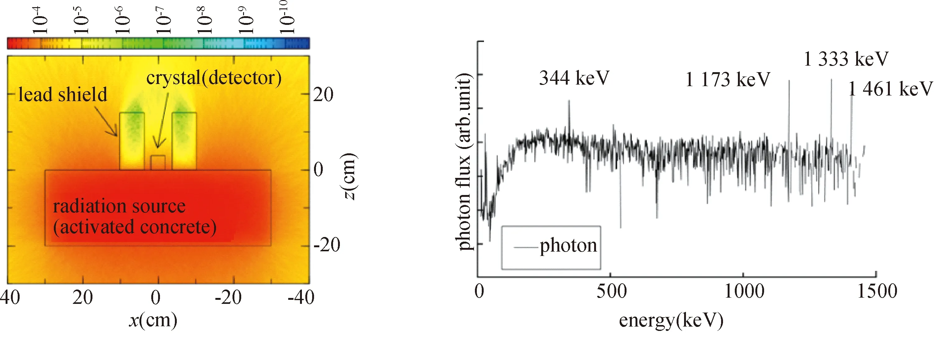
Detection limits were roughly estimated for the representative scintillation crystals of NaI,LaBr3,CeBr3,and SrI2,with the PHITS (ver.2.76)[3]and EGS5[4].The geometry is shown in Figure 1.We reproduced the situation where the detector was in close contact with the concrete.A concrete block containing60Co and152Eu,which are the nuclides for concrete decommissioning that are considered most,and40K as the common natural nuclide was employed as the radiation source.The concentrations of60Co and152Eu were varied,and the detection limit of radioactivity for each crystal was calculated.1.0 Bq/g was applied to concentrations of40K[5].The lead shield was also reproduced to be consistent with an actual experiment[1-2].One of the γ-ray spectra reproduced by calculation is shown in Figure 1.Some peaks derived from60Co,152Eu,and40K can be observed,and scattering photons are reflected as a baseline of the spectrum.Here,we defined the “peak to baseline ratio” (P/B) to determine the detection limit of each crystal,for peaks of 344 keV (152Eu) and 1333 keV (60Co).As shown in Eq.(1),the ratio of peak height value (F(n),nch) including the information of efficiency and resolution,to baseline which is the average value of 10 chs (n-14 ton-5),was calculated.In addition,3σwas derived from the standard deviation of the baseline.UnlessP/B<1,it was considered that the crystal can detect the gamma ray.
(1)

Fig.1 Calculation geometry of PHITS code (left) and an simulated γ-ray spectrum (right)
1.2 Detection limit toward activation level
The estimated detection limits (P/B) toward the activation level for various crystal sizes of NaI,LaBr3,CeBr3,and SrI2are summarized in Table 1.The total specific activities of60Co+152Eu:Dare expressed as ∑(D/C) with the clearance level of 0.1 Bq/g:C,as an index.We found that 1.5 inch LaBr3,CeBr3,SrI2,and 2.0 inch NaI crystals might be able to determine the total specific activities of60Co +152Eu at 0.1 Bq/g,which is the clearance level of60Co and152Eu.These are candidates to substitute for the Ge detector.
2 Examination for practicality of scintilla-tion detector
Evaluation for commercially available detectors installing 1.5 inch LaBr3,2.0 inch CeBr3,and 1.5 inch SrI2,respectively,was conducted at the accelerator room of the PET cyclotron facility to validate the calculation study.As in the simulation study,the detector was contacted on the concrete floor and walls,with a thick lead radiation shield,and gamma-ray spectra were obtained from the contacted concrete.

Tab.1 Estimated detection limit (P/B) toward activation level for various crystal sizes of NaI,LaBr3,CeBr3,and SrI2
2.1 Experimental conditions
2.1.1Facility
Experimental evaluations were performed in the medical and pharmacological research center,Hakui,Ishikawa,Japan.This facility had produced radio-pharmaceuticals for PET,such as18F,with the cyclotron,and also delivered them to nearby hospitals and medical institutions up to 2015.Details of the facility and the cyclotron are summarized elsewhere[1,5].Although the cyclotron was removed,the ambient dose rate of the cyclotron room was considerably higher than the background,because the concrete walls,floor,and ceiling were all strongly activated.When the experiment was conducted,short-lived nuclides had already attenuated,and only60Co and152Eu (γ-ray emitting nuclei) remained.The range of total specific activities of60Co +152Eu in the cyclotron room are 0.2~0.7 Bq/g (2.0~7.0 in ∑(D/C)).
2.1.2Detectors
As a representative of the three crystals that showed good results in the simulation,commercially available detectors,described in the following,were employed in this experiment:LaBr31.5 inch,InSpector1000 (Mirion Technologies,Canberra);CeBr32.0 inch,EMF211 (EMF Japan);SrI21.5 inch,SRI-38-PHI-38-P (RMD);and SrI21.0 inch,NHC8 (Fuji Electric).
2.1.3Radiation measurement
First,the entire room was divided into 0.5-m squares,and the entire cyclotron room was measured with a shielded survey meter and mapped contact dose rate distribution[5].From the dose rate map,four representative points for walls (E32,N23,S36) and floor (F37) were determined from the dose rate value,and they were measured using scintillation spectrometers covered with a 65-mm-thick lead shield[1].
2.2 Results and discussion
2.2.1γ-ray spectrum
A comparison of the γ-ray spectrum obtained from each detector is shown in Figure 2.Principal peaks of60Co and152Eu were confirmed in all spectra.From the peaks of 344 and 1 333 keV,P/B was derived in the same way as in the simulation.Except for 1.0 inch of SrI2,experimental values were consistent with the simulation results — that is,the detection limit estimated in Chapter 1 could reproduce the experimental value well.

Fig.2 γ-ray spectra obtained from each detector
2.2.2Specific activity of60Co and152Eu
The specific activities of60Co and152Eu at representative points are summarized in Figure 3.Detection efficiencies for 1 333 and 344 keV peaks were calculated with ISOCS[6,7]and EGS5.Radiation source information and other parameters were derived from a previous study[1].Activities of LaBr3and SrI2(1.5 in) were almost the same and consistent with the Ge,whereas that of CeBr3was higher.Although discussion is omitted here,this is considered to be caused by the incorrect calculation geometry rather than the detector problem.Despite the long measurement time required,activities could be determined correctly with SrI2(1.0 in),contrary to the simulation result in Table 1.

Fig.3 Specific activity of 60Co and 152Eu at representative points of walls (E32,N23,S36) and floor (F37)
2.2.3Making the calibration curve
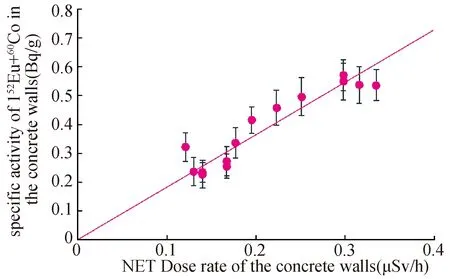
The relationship between contact dose rate and radioactivity is shown in Figure 4.At many measurement points,the results of LaBr3reproduced that of the Ge detector[2].Moreover,as in previous research[1,2],we could show the proportional relationship between the contact dose rate and the specific activity.As a result,it was shown that substitution of Ge by a scintillation spectrometer is possible for nondestructive evaluation of concrete activation.

Fig.4 Calibration curve for converting contact dose rate[1]with 6.5 cm lead shield into specific activity of 152Eu+60Co
3 Optimization for radiation shield structure
3.1 Radiation shield design
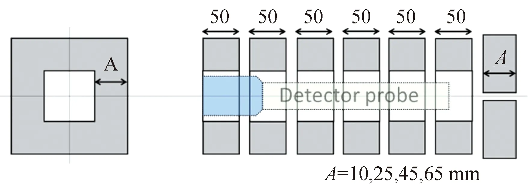
We determined the required thickness and length of the radiation shield for suppressing the background radiation from the entire room (4π direction),and the target surface was measured precisely.The lead shields for covering the detector probe shown in Figure 5 were manufactured.Four different thicknesses were available and separable every 50 mm along the axial direction.At the end of the shield,a back cover could be installed to shut the radiation from the backward direction.The thickness and length were determined by the calculation with ISOCS.

Fig.5 Schematic drawing of the lead radiation shield
3.2 Verification of the radiation shield effect
3.2.1Case of the background to be comparable with the target
We obtained the γ-ray spectra from representative points of E32,N23,S36,and F37 using an LaBr3detector with the radiation shield set shown in Figure 5.Variations of count rates of the 1 333 keV peak with shield thickness and length are summarized in Figure 6.The count rates attenuate when the shield length increases;however,they hardly change over 150 mm.It was found that there was no difference in count rate between 45 and 65 mm thicknesses—that is,the background could be sufficiently suppressed.In conclusion,the shield with a length of 150 mm and equivalent to 45 mm thick lead is optimal.It has been miniaturized compared with the prototype used previously (65 mm thickness,250 mm length).

Fig.6 Relation between relative count rate (measured at F37) and radiation shield length and thickness: filled squares mean the detector is covered with the back shield
3.2.2Case of the background being higher than the target
If the measurement target is significantly lower than the background,another evaluation is required,e.g.,a case of activated beamlines existing.A lead block (300×300×T 100 mm3) was placed at the F37 to shut off the radiation from the target surface and put the measurement set of Figure 6 on the lead block,obtaining the γ-ray spectra.It was revealed that the 45 mm thickness was not sufficient.A shield equivalent to a 65 mm lead thickness was mandatory to suppress the background radiation to 1/100 or less,as shown in Figure 7.
4 Conclusion
Every detector employed in the experiment reproduced well the values of specific activities of60Co and152Eu obtained with a Ge detector.This implies that the scintillation-type gamma-ray spectrometer can suppress the cost for concrete decommissioning considerably as a substitute for a Ge detector.

Fig.7 Relation between relative count rate and radiation shield length and thickness:the detector was put on the lead block placed on F37
The background could be sufficiently suppressed with a 150 mm length and the equivalent of a 45 mm thick lead radiation shield,in the case of the background being comparable to the target.On the contrary,a shield equivalent to 65 mm thick lead and a back shield are required in the case when the background is higher than the target found.This study is expected to make the decommissioning work easier for concrete material in a PET cyclotron room.
Acknowledgments:This research was performed with the aid of the Radiation Safety Research Promotion Fund,“Clearance of materials from the decommissioning of accelerator facilities”,by the Japan Nuclear Regulation Authority.
