Anti-inflammatory, anti-oxidative and anti-apoptotic effects of Heracleum persicum L.extract on rats with gentamicin-induced nephrotoxicity
2021-01-14MohsenAkbaribazmNaderGoodarziMohsenRahimiLeilaNaseriMozafarKhazaei
Mohsen Akbaribazm, Nader Goodarzi, Mohsen Rahimi, Leila Naseri, Mozafar Khazaei
1Students Research Committee, Kermanshah University of Medical Sciences, Kermanshah, Iran
2Department of Basic Sciences and Pathobiology, Faculty of Veterinary Medicine, Razi University, Kermanshah, Iran
3Department of Medical Parasitology and Mycology, School of Medicine, Shahid Beheshti University of Medical Sciences, Tehran, Iran
4Fertility and Infertility Research Center, Health Technology Institute, Kermanshah University of Medical Sciences, Kermanshah, Iran
ABSTRACT
KEYWORDS: Kidney; Heracleum persicum L.; Gentamicin;Stereology; Apoptosis
1.Introduction
Due to the abundance of long-chain polyunsaturated fatty acids in the composition of lipids in renal glomeruli and tubules, kidneys are very susceptible to reactive oxygen species (ROS) and free radicals[1].ROS destroy glomeruli and renal tubules by increasing the production of inflammatory cytokines such as tumor necrosis factor-α (TNF-α) and adiponectin, and promoting infiltration of kidneys by macrophages[2].Studies show that ROS enhance cell membrane permeability by altering the structure and integrity of the lipid bilayer, increasing lysosomal permeability to hydrolytic enzymes, and also impairing ion/electron oxidative phosphorylation in mitochondria.Moreover, ROS reduce glomerular filtration by degrading type Ⅳ collagen and glomerular basement membrane constituents (heparan sulfate, proteoglycans, and laminin) as well as inactivating proteinase inhibitors (such as α1-antitrypsin)[3].
Antibiotics, especially aminoglycosides, cause nephrotoxicity due to increased production of ROS mainly through inducing the INOS/NF-κB/p38MAPK signaling pathway.In particular,gentamicin employs this pathway to disrupt the activity of enzymes and mediators involved in scavenging free radicals in kidneys [i
.e
.catalase, superoxide dismutase, glutathione (GSH) and glutathione peroxidase (GPx)][4].This antibiotic induces cellular deformation and shrinkage, mitochondrial swelling, enlargement of lysosomes,and shedding of the apical brush-border villi of the proximal tubular cells in kidneys.Gentamicin also alters the uptake of ions and disrupts their blood/urine ratio by inhibiting renal brush-border ion pumps (Na/KATPase, Na/Pi co-transporter, and Na/Hexchanger)[5].Using antioxidants is the most important solution to counteract aminoglycoside-induced damages caused by ROS.Plants with polyphenolic compounds are important sources of antioxidants.These mediators have shown synergistic effects with endogenous antioxidant systems to protect cells against cytotoxic effects of ROS.Because of their anti-apoptotic, anti-inflammatory, and antioxidant activities, medicinal plants have demonstrated wide pharmacological applications, particularly nephroprotective capacities.Additionally,they have been safe and effective in reducing nephrotoxic effects of ROS[6].
Heracleum
persicum
L.(H
.persicum
) [commonly known as golpar(Persian hogweed)] belongs to the family Apiaceae and grows up to about 150 to 200 cm in height in mountainous areas.It is a flowering, herbaceous, aromatic, and perennial plant, endemic to Iran (the marginal cities bordering the Alborz and Zagros mountains).In traditional medicine, the herb has been used as a diuretic, flavoring, anti-flatulence, antiseptic, anti-helminthic agent,as well as a remedy to heal gastric ulcers, skin wounds, neuronal and memory disorders[7].Various parts of this plant are rich in flavonoids, alkaloids, terpenoids, triterpenes, and furanocoumarins.Quercetin is the most important flavonoid constituting 59.6 μg/mg of the total phenolic content ofH
.persicum
L.methanolic extract[8].Hydroalcoholic extract ofH
.persicum
was shown to suppress IL-1β-induced expression of cyclooxygenase-2[7].The plant extract also increases the activity of glutathione-S-transferase and therefore protects cells against lipid peroxidation and DNA hydroperoxidation[9].Flavonoids isolated from this plant increase the proliferation of peripheral leukocytes, the activity of immune cells such as T helper cells and macrophages involved in inflammatory processes, and the production of inflammatory cytokines such as IL-2 and γ-interferons[10].Considering the above-mentioned properties shown in this plant, the aim of the present study was to determine the effects ofH
.persicum
extracts against gentamicininduced nephrotoxicity in rats.2.Materials and methods
2.1.Extract preparation
H
.persicum
fresh leaves and stem were collected from the slopes of the Alborz Mountain (Savojblag city) and identified by an experienced botanist at Razi University of Kermanshah (voucher no.9294).After drying the leaves and stem in a dark room, they were powdered by a mill, and 700 g of the powder was then poured into 70% ethanol.After 72 h, the solution was filtered through a filter paper (Whatman, the U.K.).Then a vacuum distillation apparatus(Heidolph Collegiate, LABOROTA 4000, Germany) was used to condense the solution at 50 ℃.The final dried extract (70 g) was kept at 4 ℃ for further use[11].2.2.Liquid chromatography-electrospray ionization-tandem mass spectrometry (LC-ESI/MS) analysis
The Agilent G6410 Triple Quadrapole Mass spectrometer (Agilent,Waldbronn, Germany) along with the Agilent zorbax SB-C18 (15 cm, 3.5 μm) column was coupled to an HCTultra ion trap MS detector.The mobile phase consisted of (A) water + 0.1% acetic acid (v/v) and (B) acetonitrile containing 0.1% acetic acid (v/v).The used linear gradients included 10%, 50%, 95% (repeated twice), and 100% (repeated twice) (v/v) for the A solution and 0%, 25%, 45%,55%, 60% for the B solution.The washing time was 75 min applying 0.3 mL/min at all the times.The source conditions on a standard set of polyphenolic compounds included flavonoid aglycones and glycosylated flavonoids.The injection volume was set as 15 μL, the dwell time as 420 msec, the flow rate as 0.4 mL/min, and finally the temperature of the column as 25 ℃.The ESI-MS spectra were acquired in a negative ion mode using an electrospray ESI ion source(Bruker Daltonik GmbH, Bremen, Germany).The temperature of the drying gas (N) was 300 ℃ at a dry gas flow rate of 35 mL/min, the nebulizing pressure (N) of 30 psi, and a capillary voltage of 4 V.Quality control samples were prepared by diluting separate analytic stock solutions using the same volume of a known internal standard[11].
2.3.Experimental design and treatments
Thirty-six Wistar rats weighing (180 ± 20) g were obtained from Pastor Institute (Pastor Institute, Tehran, Iran), and fed with the standard pellet.They had free access to water and food and were kept under standard conditions [temperature of (25±3) ℃, humidity of(55±5)%, and a 12-hour light-dark cycle].The animals were divided into 6 groups, and each group consisted of 6 rats.Group 1 (healthy control) was given 0.5 mL normal saline.Group 2 (gentamicin group)was injected intravenously with 80 mg/kg gentamicin for 10 d.Groups 3-5 were treated with gentamicin (80 mg/kg) andH
.persicum
extract at doses of 250, 500, and 750 mg/kg BW, respectively.Group 6 receivedH
.persicum
extract treatment alone (750 mg/kg BW).H
.persicum
extract was dissolved in 0.5 mL distilled water and administrated by gavage for 40 d.In groups 2-5, gentamicin was injected for 10 d[12].2.4.Acute toxicity test (LD50)
Lorke’s two-phased method was used to measure the acute toxicity(LD) of the extract.In the first phase, nine additional rats were divided into 3 groups and intraperitoneally (i
.p
.) injected with the extract (10, 100, and 1 000 mg/kg).They were then monitored for 24 h to evaluate any toxicity.In the second phase, three rats (1 rat in each group) received doses of 1 500, 3 000 and 4 500 mg/kg by intraperitoneal injections and were monitored for 24 h for any sign of toxicity and probable death.The LDwas calculated using the following formula:LD= (D× D)
where Dis the highest dose in which no mortality occurred, and Dis the lowest dose that caused mortality[13].
2.5.Biochemical analysis
Twenty four-hour urine was collected using metabolic cages.Urea, creatinine, protein, and albumin levels were measured by biochemical ELISA kits following the manufacturer’s protocol (Pars Azmoon, Tehran, Iran) using an automatic analyzer (Architect c8000 Clinical Chemistry System, USA).At the end of the study (on day 41), the rats were sacrificed under general anesthesia induced by 10% ketamine (10 mg/kg,i
.p
.) and 2% xylazine (100 mg/kg,i
.p
.).Blood samples were taken from the heart, and serum samples were isolated by centrifugation at 3 000 rpm (4 ℃ for 15 min) and stored at –20 ℃.The levels of blood urea nitrogen (BUN) and creatinine in serum were measured using ELISA commercial kits (Pars Azmoon,Tehran, Iran) by a spectrophotometric method.In addition, serum sodium and potassium were measured based on a photometric method (PFP7/C Jenway Flame photometer, Jenway, U.K.).Serum cytokines (TNF-α, IL-1β, IL-6, and IL-10) and GPx activity were measured by ELISA commercial kits (BioLegend, USA).Kidney malondialdehyde (MDA) level was also assessed by a specific ELISA kit (Cayman Chemical, Ann Arbor, Mich., USA) according to the manufacturer’s protocol.Total antioxidant capacity (TAC) in renal tissue was measured by the ferric reducing ability of plasma(FRAP) method described by Ghanbariet
al
.[14,15].2.6.Determination of stereological parameters and histopathological evaluation
The left kidney was removed for histological examinations.The tissues were weighed by a digital scale (AND FA 2014, China)and preserved in 10% formalin for 24 h.The initial volume of kidneys was calculated by the immersion method.By using the isotropic uniform random (IUR) sampling method, 7 kidney slices were obtained using the Orientator method.After that, 5-μm tissue sections were prepared from each slice using a rotary microtome(Leica RM2235, USA).The tissue sections were stained by Periodic Acid–Schiff (PAS), hematoxylin and eosin (H&E), Masson’s trichrome staining, and methenamine Silver-Periodic Acid-Schiff (Jones’ Methenamine Silver) methods.A light microscope(Olympus IX71 microscope, Japan) equipped with a KE-camera(KEcam Technologies, Lekki Lagos, Nigeria) and the Top view software (Version 3.7) was used for histological examinations.Stereological parameters (length and volume of proximal and distal convoluted tubules, loop of Henle, vessels, collecting tubules,volume of interstitial tissue, and volume and number of glomeruli)were calculated applying the method described by Bazmet
al
.[14].Pathological lesions in kidneys (glomeruli and tubules) were investigated at ×400 magnification.These included replacement of the main renal parenchymal tissue with connective tissue (i
.e
.increased collagen fibers), lymphocytic infiltration, and cast formation.Renal hyperemia was defined as follows: scant (0%-25%,score: 1), mild (25%-50%, score: 2), moderate (50%-75%, score: 3)and acute (75%-100%, score: 4) based on lymphocytic infiltration,vascular hyperemia, and the presence of collagen fibers in interstitial space and casts in renal tubules[14].2.7.Total phenolic content (TPC)
The phenolic content ofH
.persicum
extract was measured using the modified Folin-Ciocalteu assay (Slinkard and Singleton method).A calibration curve was prepared using gallic acid as the standard.For this, 50 μL of 0.024, 0.075, 0.105, 0.3 and 0.4 mg/mL concentrations of gallic acid was mixed with 0.8 mL sodium carbonate (NaCO,7.5%) and 1.0 mL Folin-Ciocalteu’s reagent (diluted ten-fold).Then 50 μL of the ethanolic extract was added to the above solution.All the samples were read at 765 nm after 2 hours of incubation at 30 ℃(Shimadzu UV-Vis spectrophotometer).Results were expressed as mg of gallic acid equivalent per gram of fresh plant (mg GAE/g)[16].2.8.Total flavonoid content (TFC)
Total flavonoid content was determined using a modified method[17].Firstly, 10 mg ofH
.persicum
ethanolic extract was dissolved in 5 mL ethanol and then mixed with 1 mL methanol dissolved in a solution containing aluminum trichloride (AlCl), 0.1 mL potassium acetate (1 M), and 2.8 mL distilled water.Afterwards,the absorbance of the resulted solution was read at 415 nm after incubation at 25 ℃ for 30 min.The TFC was expressed as mg of rutin equivalent (RE) per 1 g ofH
.persicum
extract (mg RE/g).2.9.2,2-Diphenyl-1-picrylhydrazyl (DPPH) radical scavenging activity
DPPH was determined using the Brand-Williams method[18].Aliquots (25–170 μL) of theH
.persicum
extract were mixed with 3 mL of 0.06 mM methanol DPPHradical solution and incubated for 3 h.Following this, the absorbance of samples was read at 516 nm in dark.According to the Trolox calibration curve, the DPPH radical scavenging activity of the extract was expressed as μmol Trolox equivalent (TE) per 10 g of driedH
.persicum
extract (μmol TE/10 g plant).2.10.Scanning electron microscopy/energy-dispersive x-ray spectroscopy (SEM-EDS) analysis
TheH
.persicum
ethanolic extract (2 g) was coated with gold and placed on the stage of a microscope (Seron Technology’s highly competitive normal SEM, AIS2300C, South Korea) equipped with an energy dispersive X-ray spectrometer (EDS).The analysis conditions were as follows: accelerating voltage: 12 kV, image rotation (360°), vacuum: ~10Torr, beam shift & rotation: 250 μm(X, Y)[11].2.11.Quantitative real-time PCR
The right kidneys were removed and stored in a liquid nitrogen tank before being used for quantitative real-time PCR.Total RNA was isolated from 30 mg renal tissue (QIAGEN RNA purification mini kit) and qualified by Nanophotometer 2000c (Thermo Science,USA) and reading A/Aand A/Anm absorbance ratios.The cDNA was synthesized using the BioFact kit (BioFact RT Series,South Korea).Total PCR reaction volume was 20 μL including 10 μL of SYBR GreenⅠPCR Master Mix (TaKaRa, Japan), 1 μL of forward and reverse oligonucleotides (400 nM), 1 μL cDNA template, and 7 μL ddHO.The thermal cycle (38-42 rounds)consisted of 45 min at 70 ℃ followed by 30 s at 95 ℃ (denaturation),5 s at 72 ℃ (annealing and extension).Then melting curve analysis was conducted from 70 ℃ to 95 ℃ with temperature rising 1 ℃ each step.Expressions of pro-apoptotic genesp53
,caspase
-3
, andBax
and anti-apoptotic geneBcl
-2
were evaluated using High ROX BioFact2× Real-Time PCR Smart mix SYBR Green PCR master mix.Real-Time PCR light cycler device (Applied Biosystems StepOneReal-Time PCR System, U.S.) was used based on the manufacturer’s protocol.The PCR primers were designedvia
GeneRunner and Primer Express v.3.0 software (Applied Biosystems, Foster City, the USA), and the sequences were blasted in the NCBI database.The primer sequences forcaspase
-3
were 5′-GTGGAACTGACGATGATATGG-3′ (forward) and 5′-GCAAAGTGACTGGATGAACC-3′ (reverse); forBcl
-2
,5′-ATCGCTCTGTGGATGACT-3′ (forward) and 5′-CAGCCAGCAGAAATCAAACA-3′ (reverse); forp53
, 5′-AGAGACCGCCGTACAGAAGA-3′ (forward)and 5′-GCATGGGCATCCTTTAACTC-3′ (reverse);forBax
, 5′-GCTACAGGGTTTCATCCA-3′ (forward)and 5′-ACATCAGCAATCATCCTCT-3′ (reverse); for glyceraldehyde-3-phosphate dehydrogenase (GAPDH
), 5′-AAGTTCAACGGCACAGTCAAGG-3′ (forward) and 5′-CATACTCAGCACCAGCATCACC-3′ (reverse).All qRT-PCR reactions were carried out in duplicate, andGAPDH
was used as an internal reference gene.Gene expressions were measured using the below formula and Ct (2) (fold change)method.∆∆Ct = (Ct–Ct)– (Ct–Ct)
Finally, considering the primer efficiency value of ~2, gene expression level was determined as 2[19].
2.12.Statistical analysis
Data analysis was performed in SPSS-16 software.The data were presented as mean±SD.Normality and homogeneity of the data were checked by Kolmogorov-Smirnov test (P
>0.05).Significant differences were analyzed using one-way ANOVA followed by Tukey’spost
hoc
test (P
<0.05 was considered statistically significant).Data charts were designed with the Graph Pad Prism software package version 8 (Graph Pad Prism Software Inc., San Diego, California).2.13.Ethical statement
The study protocol was approved by the Ethics Committee of Kermanshah University of Medical Sciences (Ethic code: IR.KUMS.REC.1398.1153) and conducted according to the guidelines of the Animal Ethics Committee (NIH Publication 80-23, 1996).
3.Results
3.1.LC-ESI/MS analysis
The retention times and mass spectral data obtained in a negative ion mode were compared with those of standard compounds or previously reported constituents ofH
.persicum
.Tentative identification of phenolic compounds was performed based on key fragment ions and other MS observations.The obtained chromatograms of hydroalcoholic extract ofH
.persicum
are displayed in Supplementary Figure 1.In the chromatograms prepared in a negative ion mode, 18 compounds and 31 peaks(intensity > 5×10) were identified.The extracts were separated from each other based on the expected molecular weights [M−H] of their compounds, acquisition times, and counts (Mass-to-charge) ×10(Table 1).3.2.Acute toxicity test (LD50)
After 24-hour monitoring of the rats for toxicities and probable mortality, only one rat died after receiving 4 500 mg/kg of the extract.No mortality or signs of toxicity (such as ataxia, diarrhea,and numbness) were observed at other examined doses.According to the results of this study, the LDofH
.persicum
extract was 1.9 g/kg.Therefore, doses lower than 1 900 mg/kg were used for further experiments in a safe manner without any toxic signs.3.3.Biochemical results
Gentamicin significantly decreased serum levels of sodium and potassium and increased serum creatinine and urea compared with the healthy control group.Co-administration ofH
.persicum
extract at the doses of 500 and 750 mg/kg significantly reduced serum levels of creatinine and urea (P
<0.05), and increased serum sodium and potassium, compared with the gentamicin group (Table 2).Treatment withH
.persicum
extract at a dose of 250 mg/kg did not show any significant improvement in gentamicin-induced changes.Administration of gentamicin significantly decreased urine creatinine and urea and increased urinary protein and albumin compared with the healthy control group.In gentamicin-induced rats treated with 750 mg/kgH
.persicum
extract, urinary creatinine and urea were significantly increased while protein and albumin significantly decreased (P
<0.05),compared with the gentamicin group (Table 2).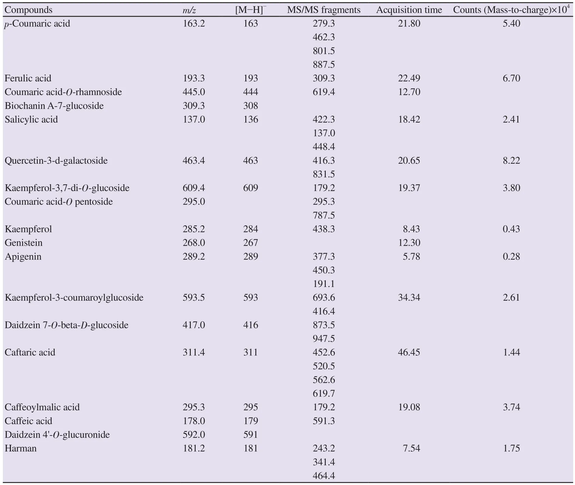
Table 1.Qualitative evaluation of Heracleum persicum flavonoids based on LC-ESI/MS analysis.

Table 2.Biochemical results in experimental groups.
3.4.Serum cytokine levels
Serum level of IL-10 was significantly decreased, while IL-6, IL-1β, and TNF-α were increased in the gentamicin group compared with the healthy control group (P
<0.05).Serum IL-6 was significantly decreased by treatment with 500 mg/kg of plant extract.In addition,H
.persicum
extract at the dose of 750 mg/kg markedly decreased serum IL-6, IL-1β, and TNF-α and increased IL-10 compared with the gentamicin group.For the group treated with 750 mg/kg extract alone, serum levels of IL-10, IL-6, IL-1β, and TNF-α were decreased compared with the healthy control group without significant difference (P
>0.05) (Figure 1A and 1B).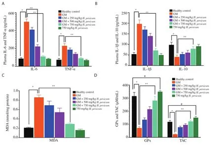
Figure 1.Serum cytokine levels of rats.A: IL-6 and TNF-α; B: IL-1β and IL-10; C: kidney MDA level; D: TAC levels and serum GPx activity.The data are expressed as mean±SD.*P<0.05, the gentamicin group vs.the healthy control group, **P<0.05, the H.persicum treated group vs.the gentamicin group, #P<0.05, the 750 mg/kg H.persicum treated group (without gentamicin) vs.the healthy control group.IL-6: interleukin-6; TNF-α: tumor necrosis factor-α; IL-1β:interleukin-1β; IL-10: interleukin-10; MDA: malondialdehyde; GPx: glutathione peroxidase; TAC: total antioxidant capacity.

Figure 2.Effect of H.persicum on the (A) volume of kidney (KV), proximal (PCTV) and distal (DCTV) convoluted tubules, loop of Henle (LHV), vessels(RVV), interstitial tissue (RIV) and renal collecting tubules (CTV) (mm3); (B) total number (TN), total volume (mm3) (TV) and mean individual volume (10-4 mm3) (MIV) of glomeruli in all groups.The data are expressed as mean±SD.*P<0.05, the gentamicin group vs.the healthy control group and **P<0.05, the H.persicum treated group vs.the gentamicin group.
3.5.Serum level of GPx and renal TAC and MDA levels
In the gentamicin treated group, TAC level in kidneys was significantly lower [(39.42 ± 3.11) μM/mL] and MDA level was significantly higher [(0.86 ± 0.05) nmol/mg protein] than the healthy control [(118.32 ± 7.12) μM/mL and (0.23 ± 0.02) nmol/mg protein,respectively] (P
<0.05).Gentamicin also significantly decreased serum GPx level [(69.33 ± 9.34) μM/mL] in comparison with the healthy control group [(307.36 ± 31.34) μM/mL] (P
<0.05).H
.persicum
extract at the dose of 750 mg/kg significantly decreased the level of renal MDA [(0.29±0.01) nmol/mg protein] and increased serum GPx levels [(283.45 ± 20.26) μM/mL] compared with the gentamicin group.Moreover, the TAC level [(129.64 ± 9.19) μM/mL] was prominently elevated in the kidney tissue of the extracttreated group (Figure 1C and D).For the group treated with 750 mg/kg extract alone, GPx and TAC levels were significantly elevated compared with the healthy control group.3.6.TPC, TFC, and DPPH free radical scavenging assay
The TPC and TFC ofH
.persicum
extract were (49.12 ± 3.31) mg GAE/g and (22.01 ± 2.16) mg RE/g, respectively.The antioxidant capacity of the ethanolic extract ofH
.persicum
leaves and stem in DPPH assay was (136.11 ± 10.21) μmol TE/10 g plant.3.7.SEM-EDS analysis
Elemental analysis using a SEM-EDS spectrum revealed the presence in order of C (621.19)> O (71.14)> K (47.19)> Cl (28.17)>Ge (9.89)> Mg (5.46)> Al (4.47)> Ca (2.43)> Zn (2.32)> N (2.19)>Co (2.08)>Ni (1.40)> Cu (1.26)> Fe (0.91)>Ag (0.87)> Mn(0.63)>Ca (0.52)> Se (0.43).The results indicated carbon (C) as the most abundant constituent (Supplementary Figure 2).
3.8.Stereological parameters
In stereological studies, gentamicin altered renal structures(proximal and distal convoluted tubules, loop of Henle, vessels,collecting tubules, and interstitial tissue) compared with the healthy control group.Gentamicin also significantly increased the volumes of total kidney, renal vessel and renal interstitial tissue, and decreased those of proximal and distal convoluted tubules, loop of Henle, and collecting tubules.H
.persicum
extract at 750 mg/kg improved renal structures (Figure 2A).On the other hand, gentamicin significantly decreased the total glomerular number and increased the total volume and mean individual volume of glomeruli compared with the healthy control group.However,H
.persicum
extract at the dose of 750 mg/kg significantly reduced mean individual volume of glomeruli compared to the gentamicin group (Figure 2B).3.9.Gene expressions of p53, Bax, Bcl-2, and caspase-3
To evaluate apoptosis in the renal tissue, expressions ofcaspase
-3
,p53
,Bax
, andBcl
-2
genes were measured.In comparison with the healthy control rats, gentamicin significantly downregulatedBcl
-2
while upregulatingcaspase
-3
,p53
, andBax
expressions.Treatment with theH
.persicum
extract at the dose of 500 mg/kg significantly downregulated the mRNA levels of pro-apoptotic genesp53
andBax
and upregulated the expression of anti-apoptoticBcl
-2
in gentamicin-exposed rats.Likewise, 750 mg/kg of the extract reversed the changes induced by gentamicin (Figure 3A and B).3.10.Histopathological findings in the kidney
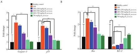
Figure 3.Effect of H.persicum on (A) caspase-3, p53; (B) Bax and Bcl-2 gene expression of the kidneys (n=6) in rats.The data are expressed as mean±SD.*P<0.05, the gentamicin group vs.the healthy control group, **P<0.05, the H.persicum treated group vs.the gentamicin group.
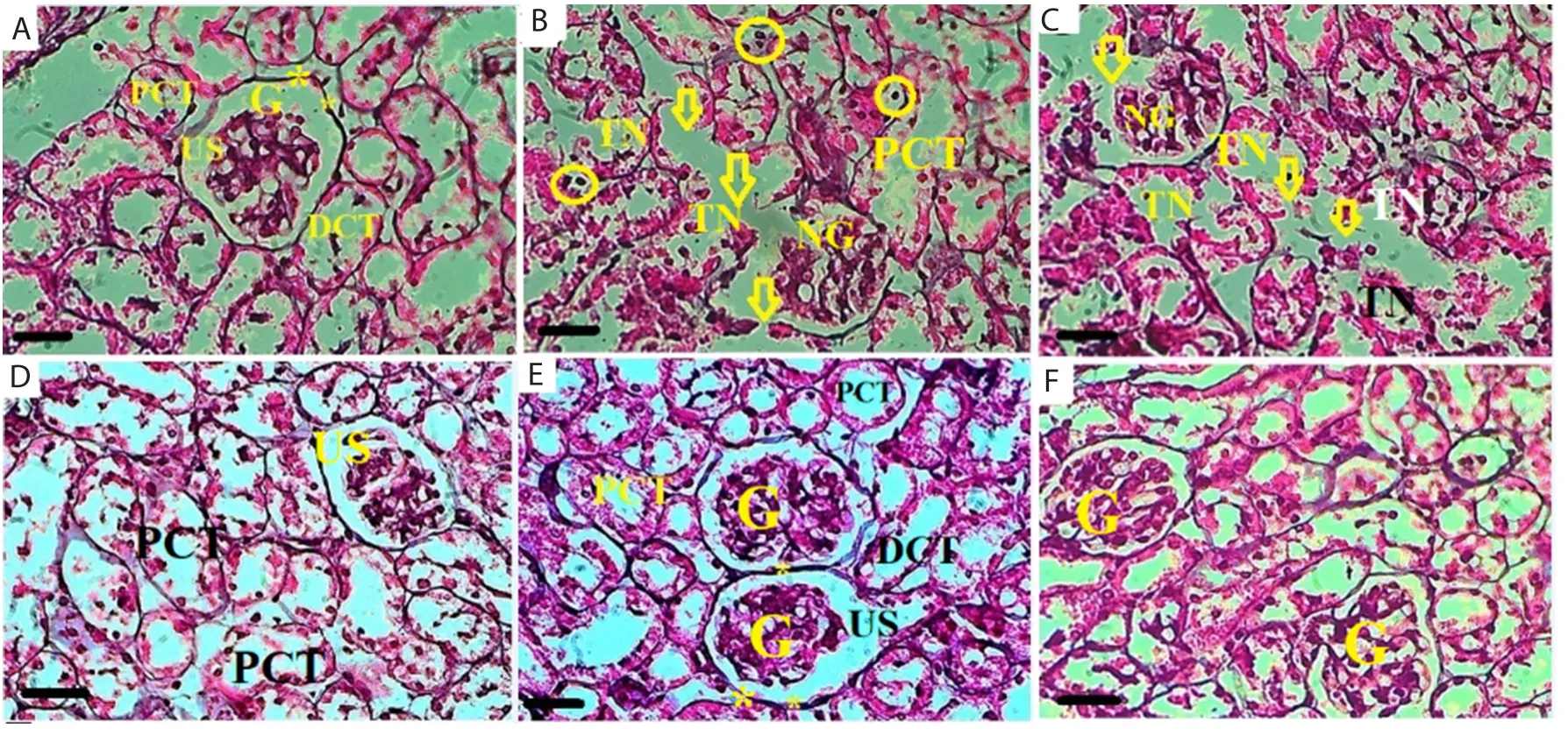
Figure 4.Histopathological changes in kidney tissue of the healthy control (A), gentamicin (B), gentamicin + 250 (C), 500 (D) and 750 (E) mg/kg H.persicum and 750 (F) mg/kg H.persicum extract alone groups (Jones’ Methenamine Silver ×400, scale bar = 30 μm).*: Normal glomerular basement membrane, Circle:Necrotic tubular cells, Arrow: Degenerated glomerular basement membrane, G: Normal glomerulus, TN: Tubular necrosis, PCT: Proximal convoluted tubule,DCT: Distal convoluted tubule and US: Urinary space.
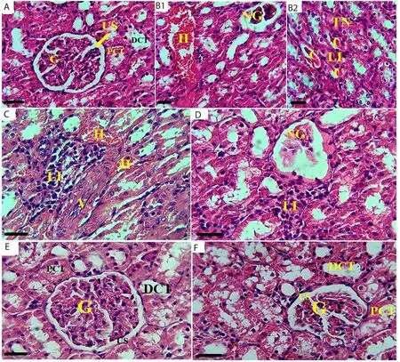
Figure 5.Histopathological changes in kidney tissue of the healthy control (A), gentamicin (B1 and B2), gentamicin + 250 (C), 500 (D) and 750 (E) mg/kg H.persicum and 750 (F) mg/kg H.persicum extract alone groups (PAS ×400, scale bar = 30 μm).H: Hyperemia and congestion, C: Hyaline cast, LI: Lymphocytic infiltration, NG: Glomerular necrosis, G: Normal glomerulus, TN: Tubular necrosis, V: Kidney vessel, PCT: Proximal convoluted tubule, DCT: Distal convoluted tubule and US: Urinary space.
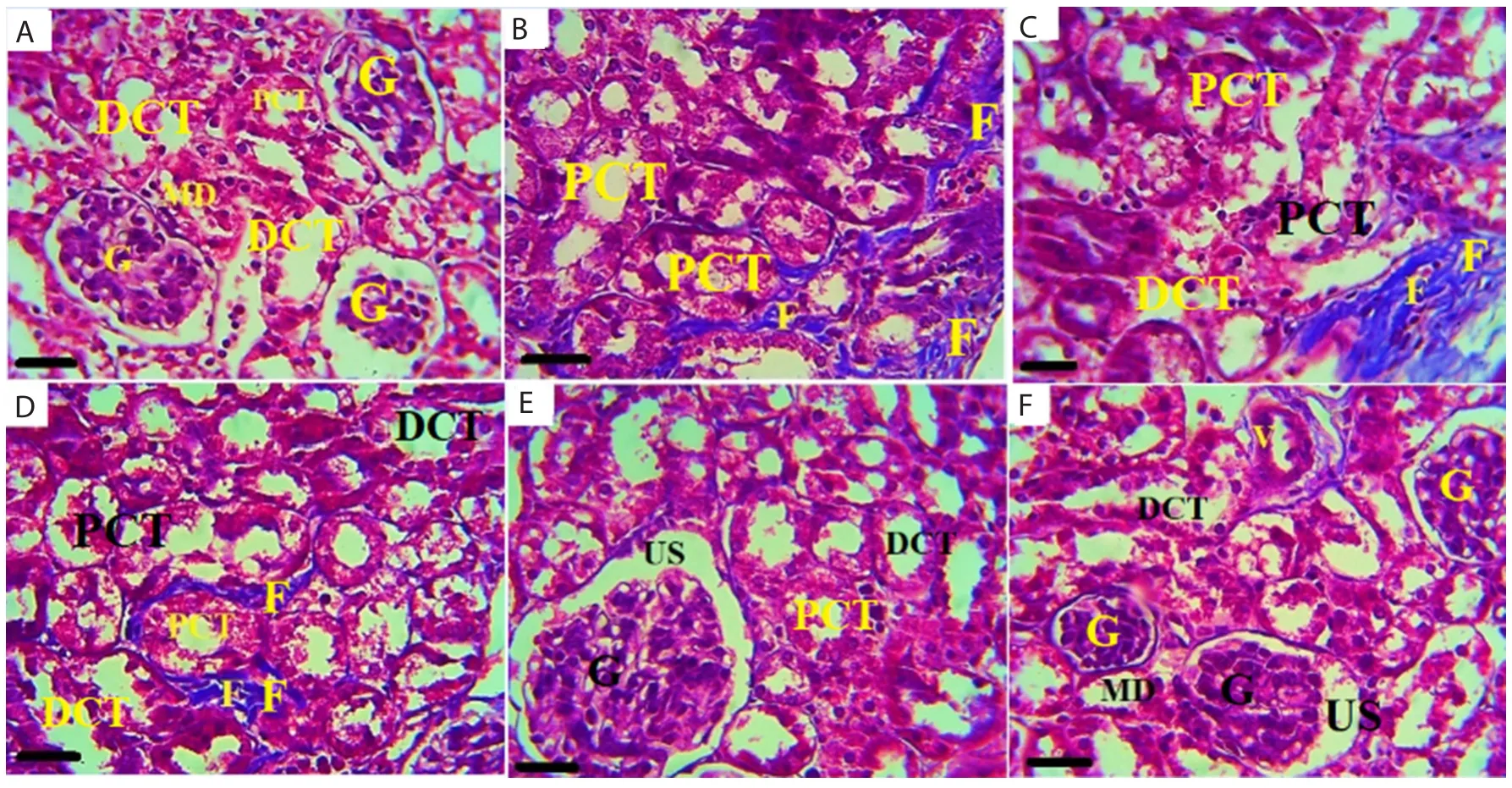
Figure 6.Histopathological changes in kidney tissue of the healthy control (A), gentamicin (B), gentamicin + 250 (C), 500 (D) and 750 (E) mg/kg H.persicum and 750 (F) mg/kg H.persicum extract alone groups (Masson’s trichrome staining ×400, scale bar = 30 μm).F: Fibrous tissue, V: Kidney vessel, G: Normal glomerulus, MD: Macula densa, PCT: Proximal convoluted tubule, DCT: Distal convoluted tubule and US: Urinary space.
In comparison with the healthy control group, gentamicin significantly increased interstitial lymphocytic infiltration, tubular hyaline cast formation, glomerular degeneration, basement membrane necrosis, and renal congestion/edema.Treatment withH
.persicum
extract preserved renal histological architecture, reduced lymphocytic infiltration, maintained the integrity of the tubular and glomerular basement membranes, decreased the formation of intratubular fibrous tissue, and prevented tubular cast formation and vascular hyperemia in gentamicin-exposed rats (Supplementary Table 1; Figures 4, 5 and 6).4.Discussion
In this study, we investigated the protective effects ofH
.persicum
extract against gentamicin-induced renal dysfunction, oxidative stress, and tissue damage in rats.The extract improved serum urea, creatinine, sodium, and potassium and preserved volume of renal tubules, blood vessels, as well as the volume and number of glomeruli in gentamicin-exposed rats.In consistent with previous studies, administration of 80 mg/kg gentamicin has been shown to impair kidney function and induce renal toxicity by inducing damages in renal tubules and glomeruli[20].Gentamicin interferes with the function of G protein-coupled receptors and prevents cAMP formation in kidneys.In this way,gentamicin reduces the expression of aquaporin-2 channelsvia
cAMP/arginine vasopressin signaling pathway resulting in decreased urinary osmolality and retention of plasma ions (i
.e
.sodium and potassium) which consequently lead to an inability to concentrate urine.In addition, gentamicin reduces urine concentration by suppressing the Na/KATPase which subsequently contributes to the edema and necrosis of renal tubular cells and a decline in urinary sodium concentration[21].In this study,H
.persicum
extract (750 mg/kg) improved serum urea, creatinine, and sodium levels, as well as renal tubular volume and increased the numbers of renal blood vessels and glomeruli.TheH
.persicum
extract contains high levels of phenolic antioxidants such as kaempferol augmenting the activity of Na-KATPase pumps and thereby regulating sodium and potassium excretion[22].The beneficial effects ofH
.persicum
extract on kidney function can be attributed to these flavonoids.Generally, inflammatory cytokines are released in response to the activation of immune response after tissue injury or during tissue repair.Inflammatory cells activated by gentamicin subsequently produce various inflammatory cytokines (such as TNF-α, IL-6,and IL-1β) leading to renal injury.Administration of 100 mg/kg gentamicin elevated serum levels of inflammatory cytokines and increased the recruitment of white blood cells to the inflicted areas in response to tissue damage.The recruited leukocytes can then release cytokines augmenting the inflammatory process[23].Other studies have also reported that gentamicin-induced toxicity against tubular and glomerular cells has been associated with elevated levels of TNF-α and IL-1α[24].In this study, administration ofH
.persicum
extract prevented gentamicin-induced elevation of IL-6, TNF-α, and IL-1β, as well as renal infiltration by inflammatory cells.In another study,H
.persicum
extract also reduced IL-6 and IL-8 levels in patients with coronary artery diseases[25].Consistent with our study,Boseet
al
.also showed thatHeracleum
nepalense
extract inhibited the production of pro-inflammatory cytokines (TNF-α and IL-6) by lipopolysaccharide-stimulated human peripheral blood mononuclear cells[26].Overall, our findings suggest protective effects ofH
.persicum
extract against gentamicin-induced inflammatory damage in renal tissue.Immunohistochemical studies have shown that gentamicin increases the levels of pro-apoptotic Bax and caspase-3 and decreases the expression of anti-apoptotic Bcl-2 in kidney tubular cells of rat[27].We here demonstrated that these effects were prevented in rats concomitantly treated with gentamicin andH
.persicum
extract.The ratio of Bax to Bcl-2 plays important roles in regulating mitochondrial release of cytochrome C and activation of caspases 3 which is the principal executioner caspase in apoptosis of renal tubules[28].The interactions among three subgroups of Bcl-2 proteins regulate the permeability of mitochondrial outer membrane and therefore apoptosis.After being released from mitochondria,cytochrome C binds to the apoptotic protease-activating factor 1 forming apoptosome complexes that activate caspases and induce apoptosis.Bcl-2 inhibits apoptosis by binding to a molecule known as BH3; the triggering factor in the mitochondria-initiated apoptotic pathway[29].In this study,H
.persicum
extract significantly increased Bcl-2 level indicating the anti-apoptotic effects of this plant.In addition to indirect actions, gentamicin can also directly affect mitochondrial function by inducing oxidative stress.Various studies have shown that gentamicin increases MDA and decreases TAC levels in kidneys.These findings confirmed the role of gentamicin in promoting oxidative stress.In the present study, we also observed elevated MDA and decreased TAC levels in kidneys of rats treated with gentamicin.In fact, these effects are resulted from elevated ROS production and decreased renal antioxidant capacity.Gentamicininduced ROS can trigger membrane phospholipid peroxidation,DNA fragmentation, and protein denaturation.As evidenced in the present study,H
.persicum
extract can protect the mitochondrial membrane against oxidative mediators by significantly reducing MDA and increasing TAC levels.Phenolic compounds ofH
.persicum
extract effectively act as hydrogen donors rendering them potent antioxidants[17].Majidiet
al
.showed thatH
.persicum
extract (400 mg/kg) increased GPx, TAC, and SOD activity and decreased MDA level in alloxan-induced diabetic rats[30].A study by Taghizabetet al
.showed that an intraperitoneal injection ofH
.persicum
(1 000 mg/kg) for 35 d reduced ROS in semen and improved sperm parameters(especially motility) in mice[12].In another study on the toxic and anticonvulsant effects of hydroalcoholic extract ofH
.persicum
(75, 150, 300, 600 and 900 mg/kg) in mice with pentylenetetrazolinduced seizure, none of the evaluated doses represented toxicities.In addition, the extracts reduced the tonic-clonic phase in a dosedependent manner and significantly decreased seizure duration[31].A study on the serum lipid profile of rabbits showed that doses of 500 and 1 000 mg/kg ofH
.persicum
extract significantly reduced the levels of cholesterol, triglyceride, and high-density lipoprotein in a dose-dependent manner.The results of this study also showed that none of the used doses ofH
.persicum
extract had toxic effects.On the other hand, flavonoids of the extract with antioxidant properties showed potential therapeutic effects on renal diseases[32].Sharififaret
al
.demonstrated that an intraperitoneal injection of aqueous extracts ofH
.persicum
up to 1 103 mg/kg/day to albino mice had no significant impacts on their general behavior and mortality[33].In the present study, acute toxicity test showed the LDvalue of 1 900 mg/kgi
.p
.for the extract ofH
.persicum
in Wistar rats.Other studies have shown that exposure to 60 and 80 mg/kg gentamicin for 10 d resulted in complete glomerular and tubular necrosis/atrophy, renal congestion/edema, formation of tubular hyaline cast, tubular epithelial necrosis, and also lymphocytic cell infiltration of interstitium leading to acute renal failure[34].These results were in line with those of our study showing that gentamicin treatment (80 mg/kg) for 10 d caused similar histopathological changes.However, the basement membrane of tubular epithelial cells, glomerular basement membrane, and plasma membrane of tubular apical cells were apparently protected against gentamicininduced oxidative damage probably due to antioxidant properties of flavonoid compounds such as quercetin, kaempferol-7-glucoside, ferulic acid, biochanin A-7-glucoside, salicylic acid,apigenin, daidzein, and caffeic acid[35,36].We also demonstrated thatH
.persicum
extract was comprised of high levels of flavonoids(22.01 mg RE/g) which preserved renal histological architecture by reducing lymphocytic infiltration, intratubular fibrosis, and tubular cast formation and maintained the integrity of the tubular and glomerular basement membranes against the destructive effects of ROS produced by gentamicin.The antioxidant effects ofH
.persicum
extract can be attributed to its important phenolic compounds includingp
-coumaric acid, quercetin, and gallic acid.These were identified by LC-ESI/MS analysis in this study.Other main compounds of the extract identified by the LC-ESI/MS analysis included ferulic acid, biochanin A-7-glucoside, salicylic acid, kaempferol, genistein, apigenin, daidzein, and caffeic acid.Additionally, SEM-EDS analysis showed the presence of minerals such as Zn, Co, Cu, Fe, Mn, and Se in this plant.In line with our study, Tunçtürket
al
.showed the presence of Mn, Fe, Cu, Zn, Cr,Co, Na, Mg, K, Ca, P, and S as the main minerals of this plant[37].These results suggest that the plant’s extract contains flavonoids and isoflavonoids, as well as important mineral elements that can play a variety of protective roles against oxidative-induced cellular damages.In the study conducted by Ekinciet
al
.,p
-coumaric acid treatment was shown to deplete lipid peroxidation products in the liver and kidney tissues of rats highlighting its anti-oxidative effects[38].Quercetin which is present inH
.persicum
extract can also stimulate the NKCC1 (Na-K-2Clcotransporter 1), a key ion transporter regulating cytosolic Clconcentration and renal Nareabsorption[39].Vijayaprakashet
al
., in their study on mercuric chloride-induced nephrotoxicity in Wistar albino rats, found that kaempferol prevented ROS-triggered glomerular damage and serum-urine electrolyte imbalance by increasing the activities of antioxidant enzymes and reducing apoptosis in tubular and glomerular cells[40].Ferulic acid and its derivatives; caffeine acid phenethyl ester and curcumin,stimulated heme oxygenase-1 which is a protective enzyme against oxidative stress-induced cellular damage[41].Ferulic acid, which can easily accept electrons from free radicals, has shown potent antioxidant properties in various studies.In addition, ferulic acid can effectively scavenge hydrogen peroxide, superoxide, as well as hydroxyl and nitrogen dioxide-free radicals[42].Apigenin (the most important isoflavonoid ofH
.persicum
), with ability to quench the lipid peroxidation chain and shield membranes from ROS-induced damage, was effective to prevent tubular and glomerular injuries[43].Daidzein, another isoflavonoid ofH
.Persicum
, mitigated cisplatininduced nephrotoxicity in micevia
modulating inflammatory process(inhibiting the production of TNF-α, IL-10, IL-18, and monocyte chemoattractant protein-1), oxidative stress (increasing GPx and SOD activities), and caspase-3 related apoptosis[44].In addition, micronutrients ofH
.persicum
extract can modulate immune and inflammatory reactions.Zinc supplementation restored renal morphological and structural changes and urine urea and creatinine levels in Wistar albino rat models with gentamicininduced nephrotoxicity.Moreover, zinc deficiency exaggerates the degradation of kidney proteins leading to renal failure[45].In this study,H
.persicum
extract, which was shown to be rich in flavonoids such as quercetin and essential minerals (as evidenced by LC-ESI/MS and SEM-EDS analyses), maintained the balance of electrolytes through regulating the Na/Kratio in serum and urine.In conclusion, this study demonstrated the protective effects ofH
.persicum
extract against gentamicin-induced nephrotoxicity through its antioxidant, anti-inflammatory, and anti-apoptotic functions.These investigations can provide insight into drug discovery studies in the future for the effective management of drug-induced nephrotoxicity.Conflict of interest statement
The authors declare that there is no conflict of interest.
Funding
This study was financially supported by Kermanshah University of Medical Science (grant number: 981004).
Authors’ contributions
MK and MAB designed the experiments and supervised their execution.MAB performed all experiments and assays.MAB and MK performed data analyses of all experimental findings.MAB,MK, NG, LN and MR contributed to manuscript writing and preparation.All the authors reviewed the data, read and approved of the manuscript.
杂志排行
Asian Pacific Journal of Tropical Biomedicine的其它文章
- Screening of phytocompounds, molecular docking studies, and in vivo antiinflammatory activity of heartwood aqueous extract of Pterocarpus santalinus L.f.
- Anti-senescence and anti-wrinkle activities of 3-bromo-4,5-dihydroxybenzaldehyde from Polysiphonia morrowii Harvey in human dermal fibroblasts
- Borassus flabellifer L.crude male flower extracts alleviate cisplatin-induced oxidative stress in rat kidney cells
- Corchorus olitorius aqueous extract attenuates quorum sensing-regulated virulence factor production and biofilm formation
- 9-Hydroxy-6,7-dimethoxydalbergiquinol suppresses hydrogen peroxide-induced senescence in human dermal fibroblasts through induction of sirtuin-1 expression
