Yiqi Huoxue traditional Chinese decoction affect ventricular remodeling in rats with heart failure after myocardial infarction by regulating osteopontin expression
2020-10-22XiaoNingSunYanZhangAiSongZhuFanDaKongWeiZhangYaoXu
Xiao-Ning Sun, Yan Zhang, Ai-Song Zhu, Fan-Da Kong, Wei Zhang, Yao Xu
1. Liaoning University of Traditional Chinese Medicine, Shenyang 110847, Liaoning, China
2. Affiliated Hospital of Liaoning University of Traditional Chinese Medicine, Shenyang 110032, Liaoning, China 3. Zhejiang University of Traditional Chinese Medicine, Hangzhou 310053, China
Keywords:Heart failure after myocardial infarction Osteopontin Yiqi Huoxue Chinese medicine Ventricular remodeling
ABSTRACT Objective: To investigate the mechanism of Yiqi Huoxue traditional Chinese medicine affecting the osteopontin (OPN) level in myocardial tissue and improving ventricular remodeling in heart failure rats after myocardial infarction. Methods: Forty SPF-grade SD male rats were prepared, and 10 rats were reserved as blank controls. The remaining rats were prepared by anterior descending coronary artery ligation combined with reduced diet and exhausted swimming to prepare a rat model of heart failure after myocardial infarction. The rats in the blank group and the model group were orally administered with distilled water, the Chinese medicine group was administered with Yiqi Huoxue Chinese medicine decoction, and the western medicine group was administered with captopril. After 6 weeks of treatment, small animal ultrasound was used to detect changes in ventricular structure and function in rats. Kill all the rats , and take myocardial tissue, observe the morphological changes of myocardial tissu under a light microscope. Use real-time quantitative PCR to detect the expression of OPN mRNA in rat myocardial tissue, and use immunohistochemical method to detect the expression of OPN protein in myocardial tissue. Results: Compared with the blank group, the left ventricular ejection fraction (EF), left ventricular short axis shortening fraction (FS) of the model group were significantly reduced, and the left ventricular end-diastolic diameter (LVEDD) and left ventricular end-systolic diameter (LVESD) were significantly increased , OPN mRNA and protein expression were significantly up-regulated (P <0.01), and myocardial structure disorder was seen under light microscope. Compared with the model group, the EF and FS of the Chinese medicine group and the Western medicine group were both significantly increased, the LVEDD and LVESD were significantly reduced, the expressions of OPN mRNA and protein were significantly reduced (P <0.01), and the myocardial structure was significantly improved under light microscopy. There was no statistically significant difference between the western medicine group and the traditional Chinese medicine group (P> 0.05). Conclusion: Yiqi Huoxue Chinese medicine may reduce the expression of OPN in myocardial tissue, improve ventricular remodeling, improve cardiac function and prevent heart failure after myocardial infarction.
1. Introduction
Heart failure is the end stage of all heart diseases, and it is one of the two major challenges of the cardiovascular world in the 21st century. It is known as "the last battlefield of heart disease" [1]. According to statistics from 2000, the prevalence of chronic heart failure in China's 35-74 year old population reached 0.9% [2]. Once sick, it seriously threatens the patient's life, reduces the quality of life, increases the financial burden, and brings great distress to the patient, family and society. Myocardial infarction is an important cause of heart failure. Hemodynamic disorders and continuous activation of the neuroendocrine system are the main pathophysiological mechanisms of heart failure, and the further ventricular remodeling it causes is an important basic factor for the occurrence and development of heart failure. [3]. Osteopontin (OPN), also known as cytokine Eta-1, is a secreted non-collagenous glycosylated phosphoprotein that is involved in bone metabolism, tumor growth and metastasis, immune regulation, cell adhesion and other processes. It is a blood vessel Cell's main adhesion and chemokines [4]. Current studies have shown that OPN expression is up-regulated when myocardial injury or increased heart load, and the level of OPN in plasma is closely related to cardiac function classification and prognosis in patients with chronic heart failure [5]. OPN may be closely related to the occurrence of chronic heart failure and the severity of the disease by participating in the process of myocardial fibrosis and ventricular remodeling [6]. Our team has confirmed in previous clinical studies that Yiqi Huoxue Chinese medicine can significantly improve the clinical symptoms of patients with chronic heart failure, improve NYHA cardiac function classification, improve quality of life, reduce mortality and readmission rates [7-8]. In this experiment, a rat model of heart failure after myocardial infarction was established by coronary ligation combined with exhaustive swimming and reduced diet. The effects of Yiqi Huoxue Chinese medicine decoction on the expression of OPN in rats with heart failure after myocardial infarction were investigated to explore its effect on improving ventricular remodeing and treatment of heart failure after myocardial infarction.
2. Materials and methods
2.1 Animal
Forty SPF SD rats, 12 weeks old, weighing 200-230 g, male. Provided by Liaoning Changsheng Biotechnology Co., Ltd., laboratory animal certificate number: SCXK (Liao) 2015-0001.
2.2 Drugs
The Yiqi Huoxue Chinese medicine is composed of astragalus 20 g, panax notoginseng 6 g, ginseng 10 g, motherwort 15 g, salvia 15 g, gardenia 15 g, safflower 10 g. It is prepared by the Department of Pharmacy, Affiliated Hospital of Liaoning University of Traditional Chinese Medicine. Captopril tablets: 12.5 mg / tablet, Beijing Jingfeng Pharmaceutical Co., Ltd., batch number: H11021517.
2.3 Main reagents and instruments
Small animal ventilator (Chengdu Taimeng Technology Co., Ltd.); Paraffin microtome (German Mecon); Color Doppler ultrasound machine (Agilnet); Real-time fluorescence quantitative PCR instrument (Bioneer); Microscope (Japan Austrian) Limbus); PCR kit (Shanghai Yuduo Biotechnology Co., Ltd.); enzyme-labeled goat anti-mouse IgG polymer (Gene Technology Shanghai Co., Ltd.).
2.4 Animal modeling, grouping and administration
After 1 week of adaptive feeding, 10 were selected as blank controls by random number table method, and the remaining 30 were modeled. The method reference [9-10]. Rats were anesthetized intraperitoneally with 10% chloral hydrate. After skin preparation, the trachea was intubated and connected to a small animal ventilator. The chest was ligated 2 mm below the opening of the left anterior descending coronary artery and sutured. The ventilator was stopped after the state was stable. After 1 week, the feed was halved and exhausted swimming once a day. Four weeks later, the left ventricular ejection fraction of rats was detected by ultrasound ≤50%, and symptoms such as atrophy, reduced eating, and weight loss appeared, suggesting successful modeling [11].
Six rats died during modeling, and the remaining 24 were divided into model group, Chinese medicine group and western medicine group by random number table method. In order to balance the number of groups, two were randomly removed from the blank group, leaving eight remaining. The blank group and the model group were orally administered with distilled water; the Chinese medicine group was orally administered with Yiqi Huoxue Chinese medicine; the western medicine group was orally administered with Captopril for 6 weeks.
2.5 Observation indicators and methods
2.5.1 General
Rat weight, mental state, activity, fur gloss, hair loss, eating, and weight changes.
2.5.2 echocardiography
Six weeks after gavage, cardiac ultrasound was performed on rats in each group at a frequency of 13 MHz, a depth of 3.5 cm, and a speed of 200 mm / s. The left ventricular end-diastolic diameter (LVEDD) and left ventricular end-systolic diameter (LVESD) were measured. The left ventricular ejection fraction (EF) and the left ventricular short-axis shortening fraction (FS) were calculated [12].
2.5.3 HE staining to observe the morphological changes of myocardial tissue
Myocardial tissue was fixed with 10% neutral formaldehyde, dehydrated, embedded in paraffin, sectioned, and subjected to hematoxylin and eosin (HE) staining. Morphological changes of myocardial tissue were observed under light microscope [13].
2.5.4 Detection of OPN mRNA expression in rat myocardial tissue by real-time fluorescent quantitative PCR
Rat myocardial tissue was taken, and RNA was extracted by Trizol method. The RNA concentration was calculated by a nucleic acid protein analyzer. After reverse transcription into cDNA, a PCR kit was used to complete the quantitative PCR reaction under primer conditions to obtain the Ct value. The relative quantification of each gene expression was calculated using the (2–△△Ct) Livak method [14].
2.5.5 Detection of OPN protein expression in rat myocardial tissue by immunohistochemistry
Tissues were fixed, paraffin-embedded sections, dehydrated, antigen repaired, 5% BSA and enzyme-labeled goat anti-mouse IgG polymer were added and incubated at room temperature. DAB staining, slight counterstaining of hematoxylin, dehydration, clear, coverslip, observation under a microscope, analysis of the positive area of the visual field with Image J software, calculation of protein expression [15].
2.6 Statistical methods
SPSS 17.0 statistical software was used for analysis. The measurement data were expressed as mean ± standard deviation (±s).
One-way analysis of variance was used for comparison. P <0.05 was considered statistically significant.
3. Results
3.1 General results of rats in each group
After the successful modeling, the model rats showed apathy, dull coat, hair loss, reduced diet and weight loss. After the intervention, the rats in the traditional Chinese medicine group and the western medicine group became better, their diet increased, their hair color recovered, their activity increased, and their weight increased.
3.2 Heart ultrasound test results of rats in each group
Compared with the blank group, EF and FS in the model group were significantly reduced, and LVEDD and LVESD were significantly increased (P <0.01). Compared with the model group, EF and FS were significantly increased, and LVEDD and LVESD were significantly decreased in the Chinese and Western medicine groups (P <0.01). There was no significant difference in the EF, FS, LVEDD, and LVESD between the western medicine group and the Chinese medicine group (P> 0.05). See Table 1.

Tab1 Comparison of cardiac ultrasound results of rats in each group (n=8,x±s)
3.3 HE staining results of myocardial tissue in each group of rats
The light microscope showed that the myocardial structure of the blank group was normal, the myocardial fibers were arranged neatly, the cell morphology and nucleus size were basically the same, and there was no change in muscle fiber structure and cell damage. Compared with the blank group, the myocardial fibers in the model group showed polymorphic changes, the fibrous structure collapsed, multiple muscle fibers were broken, atrophy and degeneration, the lateral diameter of the myocardial cells increased, and the distribution was uneven, showing a large number of free cell nuclei produced by cell necrosis. Compared with the model group, the myocardial fiber structure disorder of the Chinese medicine group was corrected, the myocardial tissues were arranged neatly, and the cell morphology returned to normal. In the western medicine group, the degree of myocardial fiber structure disorder was reduced, and the cell morphology was generally normal. A few hypertrophic and atrophic myocardial cells were still visible. see picture 1.

Fig1: Morphological observation of myocardial tissue in each group of rats (HE× 400)
3.4 Comparison of OPN mRNA and protein expression in myocardial tissue of rats in each group
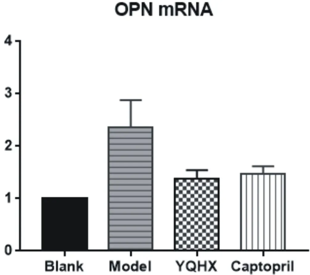
Compared with the blank group, the expression of OPN mRNA and protein in the model group was significantly up-regulated (P <0.01). Compared with the model group, the expressions of OPN mRNA and protein in the traditional Chinese medicine group and western medicine group were significantly reduced (P <0.01). There was no significant difference in OPN mRNA and protein expression between the western medicine group and the traditional Chinese medicine group (P > 0.05). See Table 2, Figure 2, and Figure 3.

Tab2 Expression of OPN mRNA and protein in myocardial tissue of rats in each group (n=8,x±s)

Fig2: OPN mRNA expression in rat cardiac tissue

Fig3: Expression of OPN protein in rat cardiac tissue
4. Discussion
According to the survey, the number of patients with coronary heart disease in China currently reaches 11 million, and the number of AMI cases in 2016 reached 4 million. It is estimated that the number of AMI cases in 2030 will reach approximately 6.1 million [16]. Myocardial infarction is the main cause of heart failure. With the advancement of interventional technology and medical conditions in recent years, most patients with myocardial infarction can be better treated and achieve higher survival rates. However, there are still many patients with varying degrees of cardiac dysfunction, such as fatigue, decreased physical strength, cough, and depression of lower limbs, within a few years after myocardial infarction. Ventricular remodeling and neurohumoral activation after acute myocardial infarction are important links in the development of heart failure
[17]. When the coronary artery is occluded, the ischemic necrosis and lysis of the cardiomyocytes release the intracellular components into the blood, which activates the body's inflammatory waterfall response. Inflammatory cells such as neutrophils and macrophages infiltrate tissues and release inflammatory mediators to clear necrotic tissue and repair damaged myocardial tissue. Cardiac fibroblasts proliferate in large quantities, secrete extracellular matrix proteins (ECM), form scars, and repair damaged tissues. Elevated ECM levels can induce myocardial fibrosis, cause cardiac morphology and hemodynamic disorders, cause abnormal myocardial contraction and diastolic function, reduce ventricular compliance, and worsen cardiac function [18-19].
OPN is a secreted non-collagenous glycosylated phosphoprotein, also known as the cytokine Eta-1. OPN can be expressed by a variety of cells such as osteoclasts, vascular endothelial cells, and vascular smooth muscle cells [20]. As a functional ECM protein, OPN is widely distributed in various tissues, cells, and serum. It mediates cell-cell and cell-matrix interactions and participates in bone metabolism, tumor growth and metastasis, tissue repair, and immunity. Regulation, inflammatory response, angiogenesis and other processes [21]. Under physiological conditions, the expression of OPN in adult myocardial tissue is low, and it increases when myocardial injury or cardiac load increases, and its plasma level is closely related to cardiac function classification and prognosis of patients with chronic heart failure [5]. In addition, increased expression of OPN is also associated with the occurrence of some diseases (such as tumors, inflammation, and immune diseases) [22]. Changes in ECM after myocardial infarction, especially collagen deposition, are important causes of ventricular remodeling. Matrix metalloproteinases (MMPs) are the main proteolytic enzymes of ECM. OPN can inhibit the activation of MMP-2 and MMP-9 caused by IL-1β, increase collagen deposition in the later stage of myocardial infarction, cause collagen distribution imbalance, and promote Myocardial fibrosis causes pathological ventricular remodeling [23]. In addition, OPN can bind to integrin α1 and β3 through integrin-dependent pathways, mediate myocardial fibrosis caused by AngII, and participate in ventricular remodeling [24]. At the same time, TGF-β, fibroblast growth factor, epidermal growth factor, connective tissue growth factor, etc. can all stimulate the large amount of OPN secretion. Increased expression of OPN is an important factor causing myocardial fibrosis, ventricular remodeling, and heart failure [25].
Yiqi Huoxue Traditional Chinese Medicine is an experience prescription of Professor Zhang Yan. It has been used clinically for many years to treat patients with chronic heart failure and has a significant effect. It can significantly improve the clinical symptoms of patients, improve cardiac function classification, improve quality of life, reduce mortality and readmission rates. Professor Zhang Yan believes that chronic heart failure is mostly evidence of qi deficiency, blood stasis, and water cessation. In this recipe, ginseng, astragalus medicinal herbs are beneficial to warming the yang, replenishing deficiency and nourishing the heart, and safflower, panax notoginseng, and salvia miltiorrhizae to promote blood circulation and blood circulation, and motherwort to promote blood circulation and water circulation; . The whole side promotes blood circulation without consuming qi and hurts yin. It corrects the deficiency and does not cause heat and fire, flattening cold and heat, treating blood and water together, and taking both specimens into consideration to achieve the effect of strengthening the heart and channeling pulses, asthma and swelling. .
EF and FS values are important indicators of cardiac function, while LVEDD and LVESD are important indicators of ventricular structural changes. Compared with the blank group, EF and FS in the model group were significantly reduced, and LVEDD and LVESD were significantly increased. This shows that after coronary ligation combined with reduced diet and exhaustive swimming for a period of time, the cardiac structure of the rat changes, and left ventricular function declines, suggesting that ventricular remodeling has occurred. Compared with the model group, the EF and FS of the Chinese medicine group and the western medicine group were significantly increased, and the LVEDD and LVESD were significantly decreased. This shows that after the intervention of Yiqi Huoxue Chinese medicine and captopril, the cardiac function of rats was significantly improved, and the cardiac structure was significantly restored, suggesting that ventricular remodeling was inhibited. The experimental results showed that the expressions of OPN protein and mRNA in the myocardial tissue of rats were significantly reduced after the intervention of Yiqi Huoxue Traditional Chinese Medicine and Captopril (P <0.01). Histomorphology confirmed that the traditional Chinese medicine for nourishing qi and activating blood and captopril can improve the myocardial fiber structure disorder, stabilize the myocardial cell morphology, and protect the myocardial tissue structure. It is suggested that Yiqi Huoxue Chinese medicine may inhibit the ventricular remodeling by down-regulating the expression of OPN, thereby preventing and treating heart failure after myocardial infarction, improving heart function and improving prognosis. Moreover, the efficacy of the traditional Chinese medicine group is comparable to that of the western medicine group, suggesting that traditional Chinese medicine has advantages in the treatment of heart failure after myocardial infarction and needs further study.
杂志排行
Journal of Hainan Medical College的其它文章
- Study of the AQP4 expression in traumatic brain edema and multimodal MRI imaging
- Analysis of two cases of patent ductus arteriosus ligation in preterm identical twins and literature review
- Role of 18F-FDG SPECT / CT imaging in the diagnosis and initial staging of lymphoma
- The value of real-time three-dimensional echocardiography in evaluating left ventricular function for different degrees of heart failure
- Association between polymorphism of MC4R rs489693 gene and disorder of glucose and lipid metabolism in schizophrenia patients treated with olanzapine
- Target prediction and mechanism of Shiyangning in treatment of perianal eczema
