Spontaneous multivessel coronary artery spasm diagnosed with intravascular ultrasound imaging:A case report
2020-09-16HaoYuWuYiWeiCaoFengJunChangLeiLiang
Hao-Yu Wu,Yi-Wei Cao,Feng-Jun Chang,Lei Liang
Hao-Yu Wu,Yi-Wei Cao,Feng-Jun Chang,Lei Liang,Department of Cardiology,Shaanxi Provincial People's Hospital,Xi'an 710068,Shaanxi Province,China
Abstract
Key words:Coronary artery spasm;Multivessel coronary;Spontaneous;Intravascular ultrasound;Case report
INTRODUCTION
Coronary artery spasm is an important cause of myocardial ischemia.It may be associated with the pathogenesis of several cardiac diseases including sudden cardiac death,angina pectoris,acute myocardial infarction,fatal arrhythmias,and other diseases[1].Coronary artery spasm is thought to result from endothelin dysfunction,which impairs vasodilatation.However,under rare conditions,it may relate to multiple coronary vessels,and medical treatments may be ineffective.Variant angina is a discrete form of angina pectoris,which typically occurs during normal activity or at rest,without evident classical triggers such as exercise.In this syndrome,episodes usually occur at night or in the early hours of the morning[2].The diagnosis of variant angina can be challenging and it can easily be mistaken for diffuse coronary artery disease.
Previous intravascular ultrasound (IVUS) and optical coherence tomography (OCT)studies have reported the morphological features of vasospastic lesions[3,4].However,most of the data were obtained in a spastic lesion that was artificially induced by the administration of acetylcholine or ergonovine.In this report,we present the case of a 66-year-old male who had chest symptoms at rest for more than 30 years.Before presenting at our institution,the patient had undergone coronary angiography 4 times and percutaneous transluminal coronary angioplasty procedures twice at other hospitals without a diagnosis of coronary artery spasm.However,his chest symptoms worsened.Spontaneous multivessel coronary artery spasm occurred during IVUS without provocation testing,and the IVUS image was recorded.
CASE PRESENTATION
Chief complaints
A 66-year-old male had chest squeezing at rest for more than 30 years.
History of present illness
Thirty years ago,the patient had chest squeezing at rest which occurred most often from midnight to early morning lasting from 5 to 15 min,and was not usually induced by exercise in the daytime.Over the preceding 13 years,he had undergone coronary angiography 4 times and percutaneous transluminal coronary angioplasty procedures twice at other hospitals without a diagnosis of coronary artery spasm.In July 2006,70% stenosis was found in the ostial left anterior descending artery and 90% stenosis was found in the mid left circumflex.The left circumflex was dilated by balloon without a stent as the left circumflex was small according to the previous angiography report.The patient was given various medications,such as beta blocker,aspirin,angiotensin-converting-enzyme inhibitor,clopidogrel,and statin,but his chestsymptoms were not relieved.Due to repeated similar complaints,angiography was performed again without percutaneous transluminal coronary angioplasty in November2014and August2015.In August2017,an electrocardiography was performed during a chest squeezing episode in the early morning at 6:08 in a hospital and ST-segment elevations in II,III,aVF and V2-V5 leads were observed.Subsequently,ST-segment depression in II,III and aVF,and ST-segment elevation in V2-V5 leads became apparent.The symptoms and ST-segment elevation or depression disappeared 3 min and 25 s after sublingual administration of nitroglycerin (Figure 1).However,the doctors did not realize that the symptoms were caused by coronary artery spasm.Coronary angiography was performed and showed severe stenosis in the proximal left circumflex which was dilated by balloon again without a stent.However,his chest symptoms occurred approximately 5 times/mo.The patient presented to the emergency room of our hospital with frequent chest squeezing episodes in March 2019.
History of past illness
The patient had a history of hypertension for 10 years.
Personal history
The patient had a history of cigarette smoking for more than 40 years.
Family history
No family history of inherited diseases,autoimmune diseases or coronary heart disease was recorded.
Physical examination
Vital signs were stable and physical examination was unremarkable.
Laboratory examinations
No elevation in high sensitivity troponin T was detected and the rest of his laboratory examinations were also completely normal.
Imaging examinations
Electrocardiography was performed during a chest pain episode and ST-segment elevations in I,aVL and V2-V3 leads were recorded which spontaneously resolved,accompanied by pain relief (Figure 2).Neither chest X-ray nor echocardiography showed any specific changes.
In order to evaluate the condition of the coronary artery,coronary angiography and IVUS were performed.An injection of acetylcholine into the coronary artery was administered to induce spasm.Coronary angiography was performedviaright radial access.Intravenous injection of diltiazem and intracoronary injection of nitroglycerin were administered to prevent coronary artery spasm induced by the catheter or guidewire.
An initial coronary angiography showed mild atherosclerotic changes from the proximal to the mid segment in the right coronary artery.There were no obvious atherosclerotic changes in the left main trunk,left anterior descending coronary artery and left circumflex (Figure 3).
After intracoronary injection of 200 μg of nitroglycerin,each segment was examined using a mechanical IVUS system.During IVUS checking of the right coronary artery and left anterior descending coronary artery,the patient felt chest squeezing without the use of acetylcholine or ergonovine.Coronary angiography showed that a local coronary spasm occurred at the distal segment of the right coronary artery and a diffuse coronary spasm occurred at the proximal-distal segment of the left anterior descending coronary artery (Figure 4).Following an intracoronary injection of nitroglycerin (right coronary artery with 400 μg nitroglycerin and left coronary artery with 1000 μg nitroglycerin),the patient’s chest symptoms and coronary spasm were relieved (Figure 5).
竖向位移的研究对钢-混凝土双面连续组合梁的起拱度、挠度有重要意义。图4与图5分别描述了组合梁边跨跨中与中跨跨中的混凝土顶板与钢底板挠度的时间历程。由图4、图5可知,挠度均随温度上升而逐渐增大,至最大值后再逐渐减小,但存在滞后现象。混凝土顶板上下表面挠度的时间历程曲线相差不大,而钢底板的挠度均大于混凝土顶板。在10:00时,边跨跨中的混凝土顶板上下表面达到最大挠度3.8 mm;而钢底板在12:00达到最大挠度5.16 mm。中跨跨中处混凝土顶板上下表面及钢底板的挠度均在15:00达到极值,挠度分别为6.8 mm与8.3 mm。
IVUS images showed that the right coronary artery and left anterior descending coronary artery demonstrated increased media thickness (Figure 6).
FINAL DIAGNOSIS
The diagnosis of multifocal spontaneous coronary artery spasm was confirmed.

Figure 1 Electrocardiographic findings in August 2017.A:Admission electrocardiography without symptoms;B:ST-segment elevation in II,III,aVF and V2-V5 leads during a chest squeezing episode in the early morning at 6:08;C:ST-segment depression in II,III and aVF,and ST-segment elevation in V2-V5 leads 83 s after the chest squeezing episode;D:ST segment depression in II,III and aVF,and ST-segment elevation in V2-V5 leads 3 min after the chest squeezing episode;and E:ST-segment elevation or depression disappeared 3 min and 25 s after sublingual administration of nitroglycerin.
TREATMENT
Beta blocker was discontinued.The patient was placed on oral diltiazem,isosorbide mononitrate,and nicorandil to suppress coronary artery spasms.All medications were given at the maximum dosages tolerated by the patient.
OUTCOME AND FOLLOW-UP
The patient was discharged after 5 d without complications.During the six-month follow-up period,the patient was symptom-free.
DISCUSSION
We present the case of a patient with angina pectoris at rest for more than 30 years.According to electrocardiography at onset of chest symptoms,ST elevations were observed in the precordial (V2-V5),inferior (II,III,aVF) and lateral (I,aVL) leads.Before being diagnosed with variant angina at our hospital,the patient had undergone coronary angiography 4 times and percutaneous transluminal coronary angioplasty procedures twice at other hospitals without IVUS,OCT or fractional flow reserve.Spontaneous multivessel coronary artery spasm occurred during IVUS withoutprovocation testing,and the IVUS image was recorded at our hospital.The patient was placed on oral diltiazem,isosorbide mononitrate,and nicorandil to suppress coronary artery spasms.The patient was free of symptoms during the six-month follow-up period.Thus,the diagnosis of multifocal spontaneous coronary artery spasm was confirmed.
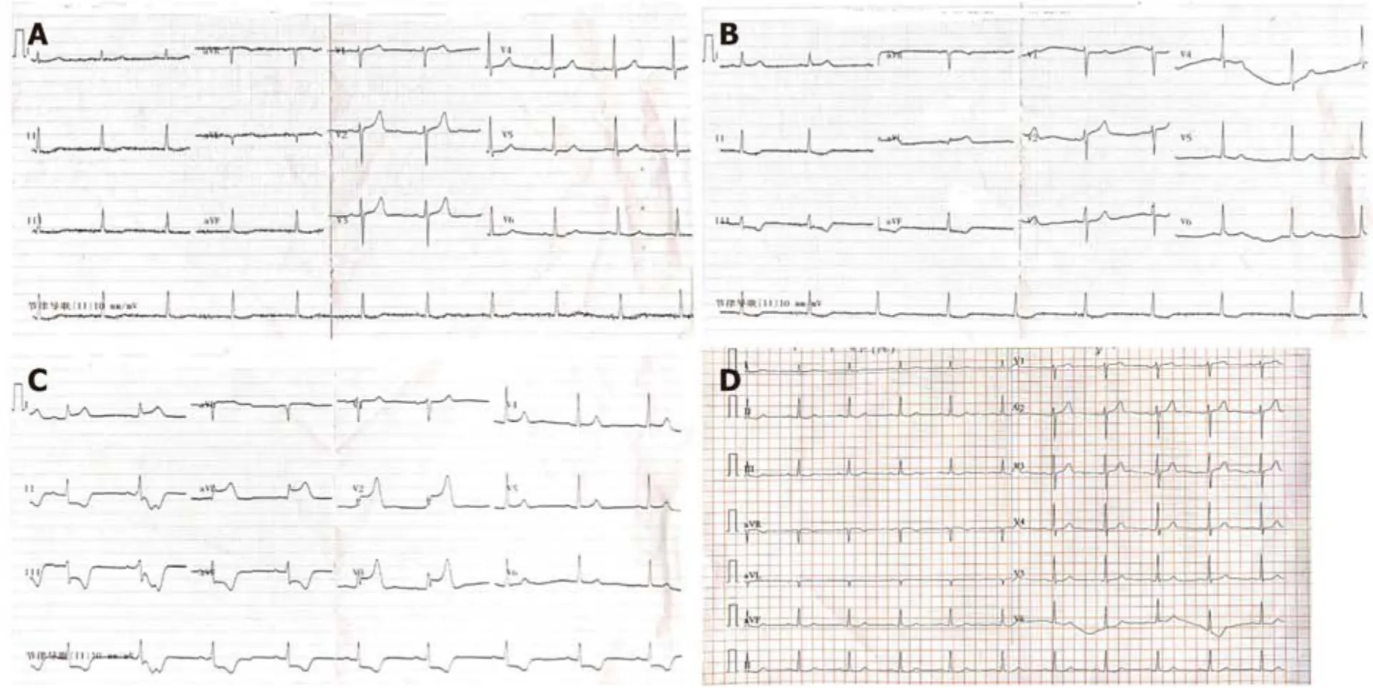
Figure 2 Electrocardiographic findings in March 2019.A:Admission electrocardiography without symptoms;B:ST-segment elevation in I and aVL leads during a chest squeezing episode;C:ST segment elevation in I,aVL and V2-V3 leads during a chest squeezing episode;and D:ST segment elevation or depression disappeared accompanied by pain relief.
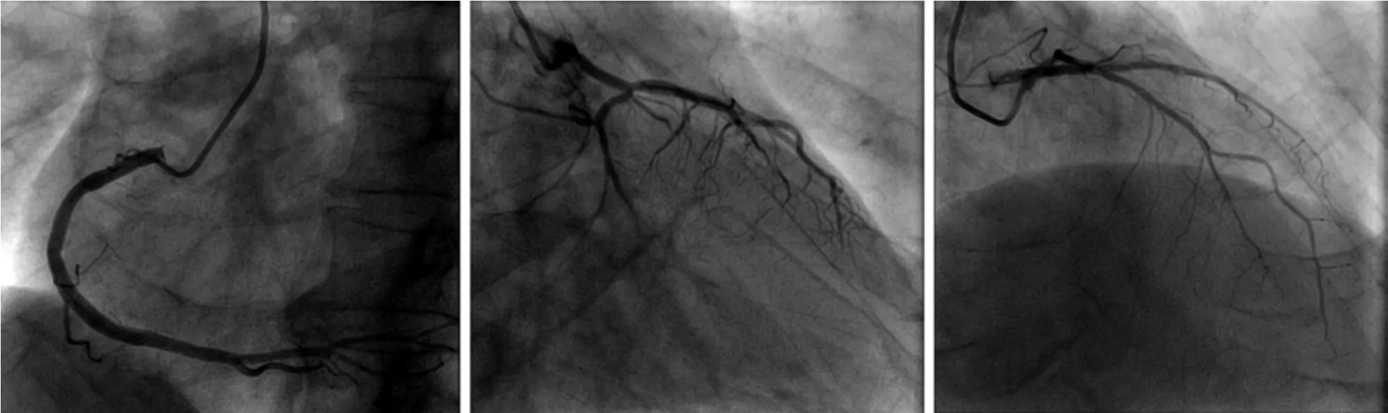
Figure 3 Initial coronary angiography before intravascular ultrasound.Mild atherosclerotic changes from the proximal to the mid segment in the right coronary artery.No obvious atherosclerotic changes in the left main trunk,left anterior descending and left circumflex coronary artery.
IVUS and OCT studies have reported the morphological features of vasospastic lesions.However,most of the data were obtained in a spastic lesion that was artificially induced by the administration of acetylcholine or ergonovine.In the present study,spontaneous multivessel coronary artery spasm occurred during IVUS without provocation testing,and the IVUS image was recorded.Tanaka A used provocation testing to assess coronary artery spasm with OCT.The results showed that the spasm lesion demonstrated an increased maximum media thickness[3].Our patient also showed increased media thickness in the spastic coronary artery.
Coronary artery spasm is mainly a disease of middle-aged and elderly men.Smoking is a significant risk factor for coronary artery spasm.Our patient with multifocal spontaneous coronary artery spasm had a history of cigarette smoking for more than 40 years.A significantly higher proportion of patients with coronary artery spasm angina are smokers than those with stable effort angina,suggesting that the pathogenesis of coronary artery spasm may be different to the pathogenesis of coronary atherosclerosis.As cigarette smoking is an important risk factor for coronaryartery spasm,a high smoking rate may be one of the factors associated with the high incidence of coronary artery spasm in Asian countries (e.g.,Japan)[5].
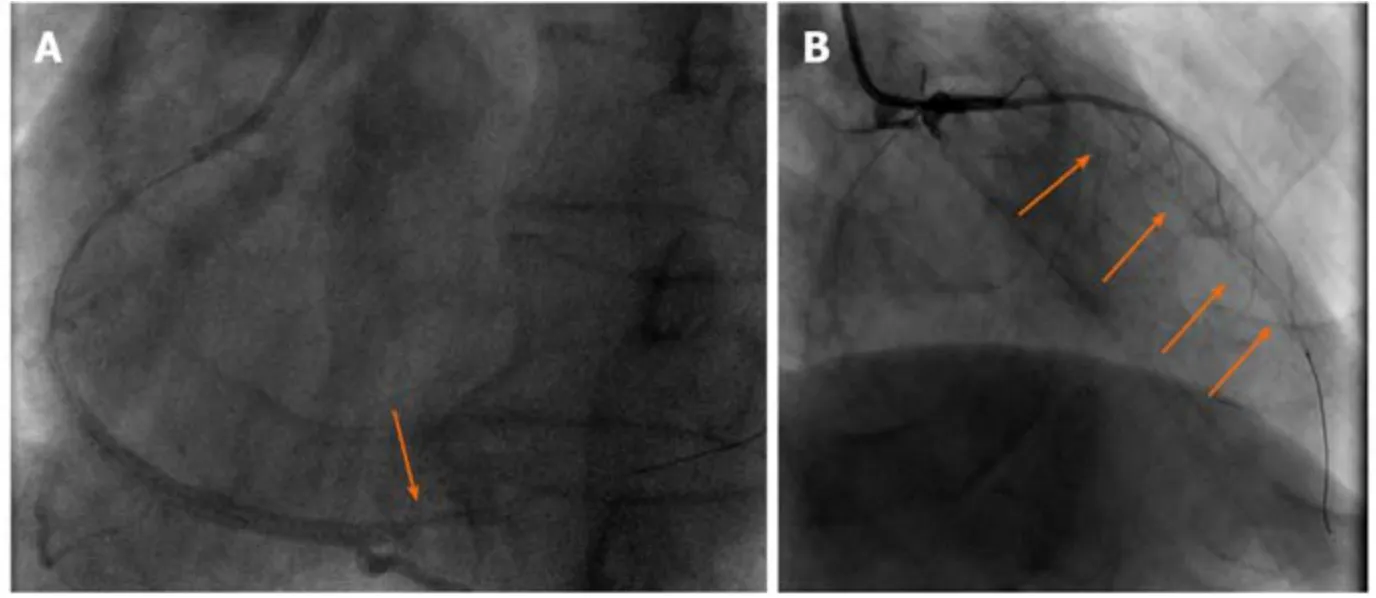
Figure 4 Coronary angiography during intravascular ultrasound.A:In the right coronary artery,a local coronary spasm occurred at the distal segment;and B:In the left coronary artery,a diffuse coronary spasm occurred at the proximal-distal segment of the left anterior descending coronary artery.The coronary spasm segments are indicated by arrows.

Figure 5 Coronary angiography after the intracoronary injection of nitroglycerin.The left anterior descending coronary artery spasm was relieved.
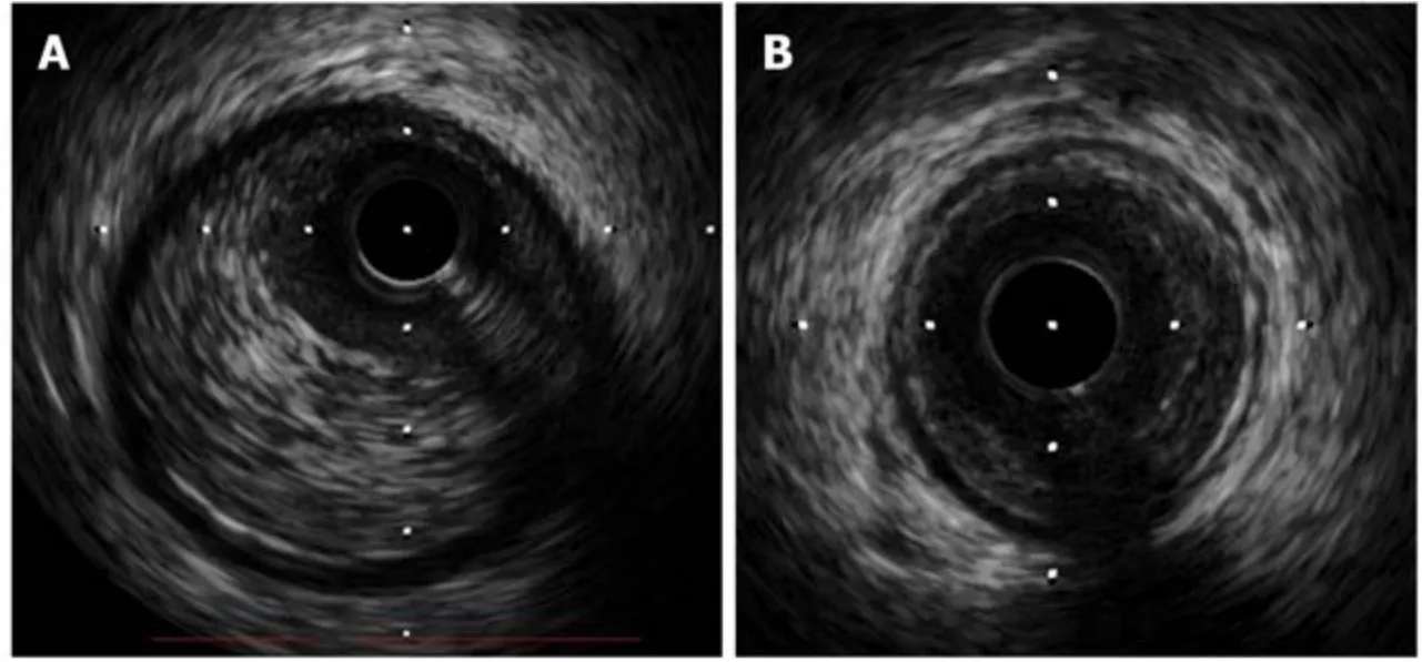
Figure 6 Intravascular ultrasound.Intravascular ultrasound images of the right coronary artery (A) and left anterior descending coronary artery (B) with increased media thickness.
CONCLUSION
Although obstructive coronary artery disease is frequently considered to be the cause of symptoms,vasospasm should be considered in some clinical settings[6].Many conventional drugs are ineffective in patients with coronary artery spasm and this may result in fatal arrhythmia or sudden death.It is essential for clinicians to be aware ofcoronary artery spasm,which may be hard to detect but can be fatal.Further studies are required to determine the pathogenesis of coronary artery spasm and develop more effective and long-lasting treatments for this disease.
猜你喜欢
杂志排行

World Journal of Clinical Cases的其它文章
- Management of cancer patients during COVID-19 pandemic at developing countries
- Liver in the limelight in the corona (COVID-19) time
- First branchial cleft cyst accompanied by external auditory canal atresia and middle ear malformation:A case report
- Delayed perforation after endoscopic resection of a colonic laterally spreading tumor:A case report and literature review
- Giant benign phyllodes breast tumour with pulmonary nodule mimicking malignancy:A case report
- Multifocal neuroendocrine cell hyperplasia accompanied by tumorlet formation and pulmonary sclerosing pneumocytoma:A case report