Rare recurrent gallstone ileus:A case report
2020-09-14HaoJiangChongJinJingGangMoLieZhiWangLeiMaKunPengWang
Hao Jiang,Chong Jin,Jing-Gang Mo,Lie-Zhi Wang,Lei Ma,Kun-Peng Wang
Hao Jiang,Chong Jin,Jing-Gang Mo,Lie-Zhi Wang,Lei Ma,Kun-Peng Wang,Department of General Surgery,Taizhou Central Hospital(Taizhou University Hospital),Taizhou 318000,Zhejiang Province,China
Abstract
Key words:Recurrent gallstone ileus;Enterolithotomy;Cholecystectomy;Cholecystolithotomy;Case report
INTRODUCTION
Gallstone ileus(GSI)is a rare complication of cholecystolithiasis that occurs in 0.15%-1.5% of cholelithiasis cases[1].It has been recognized that early diagnosis of GSI can be made by combining clinical symptoms(epigastralgia with nausea and vomiting)with abdominal computed tomography(CT)findings of Rigler's triad(ectopic gallstone,bowel obstruction,and pneumobilia)[2].However,patients often are in a critical condition,have complications due to other medical diseases,and cannot tolerate long-term surgery.Therefore,the management of GSI is still unsatisfactory.
Overall,the operative managements of GSI can be classified into two categories:Enterolithotomy,cholecystectomy,and fistula repair(single-stage surgery);and simple enterolithotomy(the most frequently reported surgical procedure)[3].Despite the lack of consensus,the vast majority of surgeons believe that enterolithotomy alone or enterolithotomy followed by a delayed two-stage treatment approach is the preferred choice,offering a low mortality rate but a high risk of recurrence[4,5].
In general,the recurrence rate of GSI is approximately 5%-8%[1],and there is no consensus on how to reduce the incidence of recurrent GSI.To highlight this,we report an interesting case of recurrent GSI,in order to elucidate and review the pathogenesis,presentation,diagnosis,and consensus recommendations regarding the management of recurrent GSI.
CASE PRESENTATION
Chief complaints
A 79-year-old man presented to the Emergency Department of our hospital complaining of right upper abdominal pain.
History of present illness
The patient's symptoms started 3 d ago and had been aggravated by abdominal distension during the last 24 h.
History of past illness
The patient underwent epididymal tuberculosis surgery 40 years ago,had of history of hypertension for 10 years,underwent colon cancer surgery 9 years ago,and developed cholecystolithiasis with chronic cholecystitis 3 mo ago.
Physical examination
The patient's temperature was 36.9 °C,heart rate was 68 bpm,respiratory rate was 18 breaths per minute,blood pressure was 113/69 mmHg,and oxygen saturation in room-temperature air was 98%;the patient had abdominal distension,whole abdominal tenderness,and no rebound pain.The frequency of bowel sounds was once per minute,and there were no other pathological signs.Based on these findings,acute intestinal obstruction or cholecystolithiasis with acute cholecystitis was considered.
Laboratory examinations
Blood analysis showed significant leukocytosis(19.1×10⁹/L),with a predominant number of neutrophils(92.1%)and a normal haematocrit level and platelet count.The prothrombin and partial thromboplastin times were normal,and serum C-reactive protein was increased to 48.1 mg/L(normal range<10.0 mg/L).The blood biochemistry and urine analyses were normal.Electrocardiogram,chest X-ray,and arterial blood gas results were also normal.
Imaging examinations
An initial imaging evaluation by abdominal CT scanning revealed cholecystolithiasis(approximately 2.6 cm in diameter)(Figure 1A),intrahepatic bile duct dilatation,gas accumulation,small intestinal obstruction,and circular high-density shadow in the intestinal cavity(approximately 3.5 cm in diameter)(Figure 1B).
FINAL DIAGNOSIS
The final diagnosis of the presented case was gallstone ileus.
TREATMENT
Based on CT findings and the patient's symptoms,emergency surgical treatment was decided considering that the gallstone intestinal obstruction could not be relieved by itself.Laparoscopic exploration during the operation revealed that the small intestine had extensive adhesions,unclear gallbladder exposure,obvious adhesions,and difficult separation,so conversion to laparotomy occurred.It was found that the obstruction was located 70 cm between the ileum and the ileocecum,which was incarcerated by gallstones,and a decision was made to carry out simple enterolithotomy;the stone diameter was approximately 3.5 cm.After the operation,the patient was given symptomatic support treatment,such as fasting,fluid replacement,anti-infection agents,pain relief,and stomach protection.
OUTCOME AND FOLLOW-UP
On the third day after the operation,the patient had passed gas and defecated and had begun a liquid diet.On the fifth day after the operation,he suddenly experienced abdominal distension and discomfort.Emergency CT examination revealed GSI.It was also revealed that cholecystolithiasis had disappeared according to the abdominal CT scan taken before the operation(Figure 2A),and that the diameter of the stone in the bowel was approximately 2.0 cm,which is consistent with the shape of cholecystolithiasis in Figure 1A(Figure 2B).Considering that the stone was less than 2.5 cm in diameter,it was possible that it could be expelled on its own.Given the fact that the patient had no obvious abdominal pain,he was given conservative treatment,such as fasting,gastrointestinal decompression,and edible oil moistening intestine,but the patient's symptoms were not significantly relieved.Re-examination of abdominal CT on the seventh and ninth days after the operation showed that the location of the stones did not move significantly.Considering that the possibility of the spontaneous excretion of stones was small,on the ninth day after the first operation,laparotomy was performed along the original surgical incision,the stone was located in the place where the intestine was sutured during the first enterolithotomy,and the diameter of the stone was close to 3 cm(Figure 2C).However,the clinical conditions are not allowed to perform careful surgical exploration(the patient was elderly with severe adhesion of the right upper abdomen and unclear exposure of the gallbladder and duodenum),therefore,the site of bilioenteric fistula was not found.On the twentieth day after the second operation,the patient fully recovered and was discharged from the hospital.
DISCUSSION
The incidence of recurrent GSI is between 2% and 5%.Approximately 57% of recurrence cases occur within 6 mo of initial surgery[6].While the mortality rate of GSI is 12%-20%[3],it is generally believed that if the diameter of cholecystolithiasis is less than 2.0 cm,GSI can generally be relieved on its own[7].When the diameter of cholecystolithiasis is greater than 2.5 cm,surgery is the most frequently adopted and the most effective treatment in such cases.

Figure 1 Imaging examinations.A:Computed tomography image revealing cholecystolithiasis(approximately 2.6 cm in diameter);B:Circular high-density shadow in the intestinal cavity(approximately 3.5 cm in diameter).
Enterolithotomy alone or enterolithotomy followed by delayed two-stage treatment seems to be the first choice,because of its lower mortality rate and lower risk of recurrence[4,5,8].Reisneret al[9]revealed that a single-stage procedure had a higher mortality rate of 16.9% compared to the rate of 11.7% with simple enterolithotomy(P<0.17).Moreover,the recurrence rates from retained stones missed during initial surgery or the formation of new gallstones were the same in both groups[9].Gallstones pass into the bowel and are cleared in 80%-90% of cases[10].The most frequent sites of communication are cholecystoduodenal region(69%-70%),cholecystocolonic region(14%),and the small intestine(6%)[10].Based on these three situations,Koichi holds a new point of view[5].For patients with duodenal incarceration,one-stage surgery is considered to be the first choice.For patients with intestinal incarceration,a two-stage operation is recommended,and for patients with colonic incarceration,a one-stage operation is recommended.
In the present case,laparoscopic exploration revealed that the small intestine had extensive adhesions,unclear gallbladder exposure,obvious adhesions,and difficult separation.In addition,the patient was in a critical condition,and one-stage cholecystectomy prolonged the operation time and increased the risk of operation.Although cholecystolithiasis remained during the operation and the choledochojejunal fistula was not closed,the choledochojejunal fistula may have closed spontaneously,and GSI usually does not recur within 2 mo[1],so cholecystectomy was not performed.Only on the fifth day after the operation did the patient experience GSI recurrence,and CT showed that the faecal stone causing the small intestinal obstruction was the residual gallstone that had not been treated during the first operation.Although it has been previously reported that GSI recurrence usually occurs within 6 mo after operation[11],and the fastest recorded time has been within 2 mo[4],there have never been reports of GSI recurrence in such a short time.
Cholecystectomy is the most effective method for preventing recurrent GSI[1].Although CT improves the diagnostic rate of GSI,some instances of non-calcified cholecystolithiasis may be missed before the operation;hence,laparotomy should involve a systematic and meticulous search for the presence of further enteric stones[12].The discovery of faceted stones during operations also often indicates the possibility of recurrent GSI.A thorough palpation of the entire bowel must be undertaken during laparotomy to avoid the possibility of further undetected gallstones,which may lead to recurrent GSI in the future[6].Small cholecystolithiasis is also a risk factor for recurrent GSI.If cholecystectomy cannot be performed,cholecystectomy for stone extraction is also an option for reducing recurrence[13].
CONCLUSION
We believe that a thorough examination of the bowel and gallbladder for gallstones based on preoperative imaging during surgery and removal of them as far as possible on the premise of ensuring the safety of patients is an effective strategy to reduce the recurrence of GSI.
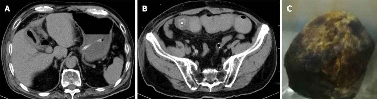
Figure 2 Postoperative images.A:On the fifth day after the operation,computed tomography(CT)examination revealed that cholecystolithiasis had disappeared according to the abdominal CT scan taken before the operation;B:The diameter of the stone in the bowel was approximately 2.0 cm,consistent with the shape of cholecystolithiasis on abdominal CT before the operation(Figure 1A);C:Gallstone was found in the small intestine during the second operation.
ACKNOWLEDGEMENTS
We are very grateful to our colleagues from the Department of Imaging for providing the computed tomography pictures.
杂志排行

World Journal of Clinical Cases的其它文章
- Needs and concerns of patients in isolation care units - learnings from COVID-19:A reflection
- Successful use of plasma exchange in fulminant lupus myocarditis coexisting with pneumonia:A case report
- Robot-assisted retroperitoneal laparoscopic excision of perirenal vascular tumor:A case report
- Ileocecal intussusception caused by two different tumors - which is the culprit lesion? A case report
- Cryptococcal pneumonia in a human immunodeficiency virusnegative patient:A case report
- Treating severe periodontitis with staged load applied implant restoration:A case report