Laparoscopic repair of complete intrathoracic stomach with iron deficiency anemia: A case report
2020-04-25DuolikunYashengWubulikasimuWulamuYiLiangLiAirexiatiTuhongjiangKelimuAbudureyimu
Duolikun Yasheng,Wubulikasimu Wulamu,Yi-Liang Li,Airexiati Tuhongjiang,Kelimu Abudureyimu
Duolikun Yasheng,Yi-Liang Li,Airexiati Tuhongjiang,Kelimu Abudureyimu,Department of Minimally Invasive Surgery,Hernia and Abdominal Wall Surgery,People's Hospital of Xinjiang Uygur Autonomous Region,Urumqi 830001,Xinjiang Uygur Autonomous Region,China
Wubulikasimu Wulamu,Department of Gastrointestinal Surgery,The University of Hong Kong-Shenzhen Hospital,Shenzhen 518053,Guangdong Province,China
Abstract
BACKGROUND
Giant paraesophageal hiatal hernias(HH) are very infrequent,and their spectrum of clinical manifestations is large.Giant HH mainly occurs in elderly patients,and its relationship with anemia has been reported.For the surgical treatment of large HH,Nissen fundoplication is the most common antireflux procedure,and the reinforcement of HH repair with a patch(either synthetic or biologic) is still debatable.
CASE SUMMARY
We report on a case of giant paraesophageal HH in a middle-aged male patient with reflux symptoms and severe anemia.After performing a series of tests and diagnostic approaches,results showed a complete intrathoracic stomach associated with severe iron deficiency anemia.The patient underwent successful laparoscopic hernia repair with mesh reinforcement and Nissen fundoplication.Postoperatively,reflux symptoms were markedly relieved,and the imaging study showed complete reduction of the hernia sac.More importantly,anemia was resolved,and hemoglobin,serum iron and ferritin level were returned to the normal range.The patient kept regular follow-up appointments and remained in a satisfactory condition.
CONCLUSION
This case report highlights the relationship between large HH and iron deficiency anemia.For the surgical treatment of large HH,laparoscopic repair of large HH combined with antireflux procedure and mesh reinforcement is recommended.
Key words:Complete intrathoracic stomach;Giant paraesophageal hiatal hernia;Iron deficiency anemia;Nissen fundoplication;Mesh reinforcement;Case report
INTRODUCTION
Classically,hiatal hernias(HH) are divided into four types according to the anatomic position of the gastric cardia.Among all types of HH,type I or sliding HH,is the most common with a prevalence of 95%,while the combination of types II,III and IV,or paraesophageal HH,account for around 5% of all HH[1].Therefore,paraesophageal HH are relatively rare and usually occur in elderly patients.A giant HH is defined as a hernia that consists of > 30% of the stomach herniating through the diaphragmatic hiatus into the thorax[2],which makes it more uncommon among paraesophageal HH.Clinical manifestations of giant HH are unspecific,making their clinical diagnosis somewhat difficult.However,the relationship between large HH and anemia has previously been reported.Likewise,the association of gastroesophageal reflux disease and HH has long been established.Yet some reported that the patients with HH may have esophagitis or Barrett's esophagus[3,4].Hence,we report a case of a middle-aged patient with complete intrathoracic stomach,or a giant paraesophageal HH,who presented with reflux symptoms and anemia.Written consent was obtained from the patient,and the study was approved by the Ethics Committee of People's Hospital of Xinjiang Uygur Autonomous Region(Protocol number: KY2018122001).
CASE PRESENTATION
Chief complaints
A 46-year-old male was admitted to our hospital with chief complaints of heartburn,regurgitation and belching for the last 5 years,and symptoms could be worsened after having a meal.The main symptoms were as follows: Dizziness,hypodynamia and occasionally with nausea and vomiting as well as chest tightness.
History of present illness
Approximately 4 mo earlier,the patient noted that the symptoms worsened even with the medicines and was referred to our hospital.
History of past illness
A diagnosis of HH and iron deficiency anemia(IDA) was made by another hospital,and the patient received omeprazole(40 mg bid) and domperidone(10 mg tid) per day.In addition,the patient received several blood transfusions with the total volume of 1200 mL(the lowest hemoglobin level was 55 g/L).After discharge,the patient took the medicines for a long period of time.His conditions improved only while consistently taking the medicine.
Physical examination
The patient was 170 cm in height and 84 kg in weight with body mass index of 29.07 kg/m2.On examination,after admission to our hospital,the temperature was 36.7 °C,the pulse was 96 beats per minute,the blood pressure was 135/83 mmHg,and the respiratory rate was 18 breaths per minute.Bowel sounds were present.The remainder of the examination was normal.He did not have carotid bruits or jugular venous distention,nor did he have cardiac or pulmonary murmur or rub on auscultation.
Laboratory examinations
Laboratory test results were significant for hemoglobin(105 g/L,normal 130-175 g/L),serum iron(6.06 μmol/L,normal range 11-30 μmol/L),serum ferritin(9.1 μg/L,normal range 15-200 μg/L),oxygen partial pressure(61 mmHg,normal range 80-105 mmHg) and oxygen saturation(91%,normal range 95%-98%).
Imaging examinations
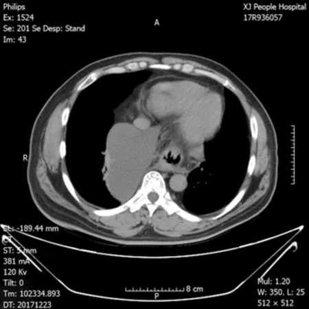
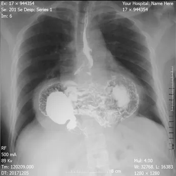
Chest X-ray demonstrated an intrathoracic gastric bubble with increased bilateral lung marking(Figure 1).Chest computed tomography with volumetric analysis demonstrated post-mediastinal location of the whole stomach along with peritoneal fat compressing both bilateral lung and heart(Figure 2).Furthermore,quantitative measurements for the size of the hernia sac,diameter of hernia port,volume of hernia sac and thoracic cavity as well as a ratio of volume of hernia sac to intrathoracic cavity were 13.4 cm × 18.6 cm,6 cm,1476.4 cm3,4025 cm3and 36.7%,respectively.Barium contrast radiography confirmed large HH and the configuration of the stomach within the hernia suggested an organoaxial volvulus(Figure 3).
Further diagnostic work-up
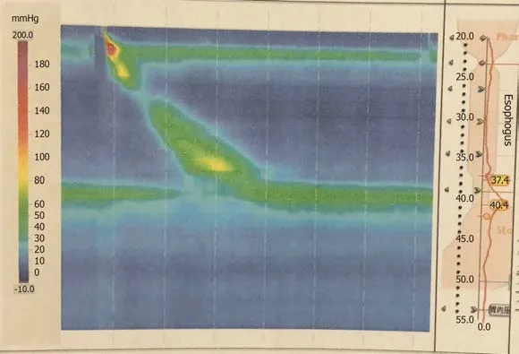
Electrocardiogram and cardiac ultrasonography did not show any abnormalities.To clarify the current and other related possible diagnosis,a series of studies were performed.However,esophageal high-resolution manometry and 24-hr multichannel intraluminal impedance-pH monitoring showed the presence of HH with an elevated level of lower esophageal sphincter of 9.6 cm high,pathological acid reflux and DeMesster score of 64.6(Figure 4).
FINAL DIAGNOSIS
The final diagnosis of the presented case is giant paraesophageal HH and IDA.
TREATMENT
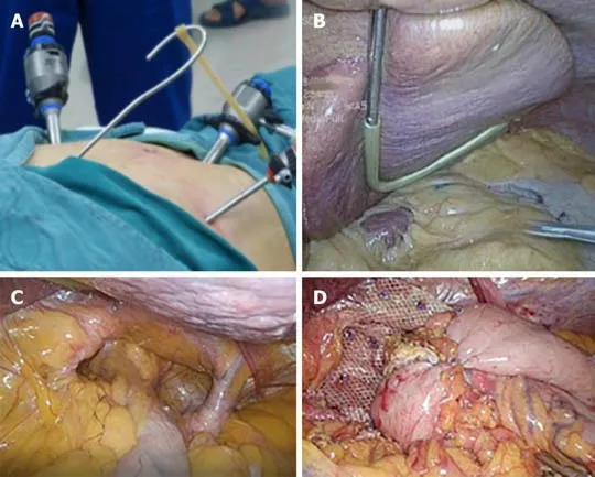
After careful preoperative evaluation for surgical repair,a successful laparoscopic hernia repair with mesh reinforcement and Nissen fundoplication was carried out in accordance with the guidelines recommended by the Society of American Gastrointestinal and Endoscopic Surgeons[5].The patient was positioned in supine,split-leg position,and the chief surgeon stood between the patient's legs,while the assistant surgeon stood on the patient's left.Four ports and a homemade liver retractor were used for surgical access.The initial port of 12 mm was placed supraumbilically for the laparoscope.After entry,the abdomen was explored looking for iatrogenic injury and the presence of intra-abdominal adhesions that would hinder subsequent port placement.A 12 mm port was then placed just below the left costal margin in the mid-clavicular line as the main working port.The other two 5 mm ports were also placed,one just below the right costal margin in the mid-clavicular line,and the other in the left flank.A separate 3 mm subxiphoid incision was made for the reverse “7” shaped,homemade liver retractor as shown in the pictures(Figure 5A and 5B).
Firstly,an atraumatic grasper was used to grasp the anterior epigastric fat pad.Then the stomach was retracted downward and toward the left lower quadrant to reposition.Subsequently,dissection was preformed until diaphragmatic crura were well displayed,along with the preservation of hepatic branch of the anterior vagus nerve.Then,3 cm of tension-free esophagus was repositioned intra-abdominally(Figure 5C).The hiatus was then repaired posteriorly with interrupted nonabsorbable sutures.As the patient's hiatal defect reached 8 cm,we preformed mesh reinforcement using Parietex™ Composite hiatal mesh provided by Medtronic(Minneapolis,MN,United States).Finally,Nissen fundoplication was completed.The 360° wrap was created by grasping the right and left portion of the mobile funds andpulling them behind the esophagus and sutured together in front of the anterior part of the abdominal portion of the esophagus.The length of the wrap was 2 cm with three sutures.At the end,a gastropexy was performed by suturing the posterior fundus to the inferior crus with three interrupted permanent sutures(Figure 5D).Estimated blood loss was 20 mL,and it took 80 min for the surgical procedure.

Figure 1 Preoperative chest X-ray.
OUTCOME AND FOLLOW-UP
For the first day after surgery,abdominal examination revealed normal bowel sounds,then the gastric tube was removed.In the following days,the patient started a liquid diet.According to the patient's statement,almost all of the preoperative discomforts were gradually resolved.After discharge from the hospital,the patient presented to our department at 1-mo post-surgery for follow-up.Neither distinctive abnormalities nor hernia recurrence were observed from the chest X-ray(Figure 6).The patient had an increase in postoperative hemoglobin,serum iron and serum ferritin level(135 g/L,18.3 μmol/L and 92.4 μg/L,respectively).
DISCUSSION
Paraesophageal HH are defined as the condition in which gastroesophageal junction and components of the abdominal cavity,most commonly the stomach,herniatedviaesophageal hiatus into the mediastinum.However,most large paraoesophageal HH occur in elderly patients with the incidence rate of > 60% above the age of 70 years and is a relatively rare condition[6].Interestingly,our case was a middle-aged male patient,and it indicates that the physicians should be aware of the presence of HH when making clinical diagnosis for the patient with atypical characteristics.Some studies have reported that the formation of HH is related to obesity[7].Therefore,high body mass index can be one of the risk factors contributing to large HH in this patient,whose body mass index(29.07 kg/m2) is close to the category of obesity.
It is crucial to distinguish between symptomatic paraesophageal HH and asymptomatic or minimally symptomatic HH.Generally,symptomatic patients are recommended for surgical repair to prevent subsequent acute complications,such as strangulation,perforation and bleeding.In addition to HH repair,laparoscopic antireflux procedure is a well-established treatment for patients suffering from reflux disease associated with HH,especially for large paraoesophageal HH.Nissen fundoplication is the most commonly used procedure for the treatment of gastroesophageal reflux disease due to its good postoperative long-term reflux control results in approximately 90% of patients[8].
In view of these facts,the patient underwent successful HH repair reinforced by a“U” shaped mesh with Nissen fundoplication.Even though the reinforcement of the HH repair with a patch(either synthetic or biologic) is still debatable,a meta-analysis reported by Targaronaet al[9]concluded that prosthetic reinforcement was beneficialwith an acceptable rate of secondary complications.Another meta-analysis of three randomized studies also reported that prosthetic reinforcement had a four-fold decrease in 1-year risk of recurrence[10].Therefore,according to related studies and our experience,HH repair with mesh reinforcement is recommended for symptomatic HH patients,especially for large paraoesophageal HH patients.

Figure 2 Chest computed tomography with volumetric analysis.
The relationship of IDA with HH has been studied,and it is reported that HH is a cause of gastrointestinal bleeding and increases the risk of subsequent IDA[11].Grayet al[12]suggested that the prevalence of Cameron lesion,which is considered to be a source of gastrointestinal bleeding,is known to vary with HH size,with the highest prevalence occurring in large HH patients.They identified large HH as a major risk factor for IDA.In our case,the patient's reflux-related symptoms completely resolved,and his hemoglobin level was returning to a normal range after the operation.Consequently,this case report complements the other studies and strengthens the evidence that large HH may be a cause of anemia.
CONCLUSION
Complete intrathoracic stomach,or giant HH,is very infrequent,and its spectrum of clinical manifestations is large.This report presented a case of giant HH in a middleaged male patient with reflux symptoms and severe anemia.Although studies have reported that large HH mainly occurs in elderly patients,one possible factor contributing to the situation in this relatively young patient might be his high body mass index.
According to the literature mentioned above and our case report,there appears to be a relationship between large HH and IDA and indicates that large HH may be a potential cause of IDA.Surgical repair of large HH relieves IDA symptoms as reported by others[13-15]and as seen in our patient.More importantly,for the patients suffering from reflux-related symptoms with large HH,antireflux procedure and mesh reinforcement are recommended.

Figure 3 Barium contrast radiography.

Figure 4 Esophageal high-resolution manometry.

Figure 5 Mesh reinforcement and Nissen fundoplication.

Figure 6 Postoperative chest x-ray at 1-mo follow up.
杂志排行
World Journal of Clinical Cases的其它文章
- Growth hormone therapy for children with KBG syndrome: A case report and review of literature
- Hepatoid adenocarcinoma of the stomach: Thirteen case reports and review of literature
- Cerebral venous sinus thrombosis following transsphenoidal surgery for craniopharyngioma: A case report
- Microscopic removal of type lll dens invaginatus and preparation of apical barrier with mineral trioxide aggregate in a maxillary lateral incisor: A case report and review of literature
- Hyoid-complex elevation and stimulation technique restores swallowing function in patients with lateral medullary syndrome:Two case reports
- Metabolic and genetic assessments interpret unexplained aggressive pulmonary hypertension induced by methylmalonic acidemia: A case report
