Displacement of peritoneal end of a shunt tube to pleural cavity:A case report
2020-04-09JieLiuMianGuo
Jie Liu,Mian Guo
Jie Liu,Mian Guo,Department of Neurosurgery,The Second Affiliated Hospital of Harbin Medical University,Harbin 150086,Heilongjiang Province,China
Abstract BACKGROUND The common treatment for hydrocephalus is insertion of a ventriculoperitoneal shunt.Shunt tube displacement is one of the common complications.Most shunt tube displacements occur in children and has a reportedly lower incidence in adults.CASE SUMMARY This study reports an adult patient(male,56 years)who suffered from intracranial aneurysm and subarachnoid hemorrhage and underwent aneurysm clipping following hospitalization.One month post onset of the disease,the patient underwent ventriculoperitoneal shunt due to hydrocephalus.The peritoneal end of the shunt tube was displaced in the peritoneal cavity 9 years after the aneurysm clipping.The peritoneal end of the shunt tube was removed and ventriculoperitoneal shunt was re-performed after anti-inflammatory treatment.CONCLUSION Shunt tube displacement has a low incidence in adults.In order to avoid shunt tube displacement,there is a need to summarize its causative factors and practice personalized medicine.
Key Words:Ventriculoperitoneal shunt;Hydrocephalus;Shunt tube displacement;Pleural cavity;Intracranial aneurysm;Case report;Subarachnoid hemorrhage
INTRODUCTION
Ventriculoperitoneal shunt is the most commonly used method for the treatment of hydrocephalus,but many complications may occur following the insertion of the shunt.The commonly reported complications include infection,shunt blockage,hypersensitivity,abdominal cyst formation,ascites,and shunt tube displacement,among others[1].Most of shunt tube displacements are reported in children.The incidence is low in adults and relatively few cases have been reported.This study reports a case of displacement of the peritoneal end of a ventriculoperitoneal shunt tube to the pleural cavity in an adult patient with hydrocephalus.
CASE PRESENTATION
Chief complaints
A 56-year-old male patient presented with fever,decline of memory,pain in the right chest,and unstable walking for five days.
History of present illness
The patient suffered from an aneurysm with subarachnoid hemorrhage and underwent aneurysm clipping in Department of Neurosurgery at The Second Affiliated Hospital of Harbin Medical University on July 27,2010.One month after the surgery,he underwent ventriculoperitoneal shunt due to hydrocephalus,and the patient recovered well.On March 25,2019,the patient reported fever,decline of memory,pain in the right chest,and unstable walking.
History of past illness
The patient had been in good health.
Physical examination
The patient had an appearance of chronic disease,body temperature around 38 ˚C,normal orientation,poor memory,and weak respiratory sound in the right lung.
Laboratory examinations
Cerebrospinal fluid(CSF)examination showed that the number of cells was 82 × 106cells/L,glucose level was 1.71 mmol/L,chloride was 98 mmol/L,and total protein was 891 mg/L.No bacteria were observed in CSF upon culture.The peritoneal end was surgically excised and the end was ligated.The patient was administered antiinflammatory treatment for the following 10 d(ceftriaxone sodium,2 g/time,Q12h;vancomycin 0.5 g/time,Q8h).On April 3,the CSF was re-examined,which showed that the number of cells was 8 × 10⁶cells/L,glucose was 2.69 mmol/L,chloride was 125 mmol/L,and total protein was 341 mg/L.
Imaging examinations
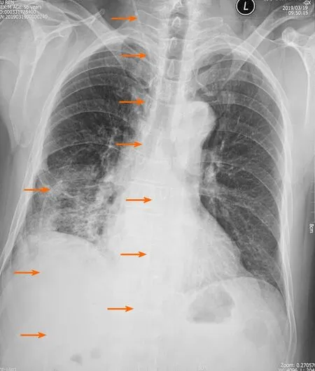
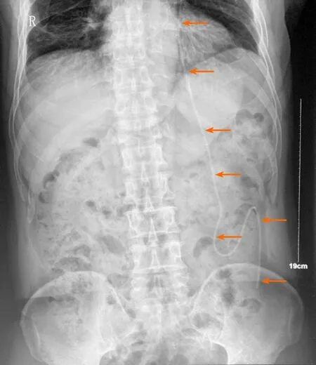
X-ray examination before operation revealed that the peritoneal end of the shunt tube had moved to the pleural cavity,resulting in pulmonary inflammation(Figure 1).An X-ray image after operation confirmed the shunt location in the abdominal cavity(Figure 2).

Figure 1 X-ray examination revealed that the peritoneal end of the shunt tube moved to the pleural cavity with the formation of pulmonary inflammation on March 25,2019(The orange arrows indicate the shunt tube).

Figure 2 X-ray examination showed that the shunt tube was located in the abdominal cavity after operation on April 4(The orange arrows indicate the shunt tube).
FINAL DIAGNOSIS
Displacement of the peritoneal end of the shunt tube to pleural cavity.
TREATMENT
Ventriculoperitoneal shunt was re-performed after inflammation subsided on April 3,2019.
OUTCOME AND FOLLOW-UP
The patient is recovering well and has no abnormal symptoms so far.
DISCUSSION
The displacement of the peritoneal end of the shunt tube after ventriculoperitoneal shunt is one of the common complications,but the reasons are still elusive.There are few hypotheses that partly explain the shunt tube displacement.Akyüzet al[2]reported that when the peritoneal end of the shunt tube is attached to the nearby organs or body wall,it will induce an inflammatory reaction,and the distal end of the catheter will gradually protrude outward.Sridharet al[3]suggested that the distal migration of the shunt tube might be caused by the use of rigid material,which was supported by the observation that the use of a softer shunt tube did reduce the incidence of shunt tube displacement[4].Additionally,some authors speculated that the distal penetration of the shunt tube through the body wall might be caused by local wound dehiscence,low patient immunity,improper surgical techniques,or dermal ischemic necrosis[5,6].Other factors that lead to the distal migration of shunt tube may include the age of the patient and the length of the shunt tube in the abdominal cavity.Most of them occur in pediatric patients.It is probably a consequence of softer organs and tissues in children which are vulnerable to rupture.
There are three types of shunt tube displacement of the peritoneal end:Internal displacement,external displacement,and mixed displacement[7].The intrathoracic migration of peritoneal shunt tube can be divided into two types:Supra-diaphragm and trans-diaphragm migration[8].The subject of this study had a 9-year history since the last ventriculoperitoneal shunt surgery.In combination with chest X-ray images,considering the end of the shunt tube at the upper edge of the liver,the long-term stimulation of the upper hepatic diaphragm may have led to the diaphragm damage.The peritoneal end of the shunt tube thus migrated to the pleural cavity,leading to an inflammatory response in the lungs.At the same time,the drainage of the shunt tube was blocked,causing hydrocephalus symptoms to reappear.
A variety of clinical manifestations can occur after the shunt is displaced.The treatment strategies,thus,need to be personalized.For patients with shunt tube displacement of the peritoneal end,it has been reported that the displaced shunt can be re-inserted directly into the abdominal cavity with the help of laparoscopic method[9].Some scholars have proved that the use of laparoscopy to treat intraperitoneal complications after intraventricular shunting of the ventricle has the advantages of shorter operation time,less trauma,and reduced intestinal damage and adhesion,among others[10,11].This patient had fever when admitted to the hospital,and the cerebrospinal fluid test showed abnormal cells,which did not rule out the possibility of pulmonary inflammation retrogradely resulting in intracranial infection.Therefore,the displaced shunt tube was not directly moved back to the abdominal cavity.The shunt tube was removed with subsequent anti-inflammatory treatment.After the number of cells in cerebrospinal fluid cells was normal,ventriculoperitoneal shunt was re-performed on the contralateral side.
CONCLUSION
Although ventriculoperitoneal shunt is a common and simple method for the treatment of hydrocephalus,its complications are not rare.For the displacement of the peritoneal end of the shunt tube,we should further explore its predisposing factors,and aim to reduce its occurrence.In events of displacement,advanced technologies such as laparoscopy to reduce pain,trauma,and infection are recommended.They also accelerate patient recovery.Infection and inflammation should be assessed and treated prior to shunt replacement surgery.Ventricular-atrial shunt can be considered for repeated displacement and blockage of the peritoneal end.
杂志排行
World Journal of Clinical Cases的其它文章
- Parathyroid adenoma combined with a rib tumor as the primary disease:A case report
- Localized primary gastric amyloidosis:Three case reports
- Bochdalek hernia masquerading as severe acute pancreatitis during the third trimester of pregnancy:A case report
- Intravesically instilled gemcitabine-induced lung injury in a patient with invasive urothelial carcinoma:A case report
- Intraosseous venous malformation of the maxilla after enucleation of a hemophilic pseudotumor:A case report
- Solitary hepatic lymphangioma mimicking liver malignancy:A case report and literature review
