Endoscopic palliative resection of a giant 26-cm esophageal tumor:A case report
2020-04-09YanLiLinJieGuoYingCaiMaLianSongYeBingHu
Yan Li,Lin-Jie Guo,Ying-Cai Ma,Lian-Song Ye,Bing Hu
Yan Li,Lin-Jie Guo,Lian-Song Ye,Bing Hu,Department of Gastroenterology,West China Hospital,Sichuan University,Chengdu 610041,Sichuan Province,China
Ying-Cai Ma,Department of Digestion,Qinghai Provincial People's Hospital,Xining 810007,Qinghai Province,China
Abstract BACKGROUND Esophageal carcinosarcoma,usually presenting as a pedunculated polypoid mass,is a rare malignancy with coexisting sarcomatoid and carcinomatous components.Its imaging and endoscopic characteristics are similar to those of leiomyosarcoma,liposarcoma and so forth.The diagnosis needs histological confirmation.Surgical resection is the traditional therapy.Endoscopic resection is minimally invasive but still controversial.This paper reports the case of a patient with a giant esophageal carsinosarcoma who underwent a palliative endoscopic resection.CASE SUMMARY A 55-year-old male patient presented with dysphagia and weight loss for 1 mo.Imaging and endoscopy showed a gray-white,polypoid,stalk-like mass,with a bulky pedicle located in the middle and lower esophagus.The mass almost filled the whole esophageal lumen,but the endoscope could still pass through.Despite the suspicion of a malignancy,repeated biopsies indicated necrosis and inflammation.After multidisciplinary team consultation,an endoscopic resection to diagnose and relieve the obstruction was recommended.The pedicle of the mass was cut off,the bleeding was stopped,and the mass was cut into pieces and pulled out.The mass was 26 cm × 5 cm × 4 cm in size.The final diagnosis was esophageal carcinosarcoma.No postoperative complications occurred.After 1 mo,the patient gained 6 kg and endoscopic reexamination revealed no obstruction.Radical surgery with lymph node dissection was carried out successfully.This lesion was the largest endoscopically resected esophageal carcinosarcoma reported to date.CONCLUSION Endoscopic palliative resection can help obtain adequate tissue for diagnosis and relieve obstructions in patients with giant esophageal carcinosarcoma.
Key Words:Case report;Dysphagia;Endoscopic resection;Esophageal carcinosarcoma;Polypoid mass
INTRODUCTION
Esophageal carcinosarcoma is a rare malignancy with coexisting sarcomatoid and carcinomatous components[1].Carcinosarcoma has also been termed as spindle cell carcinoma,sarcomatoid carcinoma,pseudosarcoma,and squamous cell carcinoma with sarcomatoid changes[1].On imaging and endoscopic examinations,the majority of esophageal carcinosarcomas present as a bulky intraluminal polypoid mass,similar to leiomyosarcoma,gastrointestinal stromal tumor,fibrovascular polyp,liposarcoma and so forth[2-5].Therefore,histological confirmation is vital for the final diagnosis.However,preoperative biopsies usually produce negative results,and consequently a more effective method is needed to accomplish diagnosis[4].
Esophagectomy with regional lymph node dissection has been the traditional therapy for esophageal carcinosarcoma,and the endoscopic procedure is recommended as a promising approach[1,3].However,a PubMed search for“esophageal carcinosarcoma”AND“endoscopic resection”,“esophageal sarcomatoid carcinoma”AND“endoscopic resection”or "carcinosarcoma" AND "endoscopic resection" returned only four case reports.Endoscopic resection has been performed for superficial esophageal carcinosarcomas or patients in poor condition[1,6].This paper reports the case of a palliative endoscopic resection of a giant esophageal polypoid mass,thus confirming the diagnosis and relieving the obstruction.This lesion was confirmed to be esophageal carcinosarcoma(T1bN1M0).The timeline of this case is shown in Figure 1.
CASE PRESENTATION
Chief complaints
A 55-year-old male patient presented to the Gastroenterology Department of West China Hospital for dysphagia and weight loss for 1 mo.
History of present illness
The patient suffered dysphagia while eating solid or semi-solid food and lost 6 kg in 1 mo.He also felt slight retrosternal pain while changing position,without reflux,vomit,fever,or cough.
History of past illness
The patient had no specific previous illness or family history.On average,the patient had a smoking history of 60 cigarettes a day for 30 years and a drinking history of 80 g/d for 20 years.
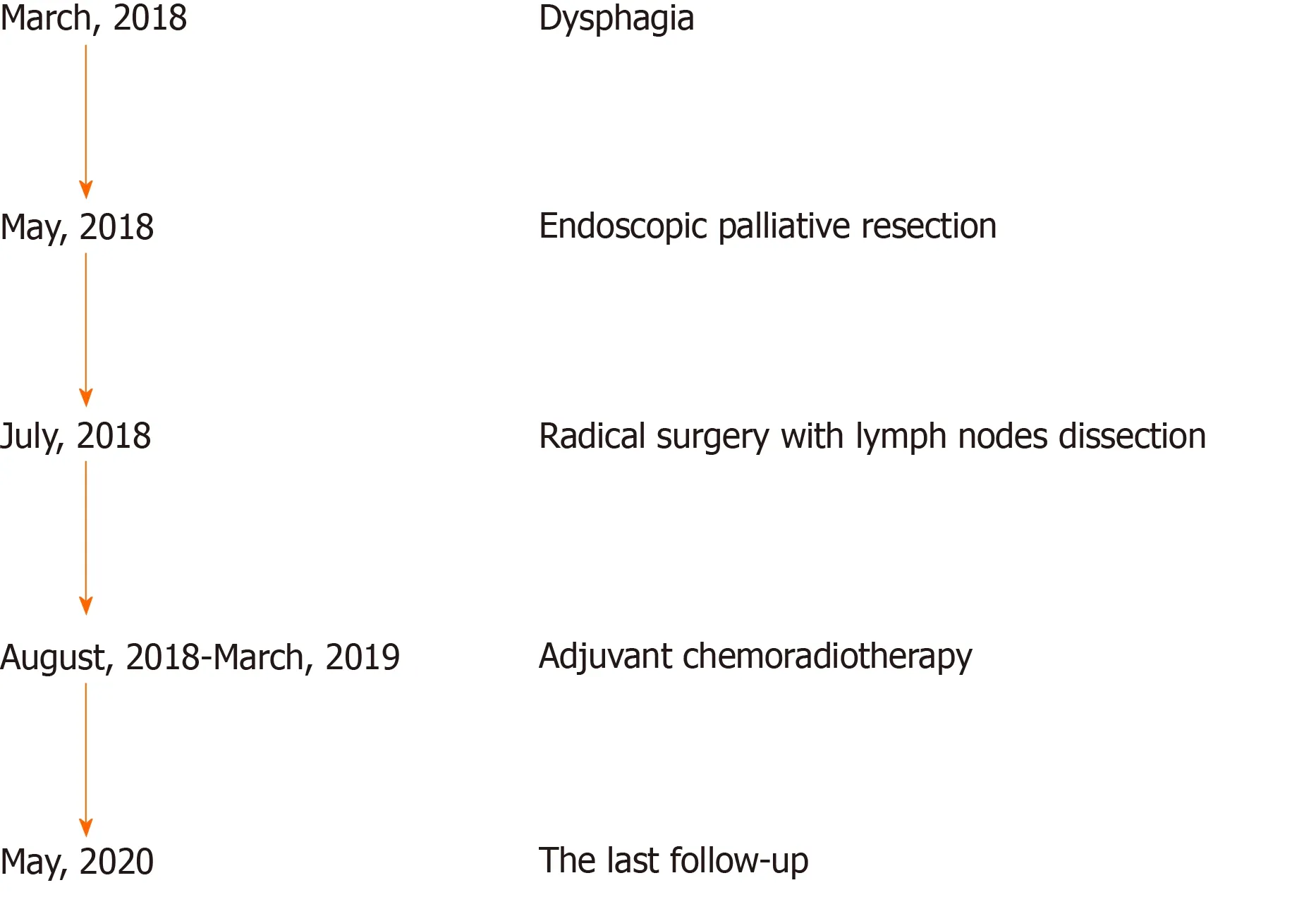
Figure 1 Timeline of the patient with esophageal carcinosarcoma.
Physical examination
At admission,the patient’s temperature was 36.5 °C,heart rate was 76 bpm,respiratory rate was 20 breaths per minute,blood pressure was 119/89 mmHg,oxygen saturation in room air was 99%,height was 172 cm,and weight was 63 kg.No other special finding was reported.
Laboratory examinations
A routine blood test indicated leukocytosis with the white blood cell count at 12.08 ×10⁹/L(normal range,3.5 × 109/L to 9.5 × 10⁹/L)and mainly neutrophils(81.9%;normal range,40%-75%).The hemoglobin concentration and platelet count were normal.The serum albumin concentration had declined to 29.9 g/L(normal range,40–55 g/L).The serum C-reactive protein concentration had increased to 74.8 mg/L(normal range,<5.0 mg/L),and the erythrocyte sedimentation rate was 60 mm/h(normal range,<21.0 mm/h).The serum tumor markers,including CEA,AFP,and CA 19-9,had normal concentrations.
Imaging examinations
An X-ray barium meal examination showed a huge intraluminal stalk-like mass along the middle and lower esophagus(Figure 2).Computed tomography(CT)showed a prominently enhanced anterior area of the mass beside the esophageal wall,and the maximum sectional area was about 5.6 cm × 3.5 cm(Figure 3).Endoscopy indicated that the mass was polypoid,gray-white,with a bulky pedicle,located in the esophagus 22-45 cm from the incisors and almost filled the whole esophageal lumen,but the endoscope could still pass through(Figure 4A-C).
PATHOLOGY
The biopsy in West China Hospital indicated necrosis and inflammation,similar to the result of the initial biopsy in the previous hospital.
MULTIDISCIPLINARY EXPERT CONSULTATION
You Lu,MD,PhD,Professor,Department of Oncology,West China Hospital,Sichuan University
Despite the suspicion of a malignancy,the diagnosis of this giant esophageal mass is still unclear.Repeated biopsy or a partial resection to diagnose should be the priority.Further surgery,chemotherapy,or radiotherapy should be done based on the final diagnosis.
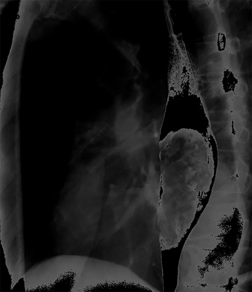
Figure 2 X-ray barium meal examination showing a huge intraluminal stalk-like mass along the middle and lower esophagus.
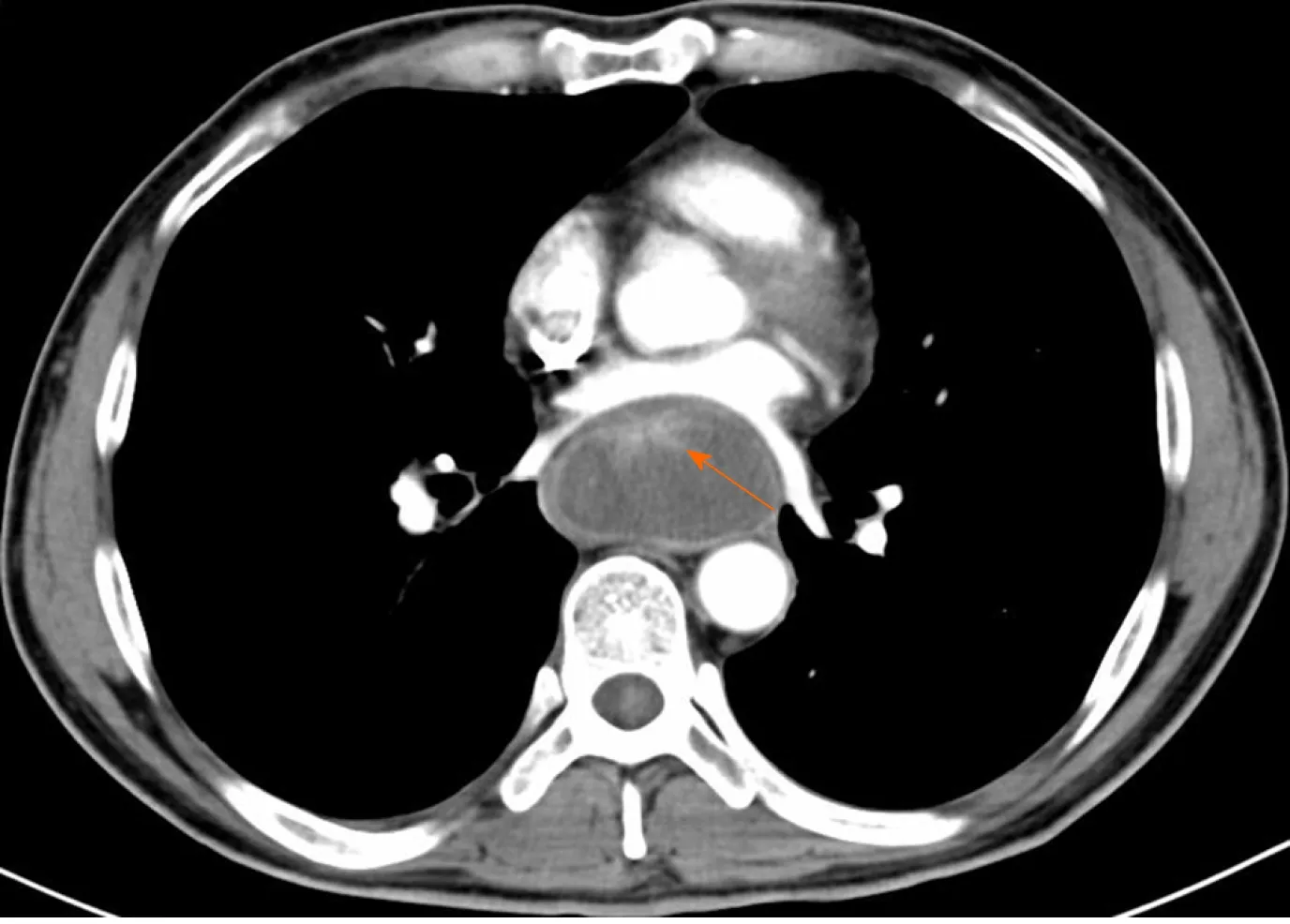
Figure 3 Computed tomography showing a prominently enhanced anterior area of the giant mass beside the esophageal wall(orange arrow),and the maximum sectional area was about 5.6 cm × 3.5 cm.
Yi-Dan Lin,MD,PhD,Professor,Department of Thoracic Surgery,West China Hospital,Sichuan University
A further biopsy might still fail due to the repeated insignificant results of the biopsy.A partial or total resection of the mass to acquire sufficient tissue for pathological diagnosis is necessary.It would also contribute to relieve the obstruction and improve nutrition.An endoscopic resection is optimal for the advantage of minimal invasiveness.If it fails,additional surgery should be performed.
Bing Hu,MD,Professor,Department of Gastroenterology,West China Hospital,Sichuan University
The mass could probably be malignant.First,an endoscopic partial resection is required to obtain adequate tissue for pathological diagnosis,and if possible,a total resection of the mass to relieve the obstruction should be the optimal choice.If needed,additional therapy should be provided.
FINAL DIAGNOSIS
The disease was diagnosed after endoscopic palliative resection.The final diagnosis was esophageal carcinosarcoma(T1bN1M0),with submucosal invasion and right superior paratracheal lymph node metastasis.Immunochemical staining indicated that the tumor cells were positive for pancytokeratin(PCK)and negative for cluster of differentiation 34(CD34),Desmin,and S-100,and Ki 67 index was 20%-30%(Figure 5).
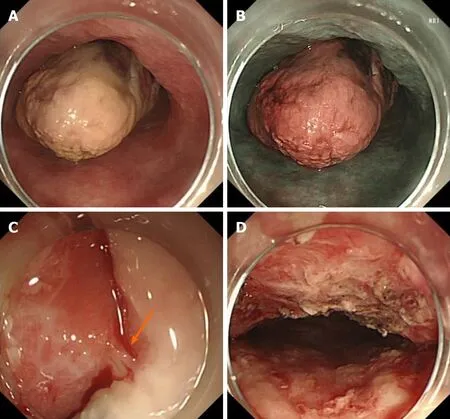
Figure 4 Endoscopy.A:The giant mass was polypoid,gray-white,almost filling the whole esophageal lumen(white light),but the endoscope could still pass through;B:The giant polypoid mass was brown,as revealed by narrow-band imaging;C:The bulky pedicle of the mass(orange arrow);D:The wound was intact after cutting off the giant mass.
TREATMENT
Endoscopic resection was conducted as follows:(1)The basal part of the mass was sought first,which was 33 cm from the incisors;(2)0.01% epinephrine saline and methylene blue were injected into the submucosa to lift the lesion from the muscle layer;(3)Dissection was performed within the submucosa beneath the mass with a Hybrid knife and an insulation-tipped diathermic knife;(4)Bleeding was stopped with hemostatic forceps;and(5)The mass was cut into pieces with a snare and pulled out.The mass was totally resected,and the wound was intact(Figure 4D).The mass was about 26 cm × 5 cm × 4 cm in size(Figure 6).
OUTCOME AND FOLLOW-UP
No postoperative complications occurred.Oral intake was resumed 2 d later.After 1 mo,the patient gained 6 kg,and endoscopic reexamination revealed no obstruction(Figure 7).Radical surgery with lymph node dissection and adjuvant chemoradiotherapy were carried out.The patient received six cycles of chemoradiotherapy(175 mg/m2of paclitaxel and 80 mg/m2of nedaplatin at first day)and 25 times of radiotherapy with a total dosage of 45 Gy(1.8 Gy per time)from August 2018 to March 2019.A 2-year follow-up revealed that the patient lived well,without recurrence or metastasis.
DISCUSSION
In 1865,Virchow initially introduced“carcinosarcoma,”a rare malignancy with coexisting carcinomatous and sarcomatoid components.Later studies demonstrated that carcinosarcoma,sarcomatoid carcinoma,spindle cell carcinoma,pseudosarcoma,and squamous cell carcinoma with sarcomatoid changes were one disease,and they could occur in the lung,thyroid,breast,uterus,esophagus and so forth[1].Esophageal carcinosarcoma accounted for about 0.5%-2.8% of all esophageal carcinomas and mainly occurred in middle-aged and elderly male patients with a heavy smoking or drinking history[7-9].The majority of esophageal carcinosarcomas presented as a bulky intraluminal polypoid mass and were located in the middle and lower esophagus[1,2,7].Sarcomatoid components formed the body of the polypoid mass,and carcinomatous components(mostly squamous cell carcinoma)surrounded the base of the tumor,with a distinct transitional area between the two components[8,10,11].The doubling time of esophageal carcinosarcoma was reported to be 2.2-5 mo,and the 5-year overall survival was 26.7%-61.9%[4,8,9,12].
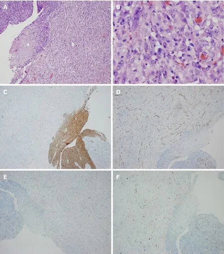
Figure 5 Postoperative pathology.A:Hematoxylin–eosin staining showing esophageal squamous cell carcinoma(a)with sarcomatoid component(b);B:Magnification of the sarcomatoid component;C:PCK staining;D:CD34 staining;E:Desmin staining;F:S-100 staining.
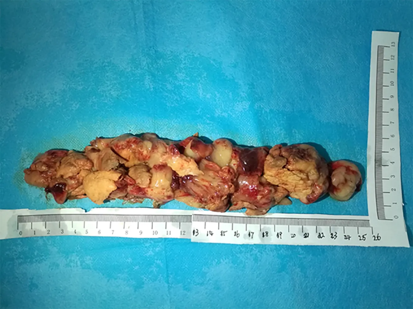
Figure 6 The giant mass was assembled after pulling out and was about 26 cm × 5 cm × 4 cm in size.
About the oncogenesis,the metaplastic hypothesis held that two components derived from a common undifferentiated stem cell,while the collision hypothesis believed that two components derived from different stem cells undergoing malignant transformation simultaneously[7,13-15].Recent studies indicated that TP53 mutation was common in patients with esophageal carcinosarcoma,consistent with positive immunohistochemical staining for p53,occurring in both sarcomatoid and carcinomatous components[16,17].These findings indicated that the two components originated from the same stem cell.
Regarding preoperative diagnosis,Zhanget al[18]and Hashimotoet al[4]reported that 14.1%(10/71)and 17.9%(5/28)of esophageal carcinosarcomas,respectively,were suspected to have sarcomatous components based on biopsy.Although imaging and endoscopy showed characteristic features,preoperative diagnosis was still difficult.A similar situation occurred in the case reported in this paper.The causes might be as follows:(1)The carcinomatous and sarcomatous components were distributed zonally;or(2)The tumor grew rapidly and the tumor size was huge,resulting in superficial necrosis.
Lymph node and distant metastases of esophageal carcinosarcoma have been frequently observed in previous studies,and metastasis of epithelial components is more common[3,4,19,20].Endoscopic ultrasonography(EUS),CT,and positron emission tomography are important for TNM staging of esophageal cancers preoperatively,although practices in esophageal carcinosarcomas are scarce.Jiet al[6]and Kuoet al[3]reported esophageal carcinosarcoma to be a hypoechoic mass with a regular margin.EUS contributed to determine the invasion depth and regional lymph node involvement.The preoperative EUS diagnosis of esophageal carcinosarcoma reached high consistency with final pathological staging(T staging:7/7,100%;N staging:5/7,71.4%)[3,6].
A surgical resection with regional lymph node dissection was the traditional treatment for esophageal carcinosarcomas without distant metastasis[1,4].Endoscopic techniques,with the advantages of minimal invasion and preservation of the esophagus,have also been applied to the resection of esophageal carcinosarcoma(Table 1).In 2004 and 2009,Pesentiet al[21]and Jiet al[6]conducted the first and second endoscopic resections of esophageal carcinosarcoma,respectively,with good tolerance and favorable prognosis.In 2013,Xuet al[22]reported the case of an 84-year-old male patient with multiple carcinosarcomas along the esophagus and stomach.He was in serious condition and received a palliative endoscopic resection of the esophageal lesion to relieve the obstruction.He resumed a normal food intake postoperatively,but succumbed 7 mo later.In 2018,Yabuuchiet al[23]reported the fourth case with a slightly elevated lesion in the middle thoracic esophagus.This patient received an endoscopic submucosal dissection but the follow-up information was unclear.The patient in our study was the fifth patient with esophageal carcinosarcoma who underwent an endoscopic resection,and the mass was much larger than those in the previous cases.A palliative endoscopic resection was performed first due to unclear diagnosis and esophageal obstruction.The patient recovered well 1 mo later.After surgical resection and adjuvant chemoradiotherapy,the patient got a favorable prognosis.This practice suggested that endoscopic resection is applicable for patients with esophageal carcinosarcoma:(1)A total endoscopic resection is achievable for small and superficial tumors;(2)A palliative endoscopic resection relieves esophageal obstruction in patients with serious condition;and(3)A partial resection could attain adequate tissue for undiagnosed tumors.Further prospective large-sampled studies are needed to verify these findings.
CONCLUSION
This paper reports the case of a patient with a giant esophageal polypoid mass who underwent a palliative endoscopic resection.This lesion turned out to be esophageal carcinosarcoma(T1bN1M0).An endoscopic resection confirmed the diagnosis and relieved the esophageal obstruction.
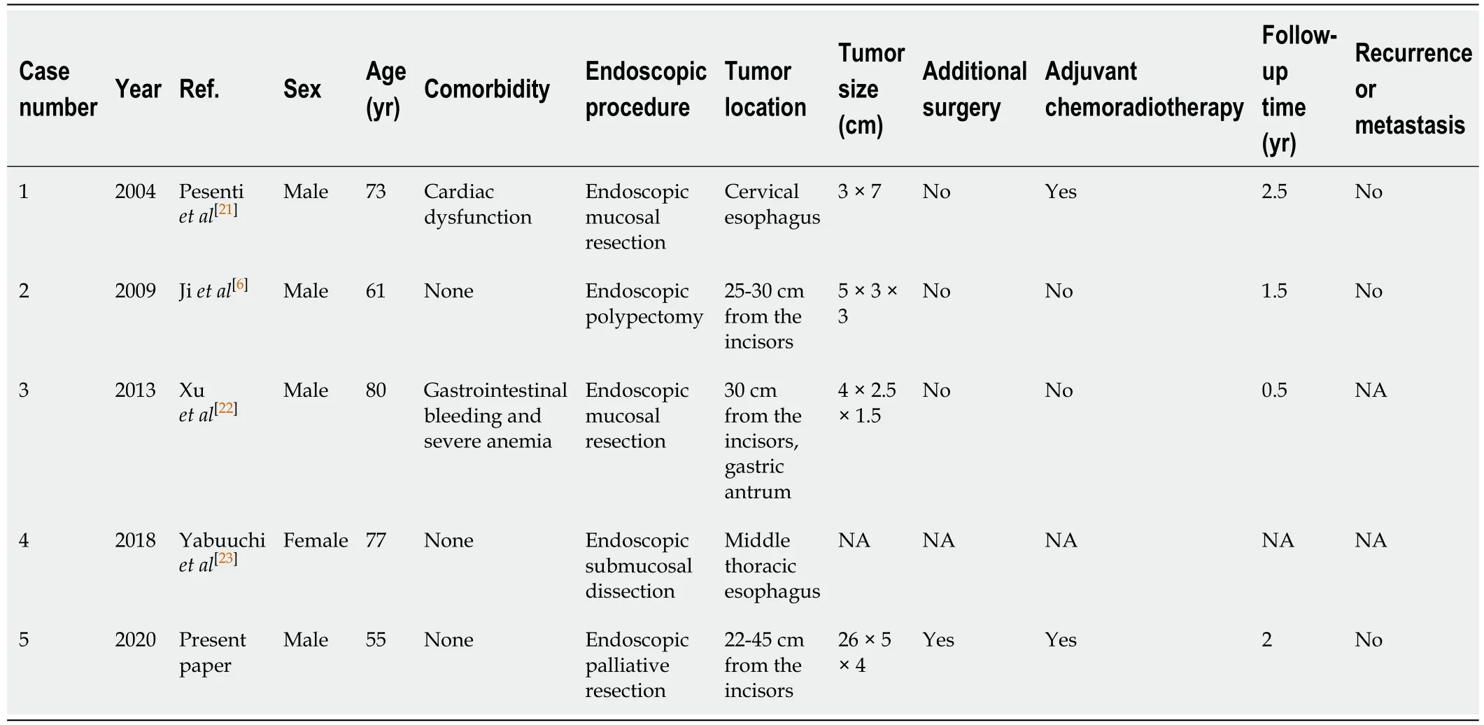
Table 1 Reported cases of endoscopic resection of esophageal carcinosarcoma
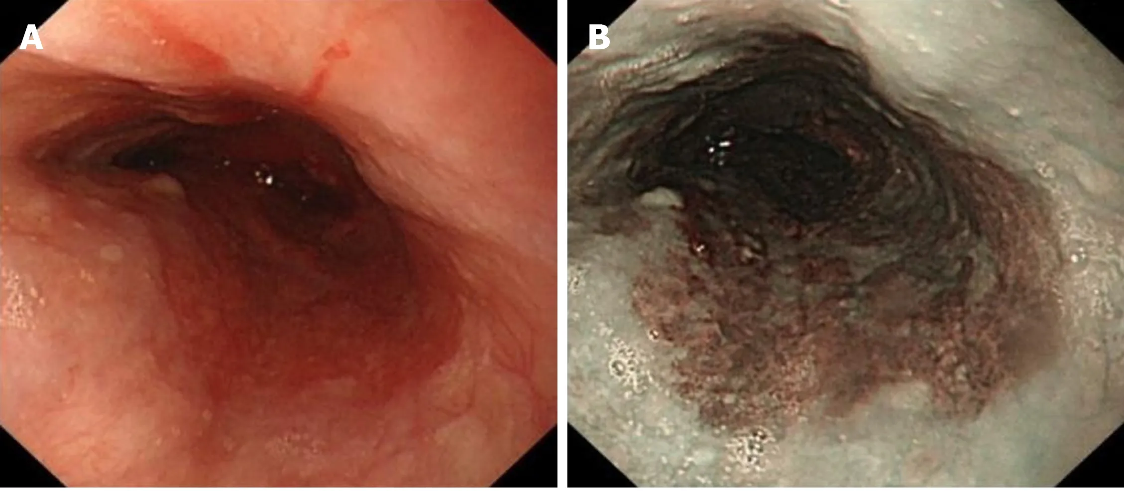
Figure 7 Endoscopic reexamination after 1 mo revealed no esophageal obstruction.A:White light;B:Narrow-band imaging.
杂志排行
World Journal of Clinical Cases的其它文章
- Role of monoclonal antibody drugs in the treatment of COVID-19
- Review of simulation model for education of point-of-care ultrasound using easy-to-make tools
- Liver injury in COVID-19:A minireview
- Transanal minimally invasive surgery vs endoscopic mucosal resection for rectal benign tumors and rectal carcinoids:A retrospective analysis
- Impact of mTOR gene polymorphisms and gene-tea interaction on susceptibility to tuberculosis
- Establishment and validation of a nomogram to predict the risk of ovarian metastasis in gastric cancer:Based on a large cohort
