Germinomas of the basal ganglia and thalamus:Four case reports
2020-04-09ZhenChaoHuangQingDongEnPengSongZhiJieChenJinHuaZhangBoHouZhengQiLuFengQin
Zhen-Chao Huang,Qing Dong,En-Peng Song,Zhi-Jie Chen,Jin-Hua Zhang,Bo Hou,Zheng-Qi Lu,Feng Qin
Zhen-Chao Huang,En-Peng Song,Zhi-Jie Chen,Jin-Hua Zhang,Bo Hou,Feng Qin,Department of Neurosurgery,The Third Affiliated Hospital of Sun Yat-Sen University,Guangzhou 510630,Guangdong Province,China
Qing Dong,Zheng-Qi Lu,Department of Neurology,The Third Affiliated Hospital of Sun Yat-Sen University,Guangzhou 510630,Guangdong Province,China
Abstract BACKGROUND The early diagnosis of basal ganglia and thalamus germinomas is often difficult due to the absence of elevated tumor markers,and atypical clinical symptoms and neuroimaging features.CASE SUMMARY Four male children aged 8 to 15 years were diagnosed with germinomas in the basal ganglia and thalamus by stereotactic biopsy from 2017 to 2019.All patients developed hemiplegia except patient 4 who also had cognitive decline,speech disturbance,nocturnal enuresis,polydipsia,polyuria,precocious puberty and abnormalities of thermoregulation.All four cases were alpha-fetoprotein and beta-human chorionic gonadotrophin(β-HCG)negative except patient 3 who had slightly elevated β-HCG in cerebrospinal fluid(CSF).No malignant cells were detected in the patients’ CSF.Brain magnetic resonance imaging findings were diverse in these patients with the exception of the unique and common characteristics of ipsilateral hemisphere atrophy,especially in the cerebral peduncle.All patients were diagnosed with germinomas of the basal ganglia and thalamus by stereotactic brain biopsy.CONCLUSION Stereotactic brain biopsy is necessary to confirm the diagnosis of ectopic germinomas.Serial neuroimaging studies can not only differentiate disease but also determine the biopsy site.
Key Words:Intracranial germinoma;Stereotactic brain biopsy;Basal ganglia;Thalamus;Tumor marker;Case report
INTRODUCTION
Intracranial germinomas account for approximately 50% of all central nervous system germ cell tumors and constitute 0.3%-3.4% of all brain cancers[1,2].They are usually located at the midline structures including the pineal and suprasellar regions.The offmidline germinomas also called ectopic germinomas are rare including those in the basal ganglia and thalamus.Germinomas in the basal ganglia and thalamus are more frequently seen in the Asian population.Intracranial germinomas have a male predominance,especially those that originate in the basal ganglia and thalamus[3].Pure intracranial germinomas are negative for alpha-fetoprotein(AFP)and beta-human chorionic gonadotrophin(β-HCG)in both body fluid and histological staining[3-6].Slightly elevated β-HCG levels in the body fluid predict syncytiotrophoblastic giant cells in germinomas[7,8].Early clinical diagnosis of a basal ganglia germinoma is more difficult than in the midline region due to unusual localization,slow clinical course and subtle or atypical neuroimaging findings.Definite diagnosis of germinoma depends on histopathological findings.The prognosis of an intracranial germinoma is usually favorable following chemotherapy and radiotherapy.However,treatment outcome of ectopic germinoma is worse if diagnosis is delayed.Here,we describe four atypical and intractable cases which were ultimately diagnosed as germinomas by stereotactic brain biopsy and histological staining.
CASE PRESENTATION
Chief complaints
All four patients were male and the age of onset ranged from 8-15 years.Patient 1 suffered from three episodes of transient numbness of his right extremities.Patient 2 and patient 3 developed slow progressive weakness of their hemilateral legs and arms.Patient 4 gradually developed walking and writing disorders,cognition decline,speech disturbance,nocturnal enuresis,polydipsia,polyuria,precocious puberty and abnormalities of thermoregulation.
History of past illness
All four patients had no particular medical history or family history.
Physical examination
Patients 1,2 and 3 had mild to moderate spastic paresis of unilateral limbs,brisk deep reflexes and the Babinski sign.Cranial nerve palsy was not observed.Sensory examinations were almost bilaterally symmetric.Patient 4 presented with mild cognitive impairment,involuntary movement of his right arm,increased muscle tone of bilateral extremities without muscle weakness and precocious puberty signs including enlarged testicles and penis,and the appearance of pubic and underarm hair.
Laboratory examinations
All four cases were AFP and β-HCG negative except patient 3 who had a slightly elevated β-HCG in cerebrospinal fluid(CSF,22.9 mIU/mL,reference value 0-5 mIU/mL)(Table 1).Serum carcinoembryonic antigen and other tumor marker levels were also within the reference range.No malignant cells were detected in CSF.No other significant abnormalities in laboratory examinations were observed.
Imaging examinations
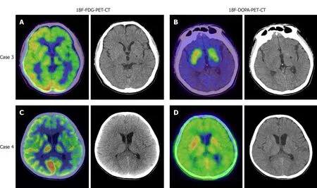
Magnetic resonance imaging(MRI)of the brain revealed local lesions in unilateral basal ganglia region in patients 1,2,and 3.Brain MRI revealed subtle and ill-defined lesions in bilateral basal ganglia and thalamus in patient 4.The characteristics of these brain lesions are shown in Table 2 and Figure 1.In addition,patient 3 and 4 underwent both 18F-fluorodeoxyglucose-positron emission tomography(18F-FDG-PET)and 18Ffluorodopa-positron emission tomography(18F-DOPA-PET).In patient 3,18F-FDGPET revealed diffuse low metabolism in the left cerebral cortex,basal ganglia and thalamus(Figure 2A).18F-DOPA-PET showed slightly low metabolism in the left basal ganglia(Figure 2B).In patient 4,18F-FDG-PET demonstrated low metabolism in the left hemisphere and left cerebral peduncle(Figure 2C).18F-DOPA-PET showed normal metabolism(Figure 2D).
FINAL DIAGNOSIS
All patients were diagnosed with germinomas of the basal ganglia and thalamus by stereotactic brain biopsy.Histopathological diagnoses were further confirmed by another hospital.
TREATMENT
All four patients received whole brain radiotherapy and chemotherapy at another hospital.
OUTCOME AND FOLLOW-UP
The brain lesions on MRI were reduced or disappeared and their symptoms remained stable without aggravation.
DISCUSSION
Germinomas in the basal ganglia and thalamus show a male predominance[3,5,6,9-11].The reason for this is unclear.They usually occur in young adolescents aged from 10 to 19 years.This may be correlated to gonad development in this age group[3].Basal ganglia germinomas usually have an insidious onset and slow progression.The clinical presentation of these tumors depends on their localization.The most common symptoms are progressive hemiparesis,mental status change and cognitive decline.In this report,patient 1 developed paroxysmal paresthesia which is very rare.The other 3 patients had hemiplegia.Patient 4 developed cognitive decline,diabetes insipidus,precocious puberty in addition to hemiplegia.Although patients 3 and 4 had longer duration than patients 1 and 2,the brain lesions shown by MRI were much smaller and more ill-defined.Hence,the size of the lesion did not correspond to the duration and severity of the clinical presentation.Symptoms and signs are valuable for localization and contribute to the identification of subtle lesions on brain MRI.
Tumor markers of pure germinomas including AFP and β-HCG were negative in these patients[3-6].β-HCG levels were slightly elevated in the body fluid of some patients which indicated syncytiotrophoblastic giant cells in the germinoma.Germinomas with elevated β-HCG in serum but not in CSF,might be associated with a poor outcome[7,8].It was reported that intracranial germinomas with serum β-HCG levels higher than 15 mIU/mL had a high recurrence rate[7].However,all the cases in that study were midline germinomas including those in the pineal region and suprasellar region or both sites.Further studies are needed to evaluate the prognosisof basal ganglia germinomas with elevated β-HCG.All our cases were AFP and β-HCG negative except patient 3 who had a slightly elevated level of β-HCG in CSF.Although these tumor markers are usually negative in germinomas,they serve to differentiate germinomas from other germ cell tumors.

Table 1 Patients’ characteristics

Table 2 Neuroimaging findings before stereotactic brain biopsy
According to the literature,typical brain MRI signs of basal ganglia germinomas are usually cystic formation,amorphous calcification,focal hemorrhage,peritumoral edema,contrast enhancement and ipsilateral cerebral and brain stem hemiatrophy[12-14].None of our four cases presented all of the above typical neuroimaging characteristics.One patient had calcification,two patients exhibited cystic formation,and three patients had contrast enhancement.All these patients developed ipsilateral hemisphere atrophy especially in the cerebral peduncle.None had intratumoral hemorrhage.Although the MR images of case 1 shared overlapping features with craniopharyngioma,the location of the tumor was useful in differentiating it from craniopharyngioma.In cases 2-4,it was easy to miss the lesions on brain MRI.The most atypical case was patient 4 who showed bilateral involvement and did not present with the above signs except bilateral hemisphere atrophy.As shown in the literature,ipsilateral hemiatrophy is the predominant feature of basal ganglia germinoma[3,15].Wallerian degeneration of the conduction tract is hypothesized to be the etiology of hemiatrophy[15].In our report,patients 1 and 3 underwent diffusion tensor imaging examination which further supported this hypothesis.Why only germinomas rather than other brain tumors lead to ipsilateral hemiatrophy requires further investigation.Previous reports revealed that susceptibility weighted imaging(SWI)might be more sensitive in detecting early basal ganglia germinoma than conventional MRI,and MR spectroscopy(MRS)was helpful for monitoring the effects of treatment[6,16].Patient 1 had similar SWI and MRS findings to those in the literature.A preoperative computed tomography scan was helpful in evaluating hemorrhage and calcification.Patients 3 and 4 underwent both 18F-FDG-PET and 18FDOPA-PET.It seems that both these techniques had limited use for germinoma.Further studies are needed to confirm the value of PET examination in the diagnosis of germinoma.The reason why patient 4 had the longest clinical course before definite diagnosis was the limited findings on radiological examination.

Figure 1 Appearance of germinomas on conventional magnetic resonance images.A-D:Case 1.A round space occupying lesion in the left basal ganglia and thalamus was hypointense on T1 and hyperintense on T2/T2-fluid-attenuated inversion recovery(FLAIR)with annular enhancement around the cystic component.Mild ipsilateral hemiatrophy appeared;E-H:Case 2.An irregular lesion in the right basal ganglia was slightly hypointense on T1 and isointense to hyperintense on T2/T2-FLAIR with mild heterogeneous enhancement and ipsilateral hemiatrophy;I-L:Case 3.An ill-defined lesion was hypointense on T1 and hyperintense on T2/T2-FLAIR in the left basal ganglia beside malacia foci.The left hemisphere showed mild atrophy.Heterogeneous enhancement was shown after gadolinium administration;M-P:Case 4.The subtle lesions were isointense on both T1 and T2/T2-FLAIR around bilateral internal capsule and thalamus.Bilateral cerebral atrophy was revealed which was predominant on the left side.No enhancement was found.T1+C:Contrast-enhanced T1-weighted imaging;FLAIR:Fluidattenuated inversion recovery.
According to the above findings,germinomas originating from atypical regions are not easy to diagnosis,especially in patients with small and ill-defined brain lesions.Stereotactic biopsy was valuable for early diagnosis.Serial neuroimaging studies are needed not only for disease differentiation but also for determining the biopsy site.
CONCLUSION

Figure 2 Appearance of germinomas on positron emission tomography-computed tomography.A:18F-fluorodeoxyglucose-positron emission tomography-computed tomography(18F-FDG-PET-CT)detected diffuse low FDG uptake in the left hemisphere in case 3;B:18F-fluorodopa-positron emission tomography-computed tomography(18F-DOPA-PET-CT)detected low uptake of DOPA in the left basal ganglia in case 3;C:18F-FDG-PET demonstrated low uptake in the left hemisphere in case 4;D:18F-DOPA-PET-CT showed normal metabolism in case 4.18F-FDG-PET-CT:18F-fluorodeoxyglucose-positron emission tomography-computed tomography;18F-DOPA-PET-CT:18F-fluorodopa-positron emission tomography-computed tomography.
The diagnosis of germinomas in the basal ganglia and thalamus is often delayed due to the absence of elevated tumor markers,and atypical clinical symptoms and neuroimaging features.The association of a focal lesion in the basal ganglia or thalamus of children with progressive hemiparesis,neuroendocrine and neuropsychiatric symptoms and ipsilateral hemiatrophy could prompt the diagnosis of ectopic germinoma.Histological examinations are necessary to confirm the diagnosis of an atypical lesion.Serial neuroimaging studies not only differentiate diseases but also determine the biopsy site.
杂志排行
World Journal of Clinical Cases的其它文章
- Role of monoclonal antibody drugs in the treatment of COVID-19
- Review of simulation model for education of point-of-care ultrasound using easy-to-make tools
- Liver injury in COVID-19:A minireview
- Transanal minimally invasive surgery vs endoscopic mucosal resection for rectal benign tumors and rectal carcinoids:A retrospective analysis
- Impact of mTOR gene polymorphisms and gene-tea interaction on susceptibility to tuberculosis
- Establishment and validation of a nomogram to predict the risk of ovarian metastasis in gastric cancer:Based on a large cohort
