Residual impact of 17α-methyltestosterone and histopathological changes in sexreversed Nile tilapia (Oreochromis niloticus)
2020-02-20DewiNurmalitaSusenoEpyMuhammadLuqmanMirniLamidAkhmadTaufiqMuktiMuhammadAgusSuprayudi
Dewi Nurmalita Suseno, Epy Muhammad Luqman, Mirni Lamid, Akhmad Taufiq Mukti, Muhammad Agus Suprayudi
1Study Programme of Biotechnology of Fisheries and Marine, Universitas Airlangga, Surabaya, Indonesia
2Department of Veterinary Anatomy, Faculty of Veterinary Medicine, Universitas Airlangga, Surabaya, Indonesia
3Department of Feed and Nutrition, Faculty of Veterinary Medicine, Universitas Airlangga, Surabaya, Indonesia
4Department of Fish Health Management and Aquaculture, Faculty of Fisheries and Marine, Universitas Airlangga, Surabaya, Indonesia
5Department of Aquaculture, Bogor Agricultural University (IPB), Bogor, Indonesia
ABSTRACT Objective: To examine sex reversal both by oral and by immersion using 17α-methyltestosterone on the methyltestosterone residual concentration and the organ histopathology of tilapia fish.Methods: This study used oral and immersion treatment methods for sex reversal of tilapia fish and used normal fish as the control and each treatment was repeated 4 times. 17α-methyltestosterone at dosages of 60 mg/kg feed and 0.5 mg/L were used for oral and immersion methods, respectively. In the first step, tilapia fry were reared at 100 L aquaria, with a density of 1 fish/L for 2 months. In the next step, male tilapias were reared at happa (net cage) of (2×1×1) m3 size in the controlled pond, with a density of 30 fish/happa for 3 months. The methyltestosterone residual concentrations were analyzed by one-way analysis of variance and Duncan’s multiple range tests, while organ histopathology was analyzed by descriptive method.Results: Residual concentrations in the serum of methyltestosteronetreated fish were significantly lower than that in normal fish,especially in 4- and 5-month-old tilapias with averages of less than 5 µg/L, while in normal fish was more than 5 µg/L. In the flesh,methyltestosterone residual concentrations showed relatively no significant differences between the oral and immersion treatment groups and methyltestosterone-treated fish remained lower compared to normal fish, except in 5-month-old tilapia.Methyltestosterone-treated tilapia exhibited histopathological changes on gill, liver, kidneys, and intestine organs.Conclusions: Sex reversal either by oral or by immersion has methyltestosterone residual concentration, but does not exceed the limits (5 µg/L or 5 µg/kg) of synthetic steroid on the fish body,although methyltestosterone causes histopathological changes on gill, liver, kidneys, and intestine.
KEYWORDS: 17α-methyltestosterone; Residue; Organ histopathology; Tilapia; Sex reversal method
1.Introduction
Sex reversal both by oral and by immersion using synthetic steroids proved to be a simple, easy, and highly effective technology[1]. Androgenic anabolic steroid hormones such as 17α-methyltestosterone (17α-MT)[2,3] is a derivative of testosterone[4], which potentially increases sexual developmental in males[3]. The 17α-MT-immersed tilapia larvae produce males of 91.6%-98.3% [5,6], however, oral treatment of 60 mg/kg feed produces males of 93.7%[7], 97.7%[8], even reaches up to 100%males[9].
Synthetic steroid hormone would enter through the blood vessels in the body and then it was modulated by the brain and pituitary hormones[10]. Steroid hormone was synthesized in either the liver or the kidneys[11], and subsequently, it would produce androstenedione which consists of 17β-estradiol and testosterone. If testosterone has increased, then the gonads would be immediately addressed to the male sex, but 17α-MT has characteristics that it is difficult to be absorbed within the body and it will also contaminate the environment[12].
The utility of hormones in aquaculture production was often debated by researchers due to the potential toxicity on human health (a carcinogenic and endocrine disorder) as well as the danger to the environment[1,3,13-15]. The group of anabolic steroids (including 17α-MT) based on the decision of the Ministry of Marine Affairs and Fisheries, Republic of Indonesia(number KEP.52/MEN/2014) has been banned because of the hormones harmful to fish, environment, and human. This study expected to prove the presumption that has been the subject of debate in the fish farmer community that the use of 17α-MT at any dosage produces dangerous and toxic residues when consumed by humans and the released into the environment, as well as the debate among researchers and to address concerns that have existed in the community and policymakers that the use of 17α-MT in certain doses is still safe and does not contain dangerous residues of concern so far. So that the regulation can be revised again for the advancement of aquaculture while maintaining a sustainable environment and human health that consumes cultured fish.Therefore, the study aimed to examine sex reversal both by oral and by immersion using 17α-MT on the MT residual concentration and the organ histopathology changes of tilapia.
2. Materials and methods
2.1. Test animal
The test animal used was Nile tilapia (Oreochromis niloticus).Tilapia fry were produced by artificial fertilization and controlled incubation.
2.2. MT treatments
MT treatment by the oral method was started 3 days after hatching with using 17α-MT (Argent) dosage of 60 mg/kg feed.The oral treatment method lasted for 28 days. Immersion method using dosage of 0.5 mg/L of 17α-MT was conducted to 10-dayold Tilapia fry and repeated in 13-day-old Tilapia fry for 3 h,respectively[8]. Treatment groups (namely MT-treated fish, both by oral and by immersion) and normal fish as control were repeated 3 times, respectively with a density of 100 fish/replicate/treatment,so the total of fish, both treated and normal were 900 fish.
2.3. Fish rearing
In the initial step, fish were reared at 100 L aquaria, with a density of 1 fish/L for 2 months, separately in each treatment group. Fish was fed on commercial pellet content of 40% crude protein, 3 times daily, at satiation. Sex was determined on 2-month-old fish through manual observation of genitalia for all fish, and gonad preparation. To verify the sex from genitalia observation, gonad was obtained from 10 fish/replicate/treatment by using the squash method with acetocarmine dye according to Mukti[8]. Based on 17α-MT hormone treatment either by oral or by immersion and verify the sex by fish genetalia observation and followed by gonad preparation shows male of 97%-98% and female of 2%-3%[8].Then, male fish of 3 treatments were selected for further study.
In the next step, a total of 360 male tilapias used in this study for 3 treatments (120 fish/treatment) were reared separately at happa(net cage) of (2×1×1) m3size in the controlled pond, with the density of 30 fish per happa or replicate, respectively for 3 months.Each treatment was repeated 4 times. Fish was fed on commercial pellet content of 32% crude protein, 3 times daily, at-satiation.
2.4. Sampling
Fish sampling was done in the 3rd, 4th, and 5th months as much 3 fish/replicate/treatment, respectively for residue test. 3-month-old fish were used for histology preparation. Fish were anesthetized by using MS222 of 1 mg/L according to Gogal et al[16]. Serum (1 mL)was collected according to Atli et al[17], and flesh (10 g) of fish was collected to do testing of residues.
2.5. Measurement of MT residue
MT residue, both the serum and the flesh were measured by the sandwich enzyme-linked immunosorbent assay method using fish MT kit (cat number E0103Fi; Bioassay Technology Laboratory,Shanghai, China). Previously, the sample and the reagents were stored at a temperature of 18 ℃-25 ℃[3].
2.6. Histology preparation
Fish was carefully dissected on the abdominal part according to Wu et al[18] and gill, liver, kidneys, intestine, and gonad organs were collected and stored in the 50 mL tubes which consisted of buffer neutral formalin, with the ratio of 1:2 at room temperature.Histology processes were conducted according to the standard operational procedure, generally with slight modification[19]. The flow chart of the study was shown in Figure 1.
2.7. Statistical analysis
Data of MT residual concentrations were analyzed statistically by using analysis of variance (ANOVA) with SPSS ver.10 software.Significant ANOVA was followed by Duncan’s multiple range test,while organ histopathology was analyzed descriptively. Data were expressed as mean±standard deviation (mean±SD). P-value < 0.05 was considered as statistical difference.
2.8. Ethical approval
The study was approved by the Animal Care and Use Committee of Brawijaya University; the protocol number was 985/8.8.2017.
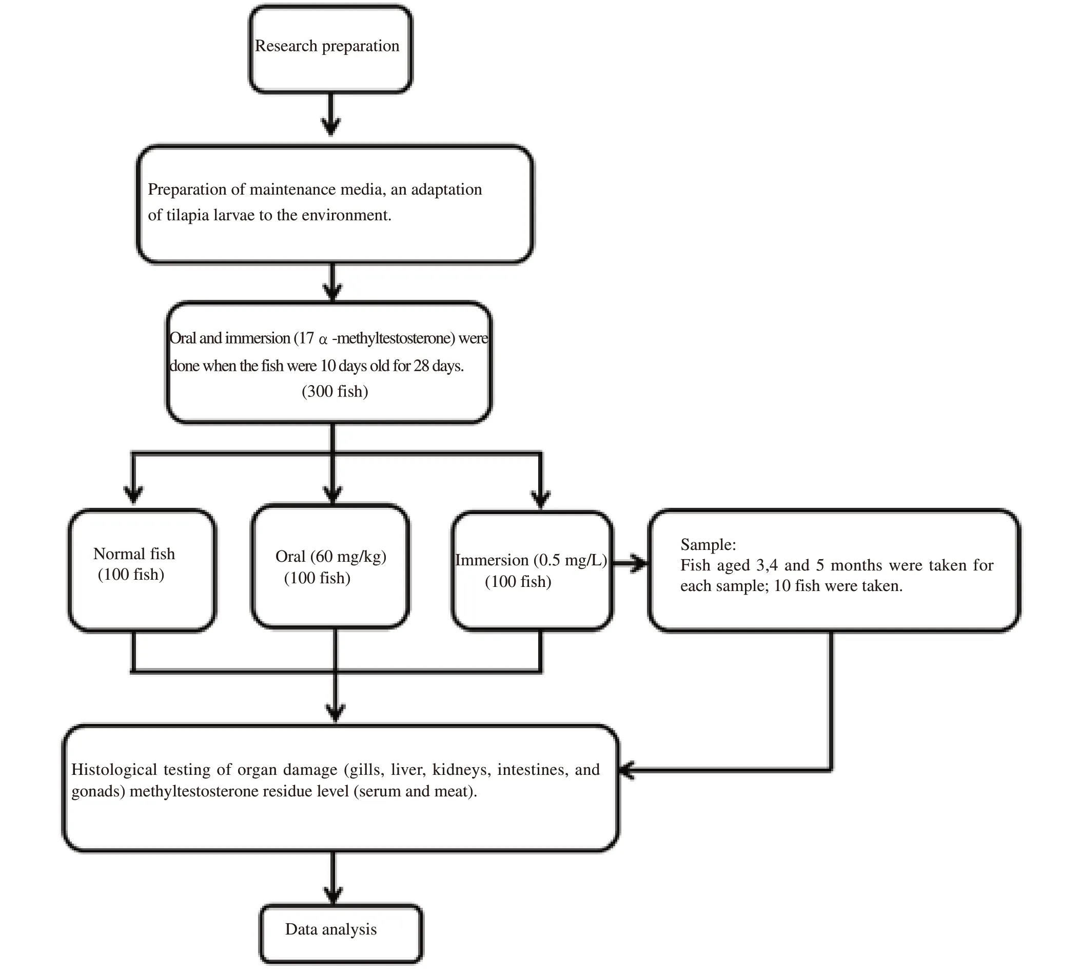
Figure 1. Flow chart of the study.
3. Results
3.1. MT residual concentrations
MT residual concentration in the serum of MT-treated male tilapia, both by oral and by immersion, was decreased on the 4th month while increased again on the 5th month, the normal male fish was increased from the 4th month while slightly decreased on the 5th month (Table 1). On the 4th and 5th months, the MT residue concentrations were lower in both oral and immersion groups comparing with that of the normal group (P both <0.05).
In the flesh, MT residual concentration showed relatively no significant difference between the oral and immersion treatment groups on 4th month, but the MT residual was significantly higher in the oral treatment group than that in the immersion group. MTtreated male tilapia remained lower than normal male tilapia in the 3rd and 4th months, except in the 5th month. However, the result showed that all males had increased MT residue in the 5th month comparing with that of the 4th month (Table 2).
3.2. Organ histopathology
MT-treated male tilapias showed histopathology changes in gill, liver, kidneys, and intestine organs (Figure 2). In the gill,such as hyperplasia was found in the bottom secondary lamella.Hypertrophy appeared on the lamella stem due to the occurrence of containment. Clubbing occurred at the end of the primary lamella, which was caused by the existence of retention, so edema appeared on the lamella (Figure 2A). The liver showed congestion, hemorrhage, and cell atrophy (Figure 2B). Congestion was redder due to contained erythrocytes. Atrophy was shown by the reduction of cell size of Kupper, which made sinusoid widen and made vacuoles degenerate. Congestion caused sinusoidal erythrocytes to wide. Degeneration of liver cells made vacuoles enlarge. Normally, the liver organ did not have damage. Kidneys seem hemorrhage, infiltration of lymphocytes, and neutrophils,inflammation, and necrosis (Figure 2C). The infiltration presence of lymphocytes and neutrophils caused inflammation. The intestine has look atrophy, intestinal villi hemorrhage, lymphoid follicles, and melanomacrophage (Figure 2D). The occurrence of hemorrhage led to the atrophy and melanomacrophage, so finally, it caused erosion and hemorrhage and necrosis of the intestinal villi.
On the other hand, testicular histology (Figure 3) used to observe spermatogenesis or testicular development and may be histopathology change in different treatment of 3-month-old fish.This study showed no difference in testicular between normal fish (Figur 3A) and MT-treated fish, both oral (Figure 3B) and immersion (Figure 3C).
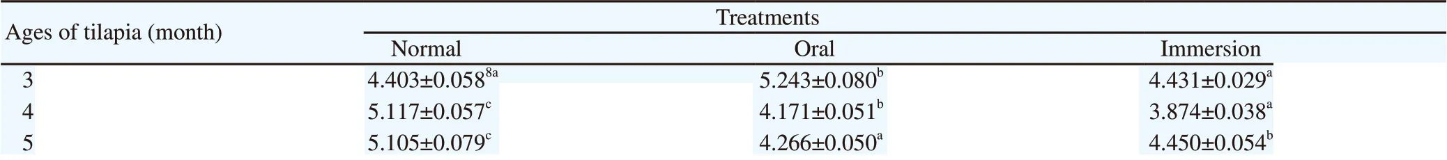
Table 1. Methyltestosterone residual concentrations (µg/L) of serum in different age of male tilapia.
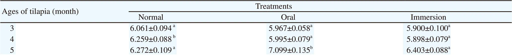
Table 2. Methyltestosterone residual concentrations (µg/kg) of the flesh in different age of male tilapia.
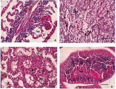
Figure 2. Organ histopathology of 3-month-old male tilapia fish (n = 3) (H & E staining, scale bar = 50 µm). (A) gill, (B) liver, (C) kidney, and (D)intestine. Note: ha = hyperplasia; hi = hypertrophy; c = clubbing; ps = bending cell; he = haemorrhage; k = congestion; dn = degeneration of nucleus; il =infiltrating lymphocytes; in = neutrophil infiltration; n = necrosis; pr = inflammation; av = intestinal villi atrophy; lf = lymphoid follicles;pr = inflammation.
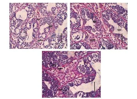
Figure 3. Testicular histology of 3-month-old male tilapia (n = 3); normal fish (A), orally methyltestosterone-treated fish (B), and methyltestosteroneimmersed fish (C), spm=spermatocytes; spt=spermatid; spz=spermatozoa. (H & E, bar scale = 50 µm).
4. Discussion
Hormonal activities are affected by three stereochemical aspects, i.e.,location of the cluster on the ring, axial and or equatorial positions,cluster, the configurationαor β, trans and/or isomer, and cyclohexane ring conformations. Testosterone is a hormone that has a short activity time due to fast absorbance in the digestive tract and rapidly undergoes hepatic degradation. It is caused by the presence of bacteria in the gastrointestinal tract that oxidizes cluster 17β-hydroxy to be inactive17β-keto. Therefore, it is necessary to add an alkyl group on 17th carbon to become C17α. This prevented the conversion of 17βhydroxy metabolism to be 17β-keto, so the 17α-MT compound has more activity in the body, but it could cause residue. The 17α-MT activity has half the strength of testosterone activity due to the length of C-chain alkyl groups, and then it would decrease androgenic activity.Otherwise, it would increase its toxicity.
The 17α-MT compounds could be transferred to live feed or water. Chemical substances had naturally incorporated into living organisms in several ways, through both the digestive and respiratory tracts[20,21]. Exposure to synthetic chemicals and their residue is a risk for human and wildlife health[3,22]. Based on serum MT concentration of males on the 3rd month, orally sex-reversed tilapia had more raising concentration than other treatments. However, in the 4th and 5th months, the MT concentration had decreased every month[3,22].The orally MT-treated fish would contain MT only in the initial 5 months[23].
MT concentration was higher in the flesh compared to in the serum. High enough MT concentration was found in the muscle and flesh[24,25], because the MT metabolite has been absorbed into the muscle and flesh of fish, thus causing the MT to accumulate in the flesh every month. In the research of Pandian and Kirankumar[25],exogenous steroid remnants of 5 µg/kg in fish were a too risk to humans. Endogenous testosterone hormone produced on the testes was 5.2 µg/kg[26], whereas tilapia had endogenous testosterone and estradiol of 3 µg/kg, respectively[25]. Normal fish have higher MT residue value than MT-treated fish as shown in this study. We suspect this is related to the reproductive cycle or period of Nile tilapia.Normally, Nile tilapia at the 4-month-old has entered the period of reproduction and spawning, so that seen an increase in hormone levels in blood serum. As is known during entering reproduction or spawning, hormone levels in the body increase and will drop back after spawning, while monosex-treated fish, although it looks the same as normal, the body's energy is preferred in increasing somatic growth compared to reproduction, so we suspect that this is one of the factors that causing male monosex-treated Nile tilapia has a larger body size than normal male tilapia.
Several limitations of this study are including: a) unable to measure specifically the MT residual concentration between introduced hormone (exogenous) and endogenously hormone by the fish, and b) unable to measure the MT residual concentration in younger fish age. Therefore, in the future, both of these limitations are our concern for further studies.
Gill layouts that were outside and directly related to water would be the first affected by the polluted water environment. The food already digested in the intestines would be circulated by blood to the liver and kidneys. The liver was the largest organ responsible for metabolism. Kidneys had functioned as a hyperosmotic regulator[27].In 3-month-old fish, the toxicity to organs is still visible.
The early stage of damage caused by gill irritation has accompanied the increasing of the mucous cells at the bottom of epithelia with causing a thickening of the secondary lamella epithelium so that the secondary lamella enlarges due to the secondary lamella attached.Gill lamella looked larger than normal which was caused by cell enlargement (hypertrophy), and it looked unclear between the primary and secondary lamellas. According to previous studies[17,28,29],hyperplasia may occur due to chemical stimuli from pollutants,environmental pollution, parasites, and bacterial infections.Contamination has characterized by a very dense accumulation of red blood cells in the blood vessels, which would block blood vessels (congestion), while edema of lamella looks like an empty white space that causes blocking. Clubbing occurred because of the thickening of epithelial tissue located near to the lamella bottom(basal hyperplasia), and then the whole room of interlamellar was filled by new cells which showed like a baseball bat[27,30].
Degeneration was the early stage of vacuole damage in the liver.Vacuole degeneration was reversible, so when exposed to toxic substances and end administration of MT, cells could be returned to normal. Necrosis could not be cured, so if it exposed the tissue activity continuously, then it would decrease cell activity, causing the cells to lose some parts even to death[31,32]. Congestion was preceded by degeneration of liver cells in which an enlarged vacuole was filled with erythrocytes that cause sinusoid to widen that accumulated blood and hemorrhage. According to the research of Robert[30],congestion occurred by the entry of toxic substances into the heart.Hemorrhage was the flow of red blood cells out of the central vein.
Sinusoidal and central venous damage occurred due to numerous blockages of blood vessels in the stomach and central intestine[33,34],which causes a greater concentration of toxic substances in this area and causes damage to the central vein. A sinusoid is a small capillary that separated the fundamental of the structural unit with tubule or trabeculae (biliary hepatocytes surrounded by central parenchyma)[33,35]. The liver had an enzyme for drug metabolism which is one of the most damaged organs but is very resistant to viral or bacterial infections and foreign substances that enter through the absorption in the intestine. It was known that nearly 80% of the liver cells were damaged. But, it was still capable of regenerating and could even be cured if the damage was lost or destroyed[34].
The infected kidneys were swelling, which was an indication of an inflammatory process that may cause necrosis[35]. Inflammation was an indication of increased lymphocytes and macrophage or neutrophil cell numbers. Kidneys were pollutant-responsive organ to indicate histopathological damage. Therefore, the kidneys were the targeted organ for the biomonitoring approach[36]. Changes that often occurred in the kidney are inflammation, necrosis, thickening of the core, hyperplasia, hypertrophy epithelial cells, hydropic vacuolation,and renal tubular regression[35-37].
The intestine damage is signed by inflammation. The inflammation or swelling of cells has a reversible characteristic that exposed to the toxic substances in a short period, the cell would return to normal,but if exposed to the toxic substances for a long time, the cell was not able to tolerate damage caused by toxin substances[38]. Melanomacrophage was caused by inflammation which was followed by erosion of the intestinal villi, hemorrhage, and atrophy leading to necrosis. Erosion and villus of the intestine with considerable damage would disturb the absorption of important substances so that that fish would suffer from malnutrition. In intestinal organs, there were cell swelling, microvillicell membrane fused, lysis, intestinal vacuum and intestinal villi erosion which suffered severe injuries to rupture caused by toxic substances[21]. Acute intestinal conditions were caused by viruses, parasites, bacteria, algae, and intestinal mucosa. Toxic chemicals could be removed by using mucous epithelial cells that coiled together with the thickening chromatin and cytoplasmic eosinophils[30]. MT concentrations of serum and flesh have not exceeded the limit (5 µg/L or 5 µg/kg) due to the estimated residual synthetic steroid in the fish body of 5 µg/kg. Influences on histopathology of gill, liver, kidneys, and intestine organs are found with varying degrees of damage because there are remaining synthetic hormones left in the body that cause organ damage. Further work is another safer natural material to replace the performance of the alkyl group as well as the histopathological figure of the 4- and 5-month-old fish to determine whether there is a recovery in the fish organ after the cessation of synthetic hormone.
Conflict of interest statement
The authors declare that there is no conflict of interest.
Authors' contributions
Dewi Nurmalita Suseno contributes to literature search, clinical and experimental studies, data analysis, and manuscript preparation.Epy Muhammad Luqman contributes to arrange the definition of intellectual content, data analysis, and manuscript review. Mirni Lamid contributes to arrange definition of intellectual content and statistical analysis. Akhmad Taufiq Mukti contributes to conceptualization, research design, arrange definition of intellectual content, data acquisition and analysis, and manuscript editing and review. Muhammad Agus Suprayudi contributes to arrange the definition of intellectual content, manuscript review, and guarantor.
杂志排行

Asian Pacific Journal of Reproduction的其它文章
- Testosterone is a surrogate and proxy biomarker for severity of late-onset preeclampsia: A cross-sectional study
- Assessment of antioxidant status of women with polycystic ovarian syndrome
- Novel genetic variants of transferrin receptor 2 exon 4 and cytokines profile of anemic and nonanemic pregnant women in Central Java, Indonesia
- Comparative proteomic analysis of mature and immature oocytes in domestic cats
- Estrogenic activity of hydroalcoholic extract of Clitoria ternatea Linn. leaves on rats
- Identification of pathogenic microorganisms of repeat breeder dairy cows and a hyperimmune treatment approach