The difference ofintraoperative free-run electromyography monitoring between percutaneous endoscopic lumbar discectomy via a transforaminal and via an interlaminal
2020-01-13JunlchiroNakamuraTomoyukiSetoueJunHara
Jun-lchiro Nakamura,Tomoyuki Setoue,Jun Hara
Orthopedic surgery,Kawasaki-saiwai hospital,Kawasaki city,Kanagawa-ken 212-0014,Japan.
Abstract
Keywords:Free-run electromyography,general anesthesia,iatrogenic nerve injury,percutaneous endoscopic lumbar discectomy
INTRODUCTION
Transforaminal percutaneous endoscopic lumbar discectomy(TF-PELD)is usually performed on an outpatient basis under local anesthesia(LA).The procedure is neurologically safe and avoids injury to the exiting nerve root(ENR)since the patient is conscious and able to notify the surgeon of any pain[1-6].
However,some patients cannot tolerate intraoperative pain and may acquire intravenous analgesia,sedative agents,or conversion to epidural or general anesthesia(GA)on another day.
PELD under GA is considered neurologically safe since the patient is unconscious.Recently,the efficacy of free-run electromyography(f-EMG)has been reported in neurosurgery and spine surgery[7-12],but not for PELD.Therefore,the aim of this study was to evaluate the efficacy of f-EMG monitoring during PELD under GA.
METHODS
Anesthesia
Propofol and remifentanil hydrochloride-based GA were administered.A muscle relaxant(rocuronium bromide)was only used at the initial stage for intubation,and sugammadex sodium was used for reversal of neuromuscular blockade.The stimulation pattern(i.e.,train-of-four)was confirmed as more than 90%before starting of f-EMG monitoring.
F-EMG
Continuous f-EMG activity was recorded using a NVM5® monitoring system(NuVasive Inc.,San Diego,CA,USA)with surface electrode patches placed at 6 positions(i.e.,the bilateral vastus mediaris,tibialis anterior,and gastrocnemius).Before affixing the patches,the skin was scraped with sandpaper to ensure impedance was less than 10 kΩ.The threshold of electrical impact was set at 80 μV.All instances of f-EMG activity were immediately reported to the surgeon.
Neurotonic,burst,and train waves that occurred in response to a surgical maneuver to the affected nerve were considered to be abnormal.A small amplitude,low frequency,and isolated discharge were not considered pathologic[10].Any of these instances automatically sounded an alarm to notify the surgeon.
Surgical procedure
TF-PELD
With the patient in the prone position on a radiolucent table,an 8-mm incision was made to the entry points of the skin at about 9-11 cm lateral to the midline and then an 18-gauge needle was inserted into the annulus fibrosus.Discography with 2 mL of liquid indigo carmine contrast agent(1:1)was performed using the same needle.A guide wire and sleeve were inserted for endoscopy with a VERTEBRIS lumbarthoracic® instrument(Richard Wolf GmbH,Knittlingen,Germany).A bipolar radio-frequency electrode system(Elliquence,Baldwin,NY,USA)was used for hemostasis and a high-speed Primado 2® drill with a super slim hand piece 200®(NSK-Nakanishi medical,Tochigi,Japan)was used for foraminoplasty.
IL-PELD
With the patient in the prone position on a radiolucent table,an 8-mm incision was made to the entry points of the skin at about 1 cm lateral to the midline.Once the entry point and the direction of the sleeve were confirmed by image intensifier,a 8-mmsleeve was inserted.A VERTEBRIS lumbar-thoracic®instrument was used for endoscopy and a bipolar radio-frequency electrode system was also used for hemostasis.The ligamentum flavum was resected piece-by-piece using a micro punch.A high-speed Primado 2® drill was used for laminotomy and medial facetectomy.Partial laminotomy was necessary in almost all cases,particularly at L4-L5,to remove the laminae and ligamentum flavum and confirm the lateral edge of the nerve root.Then,after the sleeve was rotated and thecal sac was retracted,the herniated disc was removed.

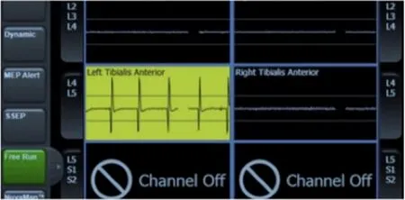
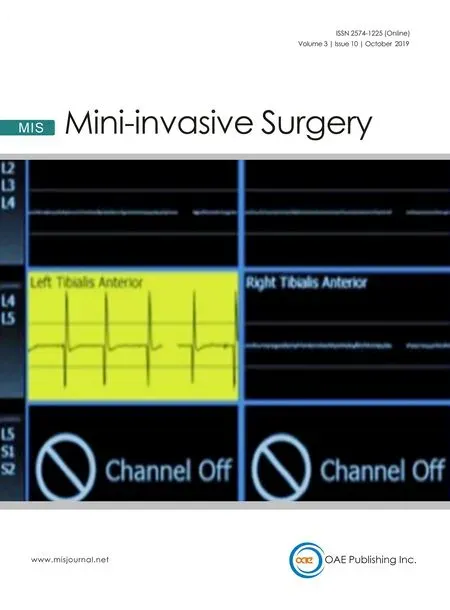
Figure 1.Train wave of free-run electromyogram.This wave was observed in TF-PELD at L4-L5 case when the sleeve was manipulated hand-down.Same waves were also observed in IL-PELD at L5-S1.TF-PELD:trans for aminal percutaneous endoscopic lumbar discectomy;IL:interlaminar
Statistical analysis
Descriptive analysis of group characteristics was performed using JMP version 11.2 software for Macintosh(SAS Institute Inc.,Cary,NC,USA).The independent two-samplet-test and Wilcoxon test were used to compare the clinical outcomes.A probability value ofP<0.05 was considered to be statistically significant.
RESULTS
There were 49 patients(43 men,6 women)with a mean age of 52.9(range,17-88)years.The mean followup period was 10(range,6-14)months.The affected level was L2-L3 in one patient(TF-PELD),L3-L4 in 9(TF-PELD),L4-L5 in 17[TF-PELD,14;interlaminar(IL)-PELD,3],and L5-S1 in 22(TF-PELD,3;ILPELD,19)patients.
The mean operative time was 63 ± 29(range,30-143)min and intraoperative blood loss was negligible in all cases.The mean hospital stay was 3.2 ± 1.5(range,1-5)days.The mean numerical rating scale score for the affected leg improved significantly from 7.7 to 1.1 at follow-up,and the mean Oswestry disability index had improved from 62.3 to 20.5.Two patients experienced recurrence of the herniated nucleus pulposus.
In all cases,single waves were observed but were not considered to be clinically significant.A truepositive was observed in one case during TF-PELD at L4-L5[Figure 1].When the sleeve was manipulated downward by hand,an alarm sounded.Careful performance of the procedure will prevent ringing of the alarm,which indicates a postoperative motor deficit.However,this patient complained of dysesthesia for 3 weeks postoperatively.The numbness gradually improved with the use of pregabalin and disappeared by the final follow-up.
False-positives were observed in 2 patients following IL-PELD at L5-S1.No alarm sound was observed when the nerve root was retracted by rotating the sleeve.Train waves appeared with the alarm several seconds after the herniated disc material was removed.At that time,no surgical maneuver was performed.In these 2 cases,no neurological deficit was observed after surgery.Including these 2 cases,no alarm sounded during IL-PELD when the nerve root and dura were retracted.
A false-negative was observed in one patient following IL-PELD at L5-S1,but no abnormal wave was observed.However,this patient complained of severe numbness for 6 months postoperatively.
The sensitivity of f-EMG for neurological deficits was 50% in all cases,100% for TF-PELD,and 0% for ILPELD.The specificity was 95.8%,100%,and 90.4%,respectively.
DISCUSSION
F-EMG's effectiveness has been reported for cranial nerve tumor resection and spinal surgery[7-11].Any irritation to the nerve,by stretching or compression,causes trains of motor unit potential discharge in the corresponding muscle[7].F-EMG is reported to have high sensitivity and relatively low specificity[8].However,monitoring in real-time would improve specificity when confirming that the related nerve was correctly manipulated during surgery[7].Therefore,application of f-EMG in PELD surgery is considered to be efficient.
One of the most important complications in TF-PELD is ENR injury,which reportedly occurs at a relatively high rate of 2%-8.9% under LA[3,4,13-16].A large LA dosage may increase the risk of ENR injury because of complete nerve blockage.Furthermore,some patients cannot tolerate surgery because of pain and,thus,had to be converted to GA on another day.Therefore,we believe that PELD under LA is not necessarily safe or comfortable for the patient.
In this study,there was one true-positive case with detectable nerve irritation during surgery.This patient complained of severe numbness without motor deficit after surgery.Train waves appeared when the sleeve was manipulated by hand.Hence,caution is required during surgery to avoid motor deficits.
Conversely,we carefully monitored f-EMG during IL-PELD while rotating the sleeve when retracting the dural sac and nerve root.No alarm sounded when performing IL-PELD.However,2 false-positives were observed during IL-PELD.An alarm sounded after herniated disc material was removed.F-EMG sensitivity during IL-PELD was low.Hence,f-EMG may be inappropriate for monitoring the nerve root during ILPELD.
F-EMG monitoring cannot detect damage to sensory nerves,but it can potentially prevent injury to motor and sensory fibers.PELD under GA,without the use of a muscle relaxant with f-EMG monitoring,can reduce neural injury.
This study was limited by the small number of patients.Therefore,future studies with larger numbers of patients will be necessary to evaluate the efficacy of f-EMG for PELD.
In conclusion,PELD was performed safely under GA using f-EMG monitoring.Patients who cannot tolerate pain are good candidates for PELD under GA.
DECLARATIONS
Acknowledgments
The authors would like to thank Enago(www.enago.jp)for the English language review.
Authors' contributions
Wrote and reviewed the manuscript:Nakamura JI,Setoue T,Hara J
Availability of data and materials
Not applicable.
Financial support and sponsorship
None.
Conflicts ofinterest
All authors declared that there are no conf l icts ofinterest.
Ethical approval and consent to participate
Not applicable.
Consent for publication
Not applicable.
Copyright
© The Author(s)2019.
