Correlation between C-reactive Protein and Morphology of Aortic Intramural Hematoma on CT Angiography
2020-01-10XinghuaZhangTaoLiLiYangXinJinJianWuRuipingChangJingZhang
Xinghua Zhang,Tao Li,Li Yang,Xin Jin,Jian Wu,Ruiping Chang,Jing Zhang
Department of Radiology,the First Medical Center,Chinese PLA General Hospital,Beijing 100853,China
Key words:CT angiography;C-reactive protein;intramural hematoma;acute aortic syndrome;morphology
Objectives To investigate the morphologic characteristics of intramural hematoma (IMH) on CT angiography(CTA),and evaluate the possible correlation of serum C-reactive protein (CRP) with morphologic characteristics of IMH.Material and Methods Forty-two patients who were initially diagnosed as IMH by aortic CTA and also had serum CRP examination on the same day of CTA were enrolled in this retrospective study,including 30 males and 12 females,with the mean age of 61 ± 14 years old.The volumetric CT data were retrospectively processed and analyzed on post-processing workstation.Based on the thickness of IMH and the length-area curve,the crosssectional area of true lumen and total vessel were measured,the hematoma-vessel ratio (HVR) was calculated.Imaging characteristics were compared between patients who had pathological elevated CRP (> 0.8 mg/dl)and those did not.Spearman correlation analyses of CRP level and morphological characteristics of IMH were performed,and the receiver operating characteristic (ROC) curve was used to evaluate the diagnostic validity of CRP.Results Of all 42 IMH patients,the mean serum CRP was 3.94 ± 4.71 mg/dl,and the mean HVR was 46.7%±14.2%.HVR in patients with elevated CRP was significantly higher than those with normal CRP (49.7% ±15.0% vs.40.7% ± 10.5 %,P=0.030).HVR was mildly correlated with CRP in all patients (r =0.48,P < 0.001).CRP levels differed neither between patients with Stanford type A and B (P=0.207),nor between patients with and without intimal disruption (P=0.230).To discriminate HVR > 47% (the mean value),the area under curve(AUC) were 0.700 (95% CI:0.535-0.865) for CRP at a cutoff point of 3.55 mg/dl,with a sensitivity of 54.5%and a specificity of 90.0%.Conclusion CRP was mildly correlated with the severity of cross-sectional hematoma area of IMH,but not with Stanford types and the presence of intimal disruption.
AORTIC intramural hematoma (IMH) is a rare subtype of acute aortic syndrome (AAS),accounting for 5%-10% of AAS based on serial studies in the International Registry of Aortic Dissection (IRAD).1-3Compared to the classical acute aortic dissection (AD),IMH shares similar clinical presentation and in-hospital mortality,but is more frequently to develop complications like pericardial effusions and periaortic hematoma.2Rapture of the vasa vasorum is considered the inciting event of IMH,causing bleeding into the aortic media.Previous study18reported the F-fluorodeoxyglucose (FDG) uptake of aortic wall in a portion of IMH patients,reflecting the activation of inflammation in the process of IMH.1,2Kitalet al.suggested that sustained elevated C-reactive protein (CRP),an inflammatory marker,might be associated with adverse events in patients with IMH.3However,the mechanism remains unclear.To our knowledge,little literature has investigated the correlation between inflammatory marker and morphology of IMH.
CT is the gold standard for diagnosis of IMH,especially in emergent scenario.4At unenhanced CT,IMH is characterized by crescent or circular hyperdensity in the aortic wall with occasionally internal displacement of intimal calcification.1,5,6At contrast enhanced CT,IMH is confirmed by the absence of an intimal flap and direct flow communication between the true lumen and hematoma area.5,6Furthermore,intimal disruption or ulcer-like projection (ULP) on CT is associated with progress of IMH.6-8The development of 3-dimensional post-processing software allows quantitative measurement of the whole aorta.9The purpose of this retrospective study was to evaluate the possible correlation between serum CRP and morphologic characteristics of IMH by a semi-quantitative analysis method on thoracic-abdominal CT angiography (CTA).
PATIENTS AND METHODS
Patient selection
This retrospective study was approved by the institutional review board,and the informed consent was waived.By searching the hospital information system,123 patients who were initially diagnosed as IMH by CTA between August 2015 and November 2017 were abtained.We retrospectively reviewed the original images and clinical data, excluded 13 patients with complication of aortic dissection,36 patients whose CT was actually a follow-up examination since onset,and 32 patients who did not have serum CRP results,or CRP examination taken after CTA over 24 hours.The threshold for pathological elevated CRP was defined as >0.8 mg/dl according to the criterion of our clinical laboratory.
CT angiography scan
All examinations were performed on 256-slice CT scanners (Brilliance iCT,Philips Medical,Cleveland,USA)with bolus tracking mode.After plain scan,60 ml-80 ml (1-1.5 ml/kg) contrast media (Ultravist 370,Shering AG,Guangzhou,China) was injected intravenously at the rate of 3.5-4.5 ml/s (usually 4 ml/s,decreased in poor venous condition),followed by a 40 ml saline bolus.The scanning parameters depend on body mass index (BMI≥24,tube voltage 120 kVp;BMI<24,tube voltage 100 kVp) and findings on plain scan.If hyperdensity confined in the descending aorta without suspected signs of involving the root,ascending segment and arch of aorta,we used non-ECG-gated scan;if hyperdensity involved the root,ascending segment and arch of aorta,or had undetermined range,we used retrospective ECG-gated scan.The Pitch and tube rotation time were 0.914 and 0.4 s respectively in non-ECG-gated scans,and 0.3 and 0.33 s respectively in retrospective ECG-gated scans.Other parameters included slice thickness 1 mm,increment 0.5 mm and matrix 512×512.Tube current modulation and iterative reconstruction were applied to all scans.Angiographic scan was triggered by the enhancement of the region of interest (ROI) reaching 150HU at the thoracoabdominal aorta junction with a delay of 5 s.
Image analyses
The volumetric CT data sets were processed on a workstation (Extended Brilliance Workspace,version V4.5.5.51035,Philips Medical,Cleveland,USA).The CT attenuation values of IMH were measured on unenhanced axial images with ROI ≥ 4 mm2,avoiding the area of aortic wall,plaque and artifacts.The thin slice angiographic images were imported into Advanced Vessel Analysis (AVA) module,a 3-dimensional post-processing software for vessels.After bone removal,seed points were displaced from aortic root to bifurcation of iliac arteries,covering the whole aorta.Manual adjustments of the center line and ROIs on axial images were applied to make sure the true lumen was appropriately selected without unnecessary aortic branches and artifact.
Morphologic characteristics of IMH were measured on curved planar reconstruction (CPR) images,including length,proximal reference area and distal reference area (Figure 1).The whole aorta was divided as (1) ascending aorta,(2) aortic arch,(3)descending thoracic aorta,(4) abdominal aorta superior to orifice of right renal artery,and (5) abdominal aorta inferior to orifice of right renal artery.The most severe segment was chosen by the maximum thickness of IMH on CPR images,and then the most severe cross-sectional slice was decided by the minimum area of true lumen according to the minimal point of Volume-Length curve.The most severe true lumen area and total vessel area were measured on the most severe cross-sectional slice with manual adjusted ROI.All measurements were completed by two observers with over 5 years of experience in CTA reconstruction independently [Z.XH.(reader 1);J.X.(reader 2)],and the average of two values was used.We calculated the hematoma area,hematoma-vessel ratio (HVR) and volume index using following formulas:
Hematoma area=total vessel area -true lumen area
Hematoma-vessel ratio (HVR)=hematoma area/total vessel area
Volume index=hematoma area × length/ 1000
IMH was diagnosed according to the criteria described above by two independent observers who were both state-certified radiologists [L.T.(reader 3);W.J.(reader 4)]with over 10 years of experience in CTA.The Stanford classification categorizes IMH by involvement of segments proximal to the left subclavian artery.Type A affects ascending aorta and aortic arch,while Type B does not.Intimal disruption was defined as a localized blood-filled pouch protruding into the thrombosed false lumen.3,6Localized contrast pooling in aortic wall,an imaging finding of pseudoaneurysm which was frequently connected to true lumen with very small orifice,was carefully differentiate from intimal disruption.7,10When two assessments differed,a consensus was made by a superior radiologist [Y.L.(reader 5)]with over 20 years of experience in CTA.
Statistical analysis
Categorical variable was expressed as counts and percentages,and continuous variable as mean and standard deviation.After testing normality,the student'sttest or non-parameter test was used for continuous variables,and the correlation between morphologic characteristics and CRP levels were assessed by Pearson or Spearman correlation.The receiver operating characteristic (ROC) curve was drawn to evaluate the diagnostic validity of CRP levels,and the area under the curve (AUC),sensitivity,and specificity were calculated.The optimal cutoff point was calculated according to the maximum of Youden index.Statistical analyses were performed using SPSS software (version 21.0,IBM,Armonk,NY).Pvalues less than 0.05 were considered statistically significant.
RESULTS
There were 42 eligible patients included in this study.The Demographics of this study cohort were shown in Table 1.The mean age was 61 ± 14 (32-91)years old,and 71.4% patients were male.The most common symptom was chest and/or abdominal pain(39/41,95.1%).History of hypertension was found in 70.7% (29/41) patients.CTA images of 34 patients were obtained within 24 hours from onset,while in 7 of 8 patients,CT images were obtained in 3 to 17(10.6±4.8) days after the onset (1 unknown).The involvement of ascending aorta was detected in 11.9%(5/42) patients,and the intimal disruption was detected in 33.3% (14/42) of all patients and in 26.5% (9/34)of patients whose onset was within 24 hours.
Of 42 patients with IMH,the mean CRP level was 3.94 ± 4.71 (0-16.6) mg/dl,while in 66.7% (28/42) of these patients,the CRP level elevated (>0.8 mg/dl).There was no significant difference in CRP between IMH Stanford type A and B (Z=-1.263,P=0.207),and between IMH patients with and without intimal disruption (Z=-1.201,P=0.230).
In measurements of 42 IMHs,the mean CT attenuation value was 68.9 ± 12.6 HU,the mean length was 24.1 ± 10.0 cm,the mean true lumen area was 455.8 ±290.6 mm2,the mean hematoma area was 424.8±427.3 mm2,the mean volume index was 101.4 ± 94.1,and the mean HVR was 46.7 % ± 14.2%.

Figure 1.Image analysis and measurement of morphologic characteristics of intramural hematoma (IMH) in a 59-year-old man who complained about chest and back pain for 5 hours.(A) Plain scan CT image shows crescent hyperdensity (white arrow,CT value:55.2 HU) in descending aorta.(B) (C) Oblique and coronal maximum intensity projection (MIP) images revealed Stanford type B IMH,involving from descending aorta to abdominal aorta inferior to orifcie of right renal artery.(D) After adjustment of center line,the area-length curve (E,upper image) shows notches along the course of IMH,and the most severe cross-sectional slice is located at the descending aorta by the thickness of IMH (E,lower image) according to the notch of the curve.(F) Axial image shows a manually drawn ROI covering the whole cross-section of aorta.This patient received thoracic endovascular aortic repair.
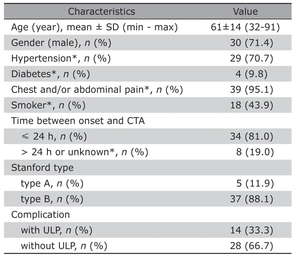
Table 1.Demographic and clinical information of enrolled IMH patients in this study (n=42)
Correlation analyses of HVR with CRP were performed with Spearman method and the result was presented in Table 2.The CRP level was mildly correlated with both HVR and hematoma area in all included patients (r=0.48,P<0.001;r=0.35,P=0.03,respectively),and also in patients whose CTA was taken ≤24 h after onset (r=0.47,P=0.005;r=0.36,P=0.04,respectively) (Figure 2).The HVR of patients with elevated CRP was significantly higher than patients with normal CRP [49.7%±15.0%(n=28)vs.40.7%±10.5% (n=14),t=-2.259,P=0.03].
In all IMH patients of this study cohort,to discriminate HVR >47% (the mean value of HVR),the area under curve (AUC) was 0.700 for CRP (95%CI:0.535-0.865),and the optimal cutoff point was 3.55 mg/dl,with a sensitivity of 54.5% and a specificity of 90.0% (Figure 3A).In patients who had CTA within 24 h of onset,to discriminate HVR>47%,the AUC was 0.706 for CRP (95%CI:0.520 -0.892),and the optimal cutoff point was 3.55 mg/dl,with a sensitivity of47.1% and a specificity of 100.0% (Figure 3B).
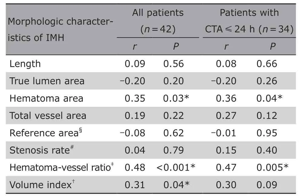
Table 2.Spearman correlation analyses between morphologic characteristics of IMH and CRP

Figure 2.Scatter diagram of CRP and hematoma-vessel ratio in 42 patients with IMH.
DISCUSSION
Our study demonstrated that CRP of patients with emergent onset of IMH is correlated with the cross-sectional HVR,a geometric character of IMH,in the most severe segment of aorta.Aortic geometry is considered as an important determinant for predicting AAS.9,11,12In previous studies,the correlation of hematoma thickness with aortic events in the progress of IMH had been discussed.13-16In the current study,we found that high CRP level was associated with high HVR,which may represent the prominent intimal separation and the increased aortic wall stress prone to aortic events.Our results suggest a potential mechanism of CPR in predicting prognosis of IMH.
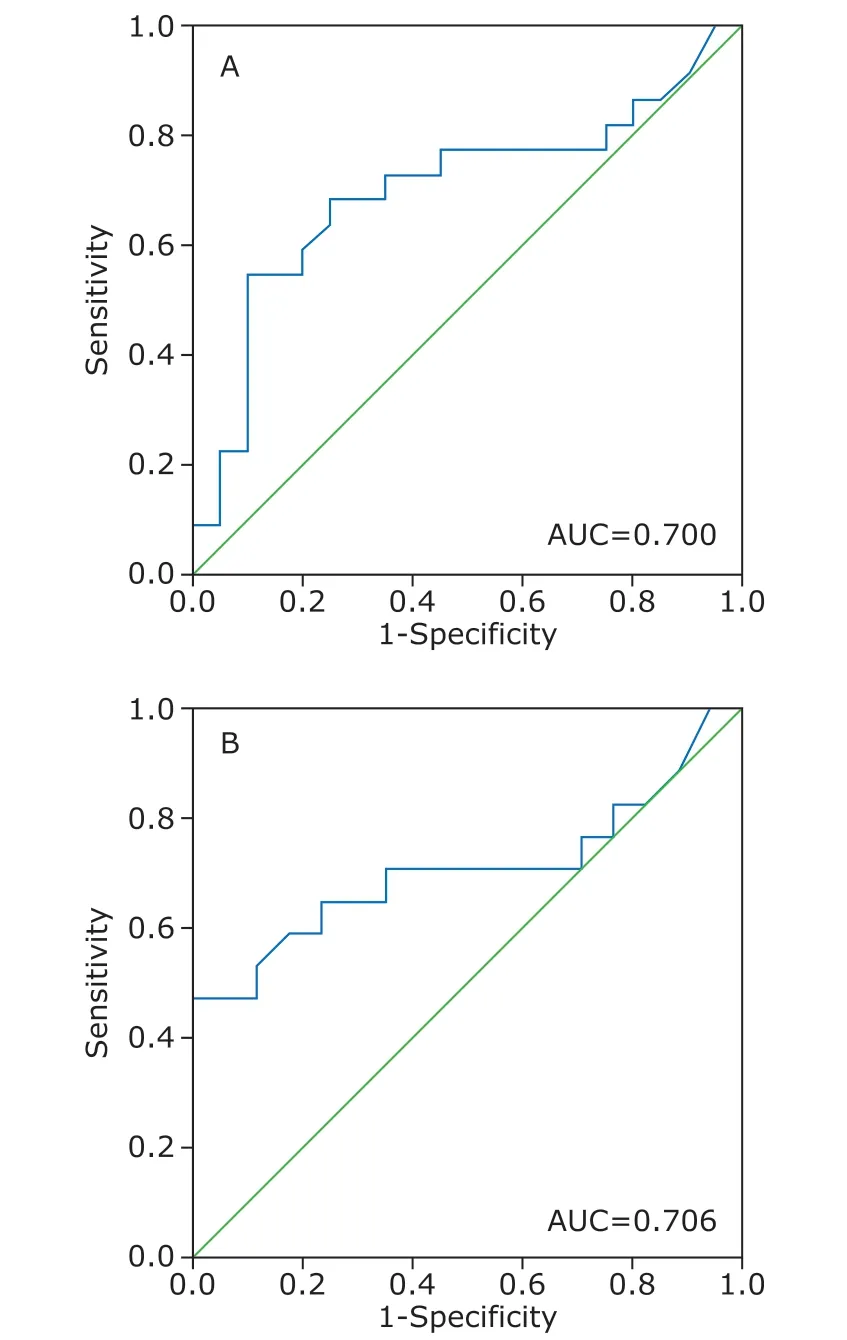
Figure 3.The ROC curves of CRP in discriminating HVR >47%.Diagonal line represents AUC of 0.50.A.in all patients (n=42);B.in patients with CTA taken within 24 h after onset (n=34).
CRP is an acute phase reactant plasma protein which increases during inflammation and after tissue damage.Elevated CRP levels detected in patients with acute aortic aneurysm or dissection was considered to be associated with poor prognosis,18,19and sustained elevation or re-elevated CRP level was considered as an independent factor of aortic events in medically treated IMH patients.3,20In our study,pathological elevated CRP was detected in 66.7% IMH patients,confirming the inflammatory activation in the process of IMH,and indicating anti-inflammatory therapy may be a choice for patients with IMH.
IMH was firstly described as“dissections without intimal tears”in 1920s.13However,recent studies have reported cases of IMH with intimal disruption,also referred to as“ULP”,“localized intimal defect”or“focal intimal disruption”,which conflicts with the conventional definition.6-8,21ULP is another independent risk factor of aortic events in patients with IMH.Moralet al.suggested that intimal disruption in acute phase (within 2 weeks) of type B IMH was associated with a poor prognosis,while chronic intimal disruption was not.8Some authors believe that IMH should be referred to as an acute arotic dissection with closed and thrombosed false lumen due to the high incidence of intimal disruption at the early stage.7,21,22Comparable with previous studies,intimal disruption was observed in 26.5%(9/34) IMH patients within 1 day after disease onset in our study.7,22Furthermore,our results suggested CRP levels did not differ in patients with and without intimal disruption,indicating the heterogeneous etiology that inflammation and intimal disruption may reflect different pathology of IMH and different prognosis as well.
CT has been the choice of imaging modality in acute scenario of AAS.23,24Measurements of aortic diameter are suggested to be perpendicular to the axis of blood flow,and detection of intimal disruption is recommended to be based on CT images with slice thickness ≤5 mm.7,24With computer-assisted analysis software,it is feasible to evaluate the areas of true lumen and hematoma perpendicular to the center line on CPR images,which runs along the direction of blood flow.The maximal diameter of hematoma area may vary depending on the position of the affected aorta.3HVR,we believe,is more accurate than hematoma thickness in evaluation of IMH.
Although CTA is recommended for acute aortic disease,there are still some patients in emergency room who cannot tolerate contrast-enhanced CT scan because of a variety of conditions,such as advanced age,poor venous condition,and allergy to contrast media.Serum CRP level may play an additional role in risk stratification for serial examinations.
This study has several limitations.First,although in the study design we defined the time interval between CRP and CTA examinations being within the same day,the two examinations were not performed precisely at the onset of IMH in each enrolled case.As there was a mild correlation in this study,our result should be testified with larger sample size and with more strict time control.Second,there exists inevitable sampling bias.The in-hospital mortality of enrolled patients in this study was zero,which we believe was mainly because most (88.1%) of cases were Stanford type B,a subtype with better prognosis.Third,CRP,as a nonspecific inflammatory marker,may be affected by concomitant inflammatory diseases such as pneumonia and pericardial effusion,but it was not controlled in this study.These limitations may weaken the results power of this study.
Our study revealed a possible association between CRP and the morphologic characteristics of IMH.Higher CRP indicates higher HVR,a feature reflecting the severity of IMH.The prognostic value of HVR still need be evaluated in further study.
Conflict of interest statement
All authors declared no conflict of interests.
杂志排行
Chinese Medical Sciences Journal的其它文章
- Bevacizumab Combined with Icotinib Overcomes Osimertinib Resistance in a Patient of Non-Small Cell Lung Cancer
- An Optimized Protocol of Azoxymethane-Dextran Sodium Sulfate Induced Colorectal Tumor Model in Mice
- Ontology:Footstone for Strong Artificial Intelligence
- Antagonistic Effects of N-acetylcysteine on Mitogenactivated Protein Kinase Pathway Activation,Oxidative Stress and Inflammatory Responses in Rats with PM2.5Induced Lung Injuries
- Physiological Variables Associated with the Development of Acute Mountain Sickness
- A Single-center Retrospective Cohort Study on Cesarean Section under General Anesthesia