Preparation of vapreotide-templated silver nanocages and their photothermal therapy efficacy☆
2018-08-18RuiyanZhuYanjiLiKexinBianZhengrongGaoDaweiGao
Ruiyan Zhu,Yanji Li,Kexin Bian,Zhengrong Gao,Dawei Gao,*
1Applied Chemistry Key Lab of Hebei Province,Department of Bioengineering,Yanshan University,Qinhuangdao 066004,China
2Hebei Province Asparagus Industry Technology Research Institute,Qinhuangdao 066004,China
3Beijing Ditan Hospital,Capital Medical University,Beijing 100015,China
Keywords:Silver nanocages Vapreotide acetate Biotemplate Photothermal therapy Nanoparticles
ABSTRACT Vapreotide acetate(Vap)was used as a biotemplate to synthesize silver nanocages through direct co-incubation of a AgNO3solution,following by reduction using fresh NaBH4.The characterized vapreotide-templated silver nanocages(Vap-AgNCs)presented a wide and red shifted absorption band with a maximum between 480 nm and 800nm and possessed a uniform structurewith a face-centeredcubic crystal structure.The biocompatibility of Vap-AgNCs was assessed using the MTT method,indicating Vap-AgNCs had better biocompatibility when its concentration was lower than 2.5 × 10⁻⁴mmol·L−1.The photothermal characteristics of Vap-AgNCs were analyzed with laser irradiation(808 nm,1.5 W·cm−2)and the results showed that the temperature of the Vap-AgNCs solution reached 45 °C starting from 25 °C within 5 min.Additionally,Vap-AgNCs with a laser led to HeLa cell death.Therefore,the prepared Vap-AgNCs is expected to be an effective photothermal therapy agent.©2017 The Chemical Industry and Engineering Society of China,and Chemical Industry Press.All rights reserved.
1.Introduction
Silver(Ag)nano particles have been reported to possess excellent antibacterial activity and have been widely used as antibacterial agents in food storage containers,plastics,textiles,etc.[1–3].Ag nanoparticles can release Ag+,which can interact with thiol groups of bacterial protein affecting DNA replication or can uncouple the respiratory chain inhibiting oxidative phosphorylation[4,5].Metal nanocages,as one type of nano particle structure,canal soactas efficient photo thermala gents by converting light to heat, leading to the rise of temperature because of a produced surface plasmon resonance (SPR)[6,7].Photothermal therapy using Au nanoparticles or Au–Ag nanoparticles on tumor cell has been reported[8,9];however,there are few reports on the photothermal therapy efficacy of Ag nanoparticles.Although Ag nanoparticles have excellent physical–chemical and biological properties,the functions of Agnanoparticles are dependent on their morphology and size,and therefore,controlling the morphology and size of Ag nanoparticles is crucial to their applications.Many methods have been reported to prepare nanoparticles[9–11],and the biological templating method has gained substantial attention to achieve controlled nanoparticle morphologies,monodispersed dimensions and abundant surface functionalities[12,13].Moreover,biological materials acting as templates endow nanoparticles with good biocompatibility[14].Vapreotide acetate(Vap)is a synthetic octapeptide somatostatin analogue containing eight amino acids(FCYWKVCW)with intramolecular disul fide bonds[15],which can link to chain structures under acidic conditions,subsequently forming specific structures.Therefore,Vap is regarded as an ideal biological molecule for preparing nanoparticles.
In this study,Vap was used as a biotemplate to prepare vapreotide-Ag nanocages(Vap-AgNCs).Vap-AgNCs were synthesized by directly co-incubating Vap and AgNO3,followed by reducing the mixture using fresh NaBH4.Then,the Vap-AgNCs were chemically characterized,and their photothermal therapy efficacy was analyzed.This study supplied both an AgNC synthesis method and a potential photothermal therapy agent against tumor.
2.Materials and Methods
2.1.Preparation of vapreotide-templated silver nanocages
Vap,AgNO3andNaBH4were of an alyticalgra de and used with out any further purification.Vap was dissolved in hydrochloric acid(pH 2.0)to produce a vapreotide solution with a concentration of 0.25 mmol·L−1.The Vap solution(200 μl)was transferred into a microtube,heated using a metal bath to 70°C for 20 min and then cooled to room temperature.The cooled Vap solution was co-incubated with 2.5 mmol·L−1AgNO3for 24 h in the dark at room temperature.After co-incubation,NaBH4was added into the co-incubated mixture to reduce AgNO3to Ag(Schematic diagram of the synthesis of Vap-AgNCs is illuminated in Fig.1).In addition,silver nanoparticles(AgNPs)were prepared by reducing 2.5 mmol·L−1AgNO3using fresh NaBH4.
2.2.Characterization of Vap-AgNCs
The prepared samples were characterized by transmission electron microscopy(TEM),UV–Vis,Fourier transform infrared(FTIR)spectroscopy,X-ray diffraction(XRD),Zeatsizer Nano and energy dispersive X-ray spectroscopy(EDS)according to Refs[16,17].
2.3.Assessment of biocompatibility
Biocompatibility of Vap-AgNCs was assessed by MTT assay[17].HeLa cell(as tumor cells)and T293 cell(as normal cells)were respectively used in MTT assay in which Vap-AgNCs was used at the following concentrations:2.5 × 10⁻¹mmol·L−1,2.5 × 10−2mmol·L−1,2.5 × 10−3mmol·L−1,2.5 × 10−4mmol·L−1,2.5 × 10−5mmol·L−1and 2.5 × 10⁻⁶mmol·L−1.Cells were cultivated in 96-well plate at a density of 106ml−1per well and then cultured at 37°C with 5%CO2to the logarithmic phase.One hundred microliters of Vap-AgNCs solution with different concentrations was added into each well and then cells were incubated at 37°C with 5%CO2for 24 h.After discarding the culture medium,200 μl of the MTT solution was putintoeachwell,andcellswereincubatedforanother4h.Aftertaking out the MTT solution,200 μl of DMSO was added into each well to dissolve the blue formazan crystals produced by live cells.An optical density(OD595)was measured by an ELISA reader,and then viabilities of the HeLa cell and T293 cell with Vap-AgNCs group at different concentrations were analyzed,respectively.
2.4.Determination of photothermal therapy efficacy of Vap-AgNCs
Photothermal therapy efficacy of Vap-AgNCs were determined according to Ref[17].Vap-AgNCs with optimal morphology and excellent biocompatibility were selected to determine their photothermal therapy efficacy.Two milliliters of HeLa cell in log-phase with optical density of 106ml−1was transferred into 96-well plates.After cultivation for 24 h,100 μl of the cell suspension was drawn out to be co-incubated with equal volume of the diluted Vap-AgNCs solution.Cells co-incubated for 4 h in each well were irradiated by a laser instrument(808 nm,1.5 W·cm−1)for 4 min.After irradiation,100 μl of cells exposed to Vap-AgNCs was added into each well of 96-well plates to culture.Continued culturing 20 h,the cells were stained by fluorescein diacetate followed by observing cells'survival status.The 96-well plate was placed under an inverted fluorescence microscope(LWD300-38LFT)to observe fluorescenceofthecells.PhotothermaltherapyefficacyofVap-AgNCswasanalyzed according to the different fluorescence of the cells.
3.Results and Discussion
3.1.Characterization of Vap-AgNCs
The morphology of Vap-AgNCs con firmed by TEM consisted of a spherical shape with an average diameter of approximately 100 nm(Fig.2A).The AgNPs displayed a relatively narrow plasmon band with a maximum at 400 nm(Fig.2B),which is similar to the previous result that AgNPs exhibited a characteristic SPR peak in the range of 400–460 nm[18].However,Vap-AgNCs presented a wide and red shifted band with a range between 480 nm and 800 nm.Most likely,Vap-AgNCs consisted of multiple bands,which could be the reason why Vap-AgNCs showed a wider band.The red shifted band of Vap-AgNCs indicated that Vap-AgNCs possess the ability of photothermal conversion property using an NIR laser.
Vap-AgNCs possessed a positive zeta potential(36.7 mV±4.5 mV),which was higher than that of Vap(19.3 mV±0.3 mV)(Fig.3A).This indicates that the higher positive charges will make the combination of Vap-AgNCs with negatively charged tumor cells easier.FTIR analysis of Vap-AgNCs was performed to identify the presence of Vap in the AgNCs structure(Fig.3B).The O--H/N--H stretching vibration band at 3400 cm−1and C--C stretching vibration band at 1620 cm−1were observed in both Vap and Vap-AgNCs.The peak of the aromatic amino acid C--H stretching vibration at 1250 cm−1in Vap disappeared in Vap-AgNCsprobablybecausePheintheVapstructurewasfoldedinside the structure in water.It is also possible that Ag interacted with the-NH group in Trp,thereby changing the C--N vibration.The elemental analysis of Vap-AgNCs was determined by EDS(Fig.3C).The EDS spectrum showed strong peaks of metallic silver in the range of 3.0–3.5 keV,and similar results were reported for metallic silver nanocrystals,which usually show strong absorption in the range of 2.5–4.0 keV[18,19].In addition,two peaks for copper(Cu)and carbon(C)were attributed to the carbon-coated grid.Then,XRD was used to determine the crystal structure of the prepared sample(Fig.3D).The results showed that four characteristic peaks for Vap-AgNCs observed at 37.93°,44.14°,64.47°and 77.54°,corresponding to the(111),(200),(220),and(311)crystal faces of face-centered cubic(fcc)Ag,respectively[19,20].Hence,Vap-AgNCs possessed a face-centered cubic structure(JCPDS Card No.01-1164).

Fig.1.Schematic diagram of the synthesis of Vap-AgNCs.
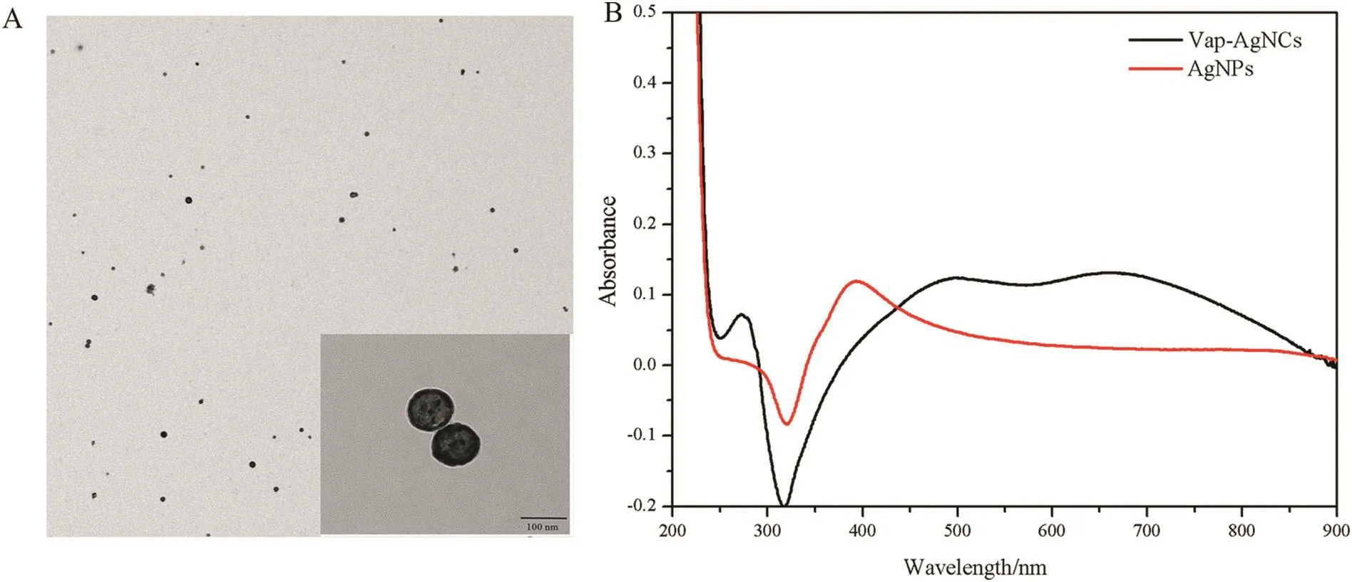
Fig.2.(A)TEM of Vap-AgNCs,the inset showed a magnified TEM image,(B)UV-vis spectra of Vap-AgNCs.
3.2.Biocompatibility of Vap-AgNCs
The biocompatibility of Vap-AgNCs was investigated by the MTT assay to analyze the survival rate of HeLa cells and T293 cell.It was found that Vap-AgNCs solutions with different concentrations had distinctInfluences on thesurvival rate of thecells,correspondingto different biocompatibilities(Fig.4).

Fig.3.Characterization of Vap-AgNCs,(A)zeta potenials,(B)FTIR spectra,(C)EDS spectrum,(D)XRD pattern.

Fig.4.Biocompatibilities of AgNCs to(A)HeLa cell and(B)T293 cell with different concentrations.*and#indicate that P<0.05 compared to Vap and AgNPs,respectively.
The biocompatibility of Vap-AgNCs was between that of Vap and AgNPs in all the experimental groups either to HeLa cells or T293 cells,indicating that Vap has a significant effect on the biocompatibility of Vap-AgNCs.With increasing concentrations,the biocompatibilities of Vap-AgNCs,Vap and AgNPs significantly decreased.When the concentration of Vap-AgNCs reached 2.5 × 10⁻⁴mmol·L−1,both HeLa cells and T293 cells shared the similar results that the viabilities of the cells exposed either to AgNPs or Vap-AgNCs decreased to about 80%indicatingthat thetoxicity of Vap-AgNCs to T293 cells was not larger than that toHeLa cells.However,theviabilityof eitherHeLacells orT293cellsexposedtoVap-AgNCswashigherthanthatofAgNPs,indicatingthatVap-AgNCs have enhanced biocompatibility comparing to AgNPs and Vap-AgNCs had better biocompatibility when its concentration was lower than 2.5 × 10⁻⁴mmol·L−1.
3.3.Photothermal characteristics of Vap-AgNCs
Vap-AgNCs were subsequently used as a photothermal agent to examine the feasibility of photothermal therapy.The temperature of Vap-AgNCs with NIR irradiation reached 45°C starting from 25°C within 5 min;however,the temperature of the AgNPs only reached 28°C within the same time(Fig.5A).Furthermore,the results of thermal images were similar to those of the photothermal properties experiments(Fig.5B),suggesting that Vap-AgNCs have excellent photothermal conversion efficiency and would be a potential material to apply in tumor treatment,as the majority of the tumor cells would die rapidly upon increasing the temperature to 40°C[8].
ToanalyzethephotothermaleffectofVap-AgNCs withNIR irradiation againsttumorcells,HeLacellstreatedwithdrugsat2.5×10−1mmol·L−1werestainedby fluoresceindiacetateasdescribedpreviously[21](Fig.6).There was no difference between the groups of phosphate buffer(PBS)with or without irradiation,indicating that the laser had no significant effect on the status of the cells.For the groups of Vap with laser irradiation,the cell morphology slightly changed compared to the PBS group.The cell morphology changed in the group of AgNPs with laser irradiation;however,no dead cells were detected.For the group of Vap-AgNCs without laser irradiation,there was no significant difference in the number of living cells compared to the PBS group;however,few living cells were detected in the area irradiated by the laser,indicating Vap-AgNCs with laser irradiation leads to HeLa cell death.The photothermal therapy effect of Vap-AgNCs against tumor cells was consistent with the results of the photothermal conversion.Thus,the prepared Vap-AgNCs could play an important role in the photothermal therapy of tumors.
4.Conclusions
In this study,Vap-Ag nanocages were synthesized using Vap as a biotemplate.Upon near infrared irradiation,Vap-AgNCs exhibited higher photothermal conversion efficiency compared to AgNPs,which can be attributed to the unique nanocage structure of Vap-AgNCs with more plasmon bands,leading to death of tumor cells irradiated by NIR laser.Excellentbiocompatibilityandhighphotothermalconversionef ficiency make Vap-AgNCs a promising treatment material against tumor cells.In this paper,photothermal therapy of Vap-AgNCs against tumor cells in vitro was carried out,whether Vap-AgNCs will exhibit excellent photothermal therapy efficacy against tumor in vivo should be confirmed in further study before Vap-AgNCs will be developed a potential photothermal therapeutic agent.

Fig.5.(A)Temperature changes and(B)Thermal imaging photographs of Vap,AgNPs and Vap-AgNCs under NIR irradiation(808 nm,1.5 W·cm−2).

Fig.6.Effect of Vap-AgNCs on HeLa cell survival irradiated by a NIR laser(808 nm),blank PBS buffer(a),Vap(b),AgNPs(c),Vap-AgNCs(d)(IA indicates their radiated area).
杂志排行

Chinese Journal of Chemical Engineering的其它文章
- High efficiency production of ginsenoside compound K by catalyzing ginsenoside Rb1 using snailase☆
- Understanding the Influence of microwave on the relative volatility used in the pyrolysis of Indonesia oil sands☆
- Degradation and mineralization of aniline by O3/Fenton process enhanced using high-gravity technology☆
- Three-liquid-phase extraction and separation of V(V)and Cr(VI)from acidic leach solutions of high-chromium vanadium–titanium magnetite☆
- Insight into the degradation mechanism of cefixime under crystallization condition☆
- Experimental and simulation of the reactive dividing wall column based on ethyl acetate synthesis☆