丹酚酸B通过mTOR/Ulk1信号通路诱导胶质瘤细胞C6自噬性死亡
2018-04-20王新成
赵 敏 张 瑜 张 岚 刘 欣 王新成
(郑州大学附属郑州中心医院神经内科,郑州 450000)
恶性胶质瘤是中枢系统最常见的原发性肿瘤,具有高致死率和高转移率的特点[1]。目前对于恶性胶质瘤的治疗仍以手术治疗为主,但由于其具有较高的复发率,使患者术后存活时间不足15个月[2]。因此,寻找新的治疗方法及药物是降低恶性胶质瘤死亡率的关键。近年来,天然药物的抗癌活性越来越受到关注。丹酚酸B(Salvianolic acid B,Sal B)是中药丹参的主要活性成分,现代研究表明其具有抗氧化、抗炎、抗凝血和免疫调节等活性[3-5],并能抑制肝癌、乳腺癌和鳞状上皮癌等癌症的恶化[6-8]。同时,有研究表明,Sal B能通过调控ROS的产生诱导人胶质瘤细胞凋亡[9]。此外,Sal B还能通过调控自噬影响直肠癌细胞和肝癌细胞的增殖[10,11]。自噬是调控癌症发展的重要机制之一,但Sal B是否能通过自噬影响胶质瘤细胞的存活还未见报道。因此,本研究将探讨Sal B对胶质瘤细胞C6自噬的影响及作用机制。
1 材料与方法
1.1试剂与仪器 DMEM培养液、胎牛血清和胰蛋白酶购自美国Gibco公司;Sal B购自成都普瑞法科技公司;酶标仪购自美国Biotek公司;CO2培养箱购自德国Binder公司;流式细胞仪购自美国ThermoFisher公司;电泳仪和转膜仪均购自美国Bio-RAD公司;CCK8试剂盒和BCA试剂盒购自南京建成生物科技公司;Ki67、天冬氨酸特异性半胱氨酸蛋白酶-3(Caspase-3)、B淋巴细胞瘤-2(B-cell lymphoma-2,Bcl-2)、Bcl-2相关X蛋白(Bcl-2 associated X protein,Bax)、雷帕霉素靶蛋白(Mammalian target of rapamycin)、UNC-51样激酶-(UNC-51-like kinase 1,Ulk1)、自噬相关蛋白5(Autophagy-related peotein 5,Atg5)、Atg7、 Atg12、 Beclin1、 p62和 LC3一抗均购自英国Abcam公司。
1.2方法
1.2.1细胞培养 大鼠神经胶质瘤细胞C6购自ATCC。用含10%胎牛血清的DMEM培养液将细胞培养于5%CO2,37℃培养箱中,每3天换液一次。
1.2.2CCK8检测细胞活性 待细胞汇合率达到80%以上时,将细胞消化接种至96孔板中,培养24 h后,加入CCK8试剂,继续孵育2 h,用酶标仪于450 nm处检测吸光度,计算细胞活性。
1.2.3流式细胞术 细胞汇合率达到80%以上后,用0.25%胰蛋白酶消化收集C6细胞,加入预冷的70%乙醇4℃固定1 h,固定完成后加入PI和FITC-Annexin V室温孵育15 min,流式细胞仪检测Sal B处理后胶质瘤细胞凋亡情况。
1.2.4Western blot 用RIPA蛋白裂解液提取细胞总蛋白,根据BCA试剂盒说明书检测蛋白质浓度并调平蛋白浓度。用12% SDS-PAGE分离蛋白后,转移蛋白至PVDF膜。用5%脱脂奶粉室温孵育含有蛋白质的PVDF膜2 h,加入适应浓度一抗4℃封闭过夜。一抗封闭完成后,用TBST缓冲液洗膜3次,加入对应二抗室温孵育1 h,最后滴加ECL于暗室曝光显影。
1.2.5免疫荧光 将C6细胞接种于提前用0.1%PLL包被的24孔板中,培养24 h后用4%多聚甲醛室温固定30 min,PBS清洗3次后,滴加0.5% TrintonX-100室温透膜15 min,加入Rabbit anti-LC3(1∶200)4℃孵育过夜,次日用PBS清洗3次,加入Goat anti-rabbit二抗室温封闭1 h后,加入DAPI即用液进行复染,最后每孔随机选取6个视野拍照统计。

2 结果
2.1Sal B对胶质瘤细胞活性的影响 为了探究Sal B对胶质瘤细胞活性的影响,我们采用CCK8法检测不同浓度Sal B处理后C6细胞的活性。实验结果表明,Sal B浓度在50 μmol/L以下时对C6细胞活性无明显影响;Sal B达到100 μmol/L时,细胞活性降至80%以下(图1)。因此,我们选择了对细胞无明显毒副作用的高中低三个剂量(10、20、50 μmol/L)进行后续实验。
2.2Sal B对胶质瘤细胞凋亡的影响 Sal B可诱导多种癌细胞凋亡。流式实验结果表明,Sal B能显著促进胶质瘤细胞凋亡,并随Sal B浓度的升高,作用逐渐增强,体现量效关系(图2);同时,与C6组比较,20 μmol/L组和50 μmol/L组细胞增殖标记蛋白Ki67的表达水平明显降低;此外,20 μmol/L组和50 μmol/L组凋亡标记蛋白Caspase-3、Bax的表达水平较C6组比较明显升高,Sal B还能明显抑制Bcl-2的表达,并体现量效关系(图3)。
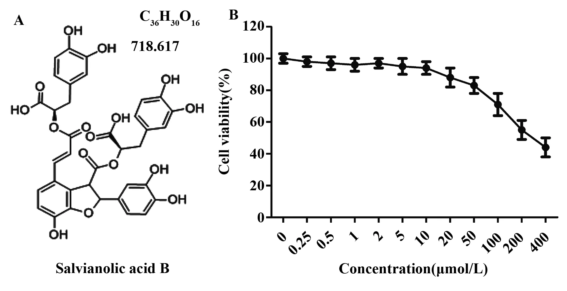
图1 Sal B对胶质瘤细胞活性的影响Fig.1 Effect of Sal B on cell viability of glioma cellsNote:Glioma cells were seed in 96-well plate and treated with Sal B with different concentrations.Cell viability was measured by CCK8 assay.A.The stracture of Sal B;B.The cell viability of C6 cells was determined by CCK8 assay.n=6,every experiment was repeated at least three times.
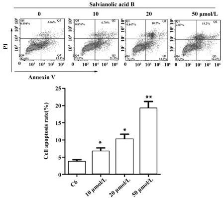
图2 Sal B对胶质瘤细胞凋亡的影响Fig.2 Effect of Sal B on apoptosis of glioma cellNote:Cell were divided into C6 10 μmol/L,20 μmol/L and 50 μmol/L group and treated with Sal B for 24 h.C6 group was treated as control.Flow cytometry was performed for cell apoptosis of C6 cell.*.P<0.05,**.P<0.01 versus C6 group.
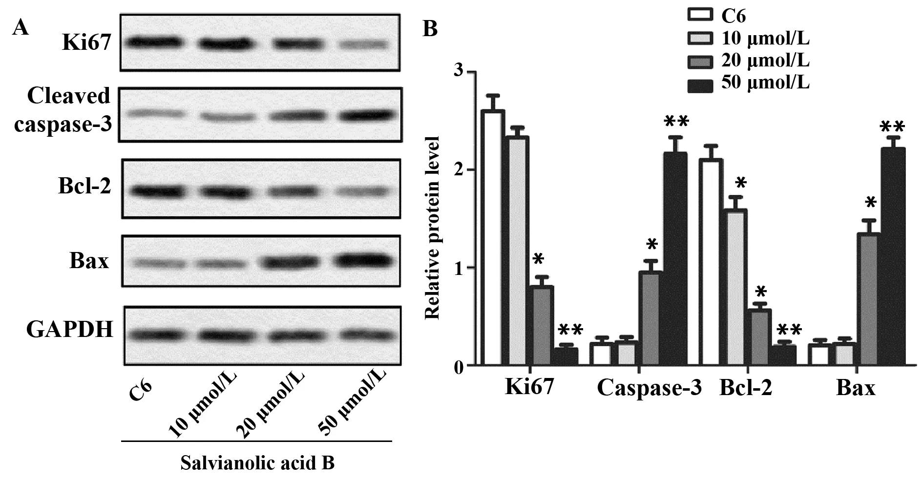
图3 Sal B对增殖、凋亡标记蛋白表达的影响Fig.3 Effects of Sal B on expressions of proliferation-,apoptosis-related proteinsNote:A.The expressions of Ki67,cleaved caspase-3,Bcl-2 and Bax were measured by Western blot;B.The quantification of protein levels of Ki67,cleaved caspase-3,Bcl-2 and Bax .
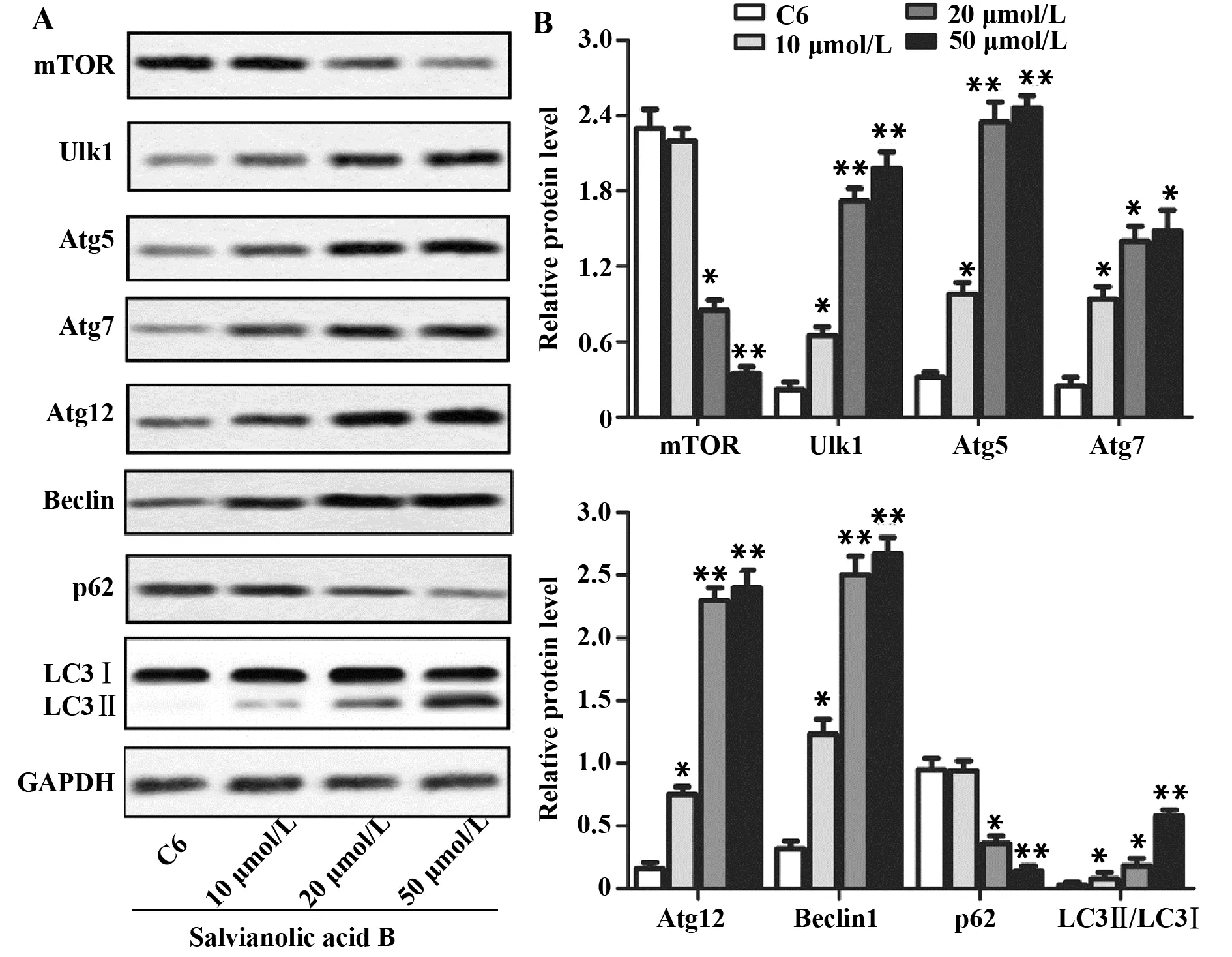
图4 Sal B对胶质瘤细胞自噬及对mTOR/Ulk1通路的影响Fig.4 Effects of Sal B on autophagy and mTOR/Ulk1 pathway of glioma cellNote:A.The expressions of mTOR,Ulk1,Atg5,Atg7,Atg12,Beclin1,p62 and LC3 were measured by Western blot;B.The quantification of protein levels.GAPDH was used as loading control.*.P<0.05,**.P<0.01 versus C6 group.
2.3Sal B对胶质瘤细胞自噬及对mTOR/Ulk1通路的影响 为了探究Sal B对胶质瘤细胞自噬的影响,我们检测了自噬相关蛋白的表达情况。如图4所示,Sal B(10、20、50 μmol/L)能显著促进自噬标记蛋白Atg5、Atg7、Atg12和Beclin1的表达,升高LC3Ⅱ/Ⅰ的比值(图4);与C6组比较,Sal B 20 μmol/L 组和50 μmol/L组p62的蛋白表达水平明显降低(图4);同时,Sal B能显著促进LC3阳性荧光斑点的形成,并随浓度增加,促进作用逐渐增强(图5),提示Sal B可诱导胶质瘤细胞发生自噬;此外,Sal B明显促进了Ulk1的表达(图4);浓度为20 μmol/L 和50 μmol/L 时,明显抑制了mTOR的表达,并体现出量效关系(图4),提示Sal B可能通过抑制mTOR/Ulk1信号通路的激活促进胶质瘤细胞自噬。
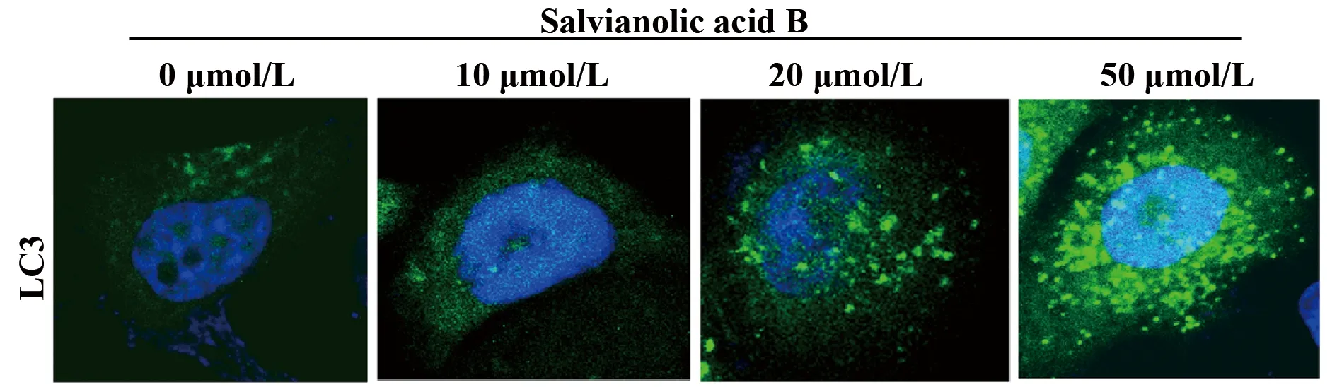
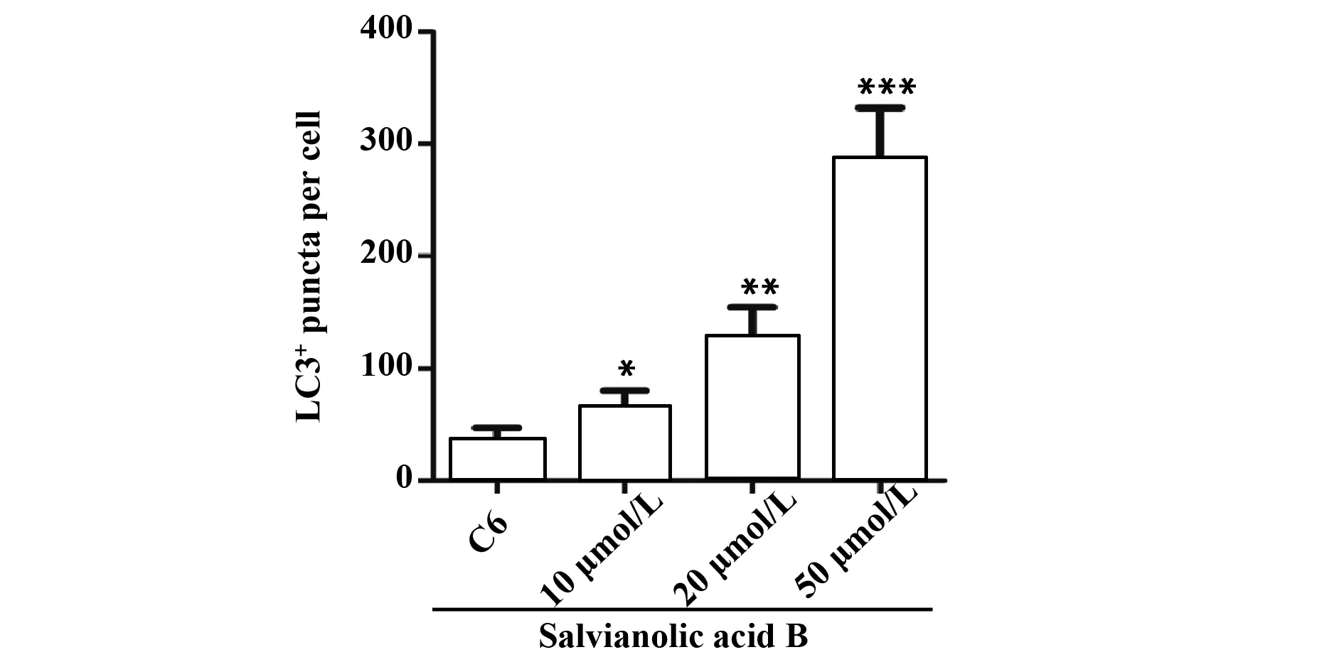
图5SalB对胶质瘤细胞LC3含量的影响
Fig.5EffectofSalBonexpressionlevelofLC3ingliomacells
Note: The level of LC3 was measured by immunofluorescence.*.P<0.05,**.P<0.01,***.P<0.001 versus C6 group.
3 讨论
胶质瘤是一类具有较高转移率和死亡率的恶性肿瘤,其发病率占神经系统原发性肿瘤的60%[12]。目前尚未有研究表明有药物可明显影响胶质瘤的预后。近年来的研究表明,天然药物在抗肿瘤方面具有较好的疗效。Sal B是中药丹参的主要活性成分之一,现代研究表明Sal B具有较强的抗肿瘤活性[9,11,13]。本文研究发现,在Sal B对胶质瘤细胞无明显细胞毒性的条件下,Sal B可明显抑制细胞增殖标记蛋白Ki67的表达,表明其能够抑制胶质瘤细胞的增殖。
细胞凋亡是指细胞程序性死亡。在癌症的发生发展过程中,细胞凋亡被明显抑制,从而促进癌细胞无限增殖[14]。研究表明,Sal B具有较强的促进癌细胞凋亡活性。Sal B可通过诱导凋亡抑制神经母细胞瘤、头颈部鳞状细胞癌和结肠癌等的细胞增殖[8,15-17]。也有研究报道称Sal B能明显促进人胶质瘤细胞U87发生细胞凋亡[9]。本文研究也发现,Sal B能显著增强胶质瘤细胞C6的凋亡程度,从而抑制C6细胞增殖。同时,Sal B还明显促进了细胞凋亡标记分子Cleaved caspase-3和Bax的表达,降低Bcl-2的蛋白表达水平。其中,Bcl-2是影响细胞凋亡的主要调控分子,其在多种癌症中均呈高表达状态[18,19]。Bcl-2过度表达可诱导癌症的发生和癌细胞的转移[20]。Bax则是Bal-2活性的主要调控分子,在调控癌细胞凋亡过程中发挥着重要作用[21]。结合实验结果表明,Sal B可通过调节凋亡调控分子的表达促进胶质瘤细胞凋亡,减缓胶质瘤的恶化进程。
自噬是人体第二类细胞程序性死亡过程[22]。研究表明,自噬在癌症的发生发展过程中扮演着“双刃剑”的作用。早期可通过抑制原癌基因激活而防止癌症的发生,癌症发生后,自噬一方面能为癌细胞提供营养,另一方面又可造成细胞的死亡[23]。有研究报道表明,在胶质瘤发展过程中,细胞自噬明显被抑制,从而促进了癌细胞增殖[1]。细胞死亡是自噬和细胞凋亡相互调控的结果。我们已经证明Sal B可促进C6细胞的凋亡,进一步研究发现,Sal B能明显促进细胞自噬标记蛋白Atg5、Atg7、Atg12和Beclin1的表达,升高了LC3Ⅱ/Ⅰ的比值,同时还降低了p62的蛋白表达水平。此外,Sal B还能促进LC3阳性荧光斑点的形成。其中Atg5、Atg12和Beclin1在自噬的起始阶段发挥着重要的调控作用,Atg7和LC3主要参与自噬小体形成,p62主要存在于细胞核,随着自噬的进行,p62会不断被降解,其表达水平与自噬程度呈负相关[24,25]。结合实验结果表明,Sal B可明显促进胶质瘤细胞自噬的发生。
mTOR/Ulk1通路是调控自噬的重要信号通路之一[26]。在自噬起始阶段,mTOR活性被上游基因抑制,从而激活下游靶基因Ulk1,Ulk1的激活会诱导Atg复合体的形成,完成自噬小体基本结构的构建。同时,复合物会促进LC3Ⅰ向LC3Ⅱ的转化,随后LC3Ⅱ在Atg7和Atg3的作用下转移至细胞膜,完成自噬体的形成[24,27]。mTOR可通过抑制Ulk1的激活抑制自噬[26]。已有研究表明,Sal B可通过调控AKT/mTOR信号通路诱导肝癌细胞自噬[10]。本文研究发现,Sal B可抑制mTOR的表达,促进Ulk1蛋白表达,表明Sal B可促进胶质瘤细胞mTOR/Ulk1信号通路的激活。上述实验结果表明,Sal B可通过调控mTOR/Ulk1信号通路的激活促进胶质瘤细胞自噬。
综上所述,Sal B可抑制胶质瘤细胞凋亡、促进癌细胞发生自噬死亡,提示调控自噬可能成为一种新的抗肿瘤途径,并且其机制与调控mTOR/Ulk1信号通路激活有关。
参考文献:
[1] Shen S,Zhang Y,Wang Z,etal.Bufalin induces the interplay between apoptosis and autophagy in glioma cells through endoplasmic reticulum stress[J].Int J Biological Sci,2014,10:212-224.
[2] Oh SY,Kim H.Molecular culprits generating brain tumor stem cells[J].Brain Tumor Res Treatment,2013,1:9-15.
[3] Lin F,Liu YY,Xu B,etal.Salvianolic acid B protects from pulmonary microcirculation disturbance induced by lipopolysac-charide rat[J].Shock,2013,39(3):317-325.
[4] Lv H,Wang L,Wang S,etal.Salvinolic acid B attuenuates apoptosis and inflammation via SIRT1 activation in experimental stroke rats[J].Brain Res Bulletin,2015,115:30-36.
[5] Ren Z,Wang X,Wang S,etal.Mechanism of action of salvianolic acid B by module-based network analysis[J].Biomed Mater Eng,2014,24(1):1333-1340.
[6] Wang QL,Wu Q,Tao YY,etal.Salvianolic acid B modulates the expression of drug-metabolizing enzymes in HepG2 cells[J].Hepatobiliary Pancreat Dis Int,2011,10:502-508.
[7] Ding L,Li J,Huang R,etal.Salvianolic acid B protects against myocardial damage caused by nanocarrier TiO2and synergistic anti-breast carcinoma effect with curcumin via codelivery system of folic acid-targeted and polyethylene glycol-modified TiO2nan-oparticles[J].Int J Nanomed,2016,11:5709-5727.
[8] Zhao Y,Guo Y,Gu X.Salvianolic Acid B,a potential chemopreventive agent for head and neck squamous cell cancer[J].J Oncol,2011,2011:534-548.
[9] Wang ZS,Luo P,Dai SH,etal.Salvianolic acid B induces apoptosis in human glioma U87 cells through p38-mediated ROS generation[J].Cell Mol Neurobiol,2013,33:921-928.
[10] Gong L,Di C,Xia X,etal.AKT/mTOR signaling pathway is involved in salvianolic acid B-induced autophagy and apoptosis in hepatocellular carcinoma cells[J].Int J Oncol,2016,49:2538-2548.
[11] Jing Z,Fei W,Zhou J,etal.Salvianolic acid B,a novel autophagy inducer,exerts antitumor activity as a single agent in colorectal cancer cells[J].Oncotarget,2016,7:61509-61519.
[12] Butowski NA.Epidemiology and diagnosis of brain tumors[J].Continuum(minneapolis,minn),2015,21:301-313.
[13] Li H,Shi L,Wei J,etal.Cellular uptake and anticancer activity of salvianolic acid B phospholipid complex loaded nanoparticles in head and neck cancer and precancer cells[J].Colloids Surfaces B Biointerfaces,2016,147:65-72.
[14] Mohammad RM,Muqbil I,Lowe L,etal.Broad targeting of resistance to apoptosis in cancer[J].Semin Cancer Biol,2015,35(Suppl):S78-S103.
[15] Zeng G,Tang T,Wu HJ,etal.Salvianolic acid B protects SH-SY5Y neuroblastoma cells from 1-methyl-4-phenylpyridinium-induced apoptosis[J].Biol Pharma Bull,2010,33:1337-1342.
[16] Zhao Y,Hao Y,Ji H,etal.Combination effects of salvianolic acid B with low-dose celecoxib on inhibition of head and neck squamous cell carcinoma growth in vitro and in vivo[J].Cancer Prev Res(Phila),2010,3:787-796.
[17] Guo P,Wang S,Liang W,etal.Salvianolic acid B reverses multidrug resistance in HCT8/VCR human colorectal cancer cells by increasing ROS levels[J].Mol Med Rep,2017,15:724-730.
[18] Ruibal A,Aguiar P,Del Rio MC,etal.Positive immunohistoch-emical expression of bcl-2 in hormone-independent breast carcinomas is associated with a greater lymph node involvement and poor outcome[J].Medical Oncol(Northwood,London,England),2014,31:105.
[19] Boonyaphiphat P,Pruegsanusak K,Thongsuksai P.The prognostic value of p53,Bcl-2 and Bax expression in laryngeal cancer[J].J Med Associ Thai,2012,95(10):1317-1320.
[20] Rostamizadeh L,Fakhrjou A,Montazeri V,etal.Bcl-2 gene expression in human breast cancers in iran[J].Asian Pac J Cancer Prev,2013,14(7):4209-4214.
[21] Sarbia M,Bittinger F,Grabellus F,etal.Expression of Bax,a pro-apoptotic member of the Bcl-2 family,in esophageal squamous cell carcinoma[J].Int J Cancer,1997,73(4):508-513.
[22] Schwartzbaum JA,Fisher JL,Aldape KD,etal.Epidemiology and molecular pathology of glioma[J].Nat Clin Pract Neurol,2006,2:494-503.
[23] Tsujimoto Y,Shimizu S.Another way to die:autophagic programmed cell death[J].Cell Death Differ,2005,12(Suppl 2):1528-1534.
[24] Jung CH,Jun CB,Ro SH,etal.ULK-Atg13-FIP200 complexes mediate mTOR signaling to the autophagy machinery[J].Mol Biol Cell,2009,20(7):1992-2003.
[25] Jiang T,Harder B,Rojo de la Vega M,etal.p62 links autophagy and Nrf2 signaling[J].Free Rad Biol Med,2015,88(Pt13):199-204.
[26] Aryal P,Kim K,Park PH,etal.Baicalein induces autophagic cell death through AMPK/ULK1 activation and downregulation of mTORC1 complex components in human cancer cells[J].FEBS J,2014,281(20):4644-4658.
[27] Funderburk SF,Wang QJ,Yue Z.The Beclin 1-VPS34 complex--at the crossroads of autophagy and beyond[J].Trends Cell Biol,2010,20(6):355-362.