宫颈癌组织中maspin、AKT、p-AKT表达变化及其与肿瘤微淋巴管密度的相关性
2017-08-09许振刘志强孟玮郭庆枝孙莉
许振,刘志强,孟玮,郭庆枝,孙莉
(滨州医学院附属医院,山东滨州256600)
宫颈癌组织中maspin、AKT、p-AKT表达变化及其与肿瘤微淋巴管密度的相关性
许振,刘志强,孟玮,郭庆枝,孙莉
(滨州医学院附属医院,山东滨州256600)
目的 观察宫颈癌组织中maspin、蛋白激酶B(AKT)、磷酸化蛋白激酶B(p-AKT)的表达变化,并分析其与肿瘤微淋巴管密度(LMVD)的相关性。方法 收集127例份宫颈石蜡组织标本,其中慢性宫颈炎组织20例份(炎症组)、CINⅠ级组织20例份(CIN1组)、CINⅡ级组织20例份(CIN2组)、CINⅢ级组织20例份(CIN3组)、宫颈癌组织47例份(宫颈癌组,其中无淋巴结转移32例为宫颈癌1组、有淋巴结转移15例为宫颈癌2组)。采用免疫组化SP法检测各组maspin、AKT、P-AKT,测算肿瘤微淋巴管密度(LMVD)。结果 宫颈癌1组、宫颈癌2组maspin蛋白阳性表达比例低于其余各组(P均<0.05)。宫颈癌1组、宫颈癌2组AKT、p-AKT阳性表达比例高于其余三组(P均<0.05)。宫颈癌2组p-AKT阳性表达比例高于宫颈癌1组(P<0.05)。宫颈癌组织中maspin与AKT表达呈负相关关系(rs=-0.472,P=0.001),maspin与p-AKT表达也呈负相关关系(rs=-0.598,P=0.000)。宫颈癌1组、宫颈癌2组LMVD分别为(19.44±2.95)、(23.24±2.46)支/mm2,淋巴结转移与LMVD呈正相关关系(rs=0.44,P=0.000)。宫颈癌组织中LMVD与maspin表达呈负相关关系(rs=-0.554,P=0.000),LMVD与p-AKT表达呈正相关关系(rs=0.39,P=0.007)。结论 宫颈癌组织中maspin表达下调,AKT、p-AKT表达上调,且maspin表达与AKT、p-AKT表达均呈负相关关系;宫颈癌组织中LMVD与maspin表达呈负相关关系,LMVD与p-AKT表达呈正相关关系。maspin表达下调可能参与了宫颈癌的发生发展,机制可能与AKT通路异常激活、促进肿瘤微淋巴管生成有关。
宫颈癌;maspin基因;蛋白激酶B;微淋巴管密度
宫颈癌是最常见的女性生殖道恶性肿瘤之一,我国每年新发病例占世界宫颈癌新发病例总数的1/3,每年死于宫颈癌的患者约5万人[1]。虽然手术、放疗及化疗的综合治疗已经明显改善了宫颈癌患者的预后,但对已经发生淋巴结转移的宫颈癌治疗效果仍不太理想。淋巴结转移是宫颈癌最常见、最重要的转移途径之一,也是影响患者预后的重要因素。maspin基因是丝氨酸蛋白酶抑制剂的一员,研究发现maspin对乳腺、前列腺等肿瘤的生长、侵袭、转移有明显抑制作用。maspin基因在宫颈鳞癌淋巴管生成及肿瘤转移中的作用机制尚不清楚。近年来学者们通过对淋巴管标志物及淋巴管生成因子进行研究发现,血管密度及微淋巴管密度(LMVD)是多种实体瘤预后的参考指标之一[2,3]。磷脂酰肌醇-3激酶(PI3K)/丝苏氨酸蛋白激酶(AKT)信号转导通路是调控细胞增殖、维持细胞恶性生物学特性的重要转导通路之一[4]。有研究[5]表明AKT通路在肿瘤淋巴管生成及淋巴结转移中起着重要作用。本研究观察了宫颈癌(鳞癌)组织中maspin、AKT、磷酸化AKT(p-AKT)的表达变化,并分析其与肿瘤LMVD的相关性。
1 材料与方法
1.1 组织标本及主要试剂 2009年2月~2016年2月滨州医学院附属医院妇产科宫颈活检或手术中切取的宫颈石蜡组织标本127例份,其中慢性宫颈炎组织20例份(炎症组)、CINⅠ级组织20例份(CIN1组)、CINⅡ级组织20例份(CIN2组)、CINⅢ级组织20例份(CIN3组)、宫颈癌组织47例份(宫颈癌组,其中无淋巴结转移32例为宫颈癌1组、有淋巴结转移15例为宫颈癌2组)。炎症组、CIN1组、CIN2组、CIN3组、宫颈癌组患者的年龄分别为(35.3±6.7)、(36.2±5.7)、(38.9±7.5)、(42.3±6.3)、(48.4±6.8)岁。各组标本来源患者均无其他恶性肿瘤病史,获取标本前均未接受过放疗及化疗,均经病理检查证实诊断。兔抗人AKT单克隆抗体购自美国Cell Signaling公司。兔抗人p-AKT(Ser473)单克隆抗体购自美国Cell Signaling公司。即用型鼠抗人D2-40单克隆抗体购自北京中杉金桥生物技术有限公司。兔抗人maspin单克隆抗体购自北京博奥森生物技术有限公司。
1.2 不同宫颈病变组织中maspin、AKT、p-AKT检测 将标本进行4 μm厚石蜡切片,免疫组化SP法染色,用PBS代替一抗作为阴性对照,用已知阳性片做阳性对照。maspin、AKT、p-AKT定位于细胞核与细胞质,以细胞核与细胞质中出现棕黄色颗粒为阳性细胞。按照着色强度和阳性细胞百分比判定结果:着色强度为无色计0分、浅黄色计1分、黄色及更深颜色计2分,阳性细胞百分比≤50%计0分、>50%~75%计1分、>75%计2分[6];上述两项得分相乘,≥2判定为目的蛋白表达阳性。
1.3 宫颈癌组织中LMVD观察及计算方法 利用D2-40单克隆抗体标记宫颈癌组织中的微淋巴管并计算LMVD。参考文献[7]方法:D2-40表达定位于淋巴管内皮细胞,呈棕黄色或棕褐色颗粒。先在40倍镜下选择4个微淋巴管高密度区即所谓的“热点”,然后在400倍镜视野(0.16 mm2/视野)下记数,取4个视野的平均数(单位为支/mm2)。含有平滑肌或大于8个内皮细胞的管腔及未染色的管腔不记数。

2 结果
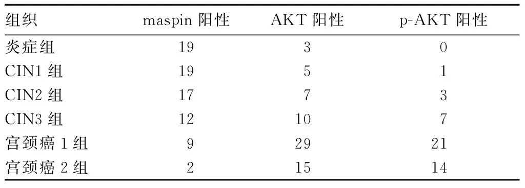
2.1 各组Maspin、AKT、p-AKT表达比较 宫颈癌1组、宫颈癌2组maspin蛋白阳性表达比例低于其余各组(P均<0.05)。宫颈癌1组、宫颈癌2组AKT、p-AKT阳性表达比例高于其余三组(P均<0.05)。宫颈癌2组p-AKT阳性表达比例高于宫颈癌1组(P<0.05)。详见表1、图1~3。

表1 各组maspin、AKT、p-AKT阳性表达情况(例)

注:A为炎症组;B为CIN3组;C为宫颈癌组。
图1 炎症组、CIN3组、宫颈癌组中mapsin表达情况

注:A为炎症组;B为CIN3组;C为宫颈癌组。
图2 炎症组、CIN3组、宫颈癌组中AKT表达情况

注:A为炎症组;B为CIN3组;C为宫颈癌组。
图3 炎症组、CIN3组、宫颈癌组中p-AKT表达情况
2.2 宫颈癌组maspin、AKT、p-AKT表达的相关性 47例宫颈癌组织中maspin、AKT同时阳性表达的有0例、同时阴性表达的有3例,maspin与AKT表达呈负相关关系(rs=-0.472,P=0.001);宫颈癌组织中maspin、p-AKT同时阳性表达的有3例、同时阴性表达的有4例,maspin与p-AKT表达呈负相关关系(rs=-0.598,P=0.000)。
2.3 宫颈癌组LMVD表达变化及其与maspin、AKT、p-AKT表达的相关性 宫颈癌1组、宫颈癌2组LMVD分别为(19.44±2.95)、(23.24±2.46)支/mm2,淋巴结转移与LMVD呈正相关关系(rs=0.44,P=0.000)。宫颈癌组中,maspin阳性组、maspin阴性组的LMVD分别为(17.45±2.88)、21.91±3.70)支/mm2,maspin阳性组LMVD低于maspin阴性组(P<0.05);AKT阳性组、AKT阳性组LMVD分别为(21.14±3.19)、(17.00±3.00)支/mm2,AKT阳性组LMVD高于AKT阴性组(P<0.05);p-AKT阳性组、p-AKT阳性组LMVD分别为(21.41±3.17)、(18.62±2.79)支/mm2,p-AKT阳性组LMVD高于p-AKT阴性组(P<0.05)。Spearman相关分析显示,宫颈癌组织中LMVD与maspin表达呈负相关关系(rs=-0.554,P=0.000),LMVD与p-AKT表达呈正相关关系(rs=0.39,P=0.007)。
3 讨论
maspin基因是一种丝氨酸蛋白酶抑制剂基因,其不但对乳腺癌、前列腺癌等肿瘤的生长、侵袭、转移有明显抑制作用,而且能上调乳腺癌、前列腺癌细胞中凋亡前蛋白Bax的表达,增强肿瘤细胞对凋亡诱导的敏感性[8]。Nam等[9]将含有maspin基因的质粒注入植有NCIH157肺癌细胞株的裸鼠瘤体中,发现含有maspin基因质粒的实验组成瘤体积远小于对照组,预示着maspin基因在肿瘤基因治疗方面有着广阔前景。本研究结果显示,宫颈癌1组、宫颈癌2组maspin蛋白阳性表达比例低于CIN3组,提示maspin蛋白在宫颈CINⅢ发展到宫颈癌的过程中起着重要作用。大量临床研究及动物实验证实maspin基因表达具有抑制侵袭和迁移、抑制肿瘤血管生成的特性[10~12],maspin是一个多功能的抑癌基因。随着肿瘤微淋巴管的相关研究不断增多,有学者研究证实maspin基因可调节肿瘤微淋巴管的生成[13,14]。Wang等[13]发现在非小细胞肺癌患者中,maspin基因表达与VEGF-C表达呈负相关,maspin基因表达与淋巴结转移情况有关。但本研究中宫颈癌1组和宫颈癌2组的maspin阳性表达比例差异无统计学意义,考虑与样本量较少有关。
PI3K/AKT通路在肿瘤细胞增殖、迁移、侵袭及抗凋亡中都起到举足轻重的作用[4,15]。PI3K/AKT通路在肿瘤细胞G1期到S期转变时变得非常活跃,可以调节细胞周期调控关键因子P21CIP1、Cyclin D、p27Kip1的蛋白质稳定性[16]。PI3K/AKT通路分子高表达使得肿瘤细胞处于增殖活跃期并具有明显的抗凋亡作用。有研究[17]显示人乳头瘤病毒16(HPV16)分泌的E7蛋白可激活AKT通路,调控肿瘤细胞的增殖和转移,这与宫颈癌的发生发展关系密切。Chen等[18]利用免疫组化法检测了55例胃腺癌患者石蜡手术标本中的mTOR、AKT、VEGF-C、VEGF-D,并计算了淋巴管密度,结果表明淋巴管密度与mTOR、AKT、VEGF-C、VEGF-D表达呈正相关关系,提示AKT通路的异常激活可能是众多恶性肿瘤发生发展及淋巴结转移的重要因素。本研究结果显示,宫颈癌1组、宫颈癌2组AKT、p-AKT阳性表达比例高于其余三组,宫颈癌2组p-AKT阳性表达比例高于宫颈癌1组,提示AKT、p-AKT高表达与宫颈癌的发生和进展密切相关,与上述研究结果一致。
淋巴转移是宫颈癌最常见、最重要的转移途径之一。Birner等[19]报道在宫颈癌早期,淋巴管未受侵组5年无病生存率达84.1%,而淋巴管受侵组仅48.2%。肿瘤细胞侵入淋巴管与微淋巴管的超微结构有着密切关系[20]。微淋巴管具有以下特征:管壁很薄,仅由一层连续的内皮细胞和少量结缔组织构成;无基膜或基膜不连续;内皮细胞常相互重叠、连接处常存在间隙,内皮外有锚丝等。这些超微结构特点为肿瘤细胞的侵入提供了结构形态基础。本研究发现,宫颈癌淋巴结转移与LMVD呈正相关关系,证实了上述结论。我们发现,在宫颈癌组织中,maspin与AKT、p-AKT表达均呈负相关关系,LMVD与maspin表达呈负相关关系,LMVD与p-AKT表达呈正相关关系,推测maspin蛋白可能有抑制AKT通路激活的作用,maspin基因失表达可能与AKT通路异常激活有着密切联系,而AKT通路与肿瘤微淋巴管生成有关。已有研究[5]表明,AKT通路可进一步促进VEGF-C、VEGF-D、VEGF-3、FGF-2、HGF、IGF、ANG-1等因子表达,促进淋巴管生成,最终导致肿瘤细胞发生淋巴结转移。
结合上述研究结果,我们推测,maspin表达下调可能参与了宫颈癌的发生发展,机制可能与AKT通路异常激活、促进肿瘤微淋巴管生成有关。但本研究实验方法较为单一,maspin基因对PI3K/AKT通路的具体作用机制以及maspin蛋白的功能靶点仍有待进一步研究。
[1] 赫捷,陈万青.2011中国肿瘤登记年报[M].北京:军事医学科学出版社,2012:26-37.
[2]Schweiger T, Nikolowsky C, Graeter T, et al. Increased lymphangiogenesis in lung metastases from colorectal cancer is associated with early lymph node recurrence and decreased overall survival[J]. Clin Exp Metastasis, 2015,33(2):1-9.
[3] Zhang N, Xie F, Gao W, et al. Expression of hepatocyte growth factor and c-Met in non-small-cell lung cancer and association with lymphangiogenesis[J]. Mol Med Rep, 2011,11(4):2797.
[4] Yu JS, Cui W. Proliferation, survival and metabolism: the role of PI3K/AKT/mTOR signalling in pluripotency and cell fate determination[J]. Development, 2016,143(17):3050.
[5] 赵静静,戴小军,张晓春.Akt介导的信号通路在肿瘤淋巴管生成中的作用[J].肿瘤防治研究,2016,43(5):422-426.
[6] Khor TO, Gul YA, Ithnin H, et al. Positive correlation between overexpression of phospho-BAD with phosphorylated Akt at serine 473 but not threonine 308 in colorectal carcinoma[J]. Cancer Let, 2004,210(2):139-150.
[7] Weidner N, Carroll PR, Flax J, et al. Tumor angiogenesis correlates with metastasis in invasive prostate carcinoma[J]. Am J Pathol,1993,143(2):401-409.
[8] Liu J, Yin S, Reddy N, et al. Bax Mediates the apoptosis-sensitizing effectof maspin[J]. Cancer Res, 2004,64:1703-1711.
[9] Nam E, Park C. Maspin suppresses survival of lung cancer cells through modulation of Akt pathway[J]. Cancer Res Treat, 2010,42(1):42-47.
[10] Al-Mamun MA, Farid DM, Ravenhil L, et al. An in silico model to demonstrate the effects of maspin on cancer cell dynamics[J]. J Theor Biol, 2016,388(4):37-49.
[11] Ciortea D, Jung I, Gurzu S, et al. Correlation of angiogenesis with other immunohistochemical markers in cutaneous basal and squamous cell carcinomas[J]. Rom J Morphol Embryol, 2015,56(2 Suppl): 665-670.
[12] Chen WS, Yen CJ, Chen YJ, et al. miRNA-7/21/107 contribute to HBx-induced hepatocellular carcinoma progression through suppression of maspin[J]. Oncotarget, 2015,6(28):25962-25974.
[13] Wang X, Wang Y, Li S, et al. Decreased maspin combined with elevated vascular endothelial growth factor C is associated with poor prognosis in non-small cell lung cancer[J]. Thor Cancer, 2014, 5(5):383-390.
[14] Strien L, Joensuu K, Heikkila P, et al. Different Expression Patterns of CXCR4, CCR7, Maspin and FOXP3 in Luminal Breast Cancers and Their Sentinel Node Metastases[J]. Anticancer Res, 2017,37(1):175-182.
[15] Zhang L, Wu J, Ling MT, et al. The role of the PI3K/Akt/mTOR signalling pathway in human cancers induced by infection with human papillomaviruses[J]. Molecul Cancer, 2015,14(1):87.
[16] Liang J, Slingerland JM. Multiple roles of the PI3K/PKB (Akt) pathway in cell cycle progression[J]. Cell Cycle, 2003,2(4):339-345.
[17] Pickard A, McDade SS, McFarland M, et al. HPV16 down-Regulates the insulin-Like growth factor binding protein 2 to promote epithelial invasion in organotypic cultures[J]. PLoS Pathog, 2015,11(6): e1004988.
[18] Chen H, Guan R, Lei Y, et al. Lymphangiogenesis in Gastric Cancer regulated through Akt/mTOR-VEGF-C/VEGF-D axis[J]. BMC Cancer, 2015,15(1):103.
[19] Birner P, Obermair A, Schindl M, et al. Selective immunohistochemical staining of blood and lymphatic vessels reveals independent prognostic influence of blood and lymphatic vessels invasion in early-stage cervical cancer [J]. Clin Cancer Res, 2001,7(1):93-97.
[20] Rovenska E. Importance of lymphangiogenesis and ultrastructure of lymphatic capillaries in metastasis of malignant melanoma[J]. Vnitr Lek, 2014,60:582-585.
Expression of maspin, AKT, and p-AKT in cervical carcinoma and its correlation with tumor lymphatic microvessel density
XUZhen,LIUZhiqiang,MENGWei,GUOQizhi,SUNLi
(BinzhouMedicalUniversityHospital,Binzhou256600,China)
Objective To observe the expression changes of maspin, protein kinase B (AKT), and phosphorylated protein kinase B (p-AKT) in the cervical cancer tissues and to analyze its correlation with tumor lymphatic microvessel density (LMVD). Methods Twenty-seven cases of cervical paraffin tissues were collected, including 20 cases of chronic cervicitis (inflammation group), 20 cases of cervical intraepithelial neoplasia (CIN) Ⅰ (CIN1 group), 20 cases of CIN Ⅱ (CIN2 group), 20 cases of CIN Ⅲ (CIN3 group), and 47 cases of cervical cancer tissues (cervical cancer group, including 32 cases without lymph node metastasis of cervical cancer group 1 and 15 cases with lymph node metastasis of cervical cancer group 2). The levels of maspin, AKT, and P-AKT were measured by immunohistochemical SP method, and the LMVD was measured. Results The positive rate of maspin protein in the cervical cancer group 1 and cervical cancer group 2 was lower than that in the CIN3 group (allP<0.05). The positive expression rates of AKT and p-AKT in the cervical cancer group 1 and cervical cancer group 2 were higher than those in the other three groups (allP<0.05). The positive expression rate of p-AKT in the cervical cancer group 1 was higher than that in the cervical cancer group 2 (P<0.05). There was a negative correlation between the expression of maspin and AKT in the cervical cancer tissues (rs=-0.472,P=0.001). There was also a negative correlation between maspin and p-AKT expression (rs=-0.598,P=0.000). LMVD in the cervical cancer groups 1 and 2 were (19.44±2.95) and (23.24±2.46) vessels/mm2, respectively. There was a positive correlation between lymph node metastasis and LMVD (rs=0.44,P=0.000). There was a negative correlation between LMVD and the expression of maspin in the cervical cancer tissues (rs=-0.554,P=0.000). There was a positive correlation between LMVD and p-AKT expression (rs=0.39,P=0.007). Conclusions The expression of AKP and p-AKT is up-regulated in the cervical cancer tissues, and the expression of maspin is down-regulated, which is negatively correlated with both AKT and p-AKT expression. In the cervical cancer tissues, LMVD is negatively correlated with maspin expression, but is positively correlated with AKT expression. The down-regulation of maspin expression may be involved in the development of cervical cancer, and the mechanism may be related to the abnormal activation of AKT pathway and the promotion of tumor micro-lymphangion genesis.
cervical cancer; maspin gene; protein kinase B; lymphatic microvessel density
山东省自然科学基金资助项目(ZR2012HL01)。
许振(1989-),男,硕士,主要研究方向为妇科肿瘤。E-mail:2005891282qq.com
刘志强(1971-),男,博士,教授,主任医师,主要研究方向为妇科肿瘤。E-mail:2005891282qq.com
10.3969/j.issn.1002-266X.2017.27.006
R737.33
A
1002-266X(2017)27-0023-04
2017-02-27)
