人颞叶内侧癫痫海马组织星形胶质细胞Syntrophin的表达变化
2017-06-01王晓璇孙振荣吴敏师忠芳闫旭徐立新董丽萍杨少华袁芳
王晓璇,孙振荣,吴敏,师忠芳,闫旭,徐立新,董丽萍,杨少华,袁芳
人颞叶内侧癫痫海马组织星形胶质细胞Syntrophin的表达变化
王晓璇1,孙振荣2,吴敏1,师忠芳1,闫旭1,徐立新1,董丽萍1,杨少华3,袁芳1
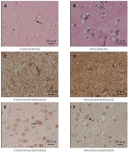
目的观察人颞叶内侧癫痫(MTLE)海马组织星形胶质细胞syntrophin的表达变化。方法2015年4月至2016年7月,17例癫痫患者手术切除的海马组织,依据苏木素-伊红(HE)染色及胶质原纤维酸性蛋白、神经元核抗原免疫组织化学染色结果,分为MTLE组13例和非MTLE组4例。免疫荧光双重组织化学法及免疫荧光组织化学法观察syntrophin的表达。结果MTLE组星形胶质细胞大量增生,神经元明显减少。syntrophin在星形胶质细胞膜及足突处表达。与非MTLE组相比,MTLE组syntrophin在血管周围星形胶质细胞足突处分布减少,在其他部位分布增加。图像分析显示,与非MTLE组相比,MTLE组syntrophin蛋白量显著增多(t=5.421,P<0.001)。结论海马组织syntrophin在星形胶质细胞上的分布及其整体表达变化可能与MTLE发生相关。
颞叶内侧癫痫;海马;星形胶质细胞;syntrophin;抗肌萎缩蛋白糖蛋白复合物
[本文著录格式]王晓璇,孙振荣,吴敏,等.人颞叶内侧癫痫海马组织星形胶质细胞Syntrophin的表达变化[J].中国康复理论与实践,2017,23(3):292-297.
CITED AS:Wang XX,Sun ZR,Wu M,et al.Distribution of astrocytic syntrophin in hippocampus from human mesial temporal lobe epilepsy[J].Zhongguo Kangfu Lilun Yu Shijian,2017,23(3):292-297.
syntrophin是抗肌萎缩蛋白糖蛋白复合物(dystrophin-associated glycoprotein complex,DGC)中的一个亚单位,主要起锚定蛋白的作用[1]。研究提示,水通道蛋白4(aquaporin 4,AQP4)和内向整流性钾离子通道
4.1(inward rectifier potassium channel 4.1,Kir4.1)通过syntrophin锚定到星形胶质细胞上[2-3]。我们以前的研究发现,星形胶质细胞上的AQP4和Kir4.1与颞叶内侧癫痫(medial temporal lobe epilepsy,MTLE)的发生有密切关系[4]。动物实验发现,敲除α-syntrophin基因的小鼠惊厥易感性增加,提示syntrophin可能参与癫痫的发生[5]。MTLE是难治性癫痫中最常见的类型,临床常采用切除硬化海马来控制癫痫发作[6]。本研究观察手术切除MTLE及非MTLE患者海马组织,运用免疫荧光染色技术观察syntrophin的分布及表达情况,探讨syntrophin参与MTLE发生的可能分子机制。
1 材料与方法
1.1实验样本
收集2015年4月至2016年7月首都医科大学附属北京天坛医院神经外科行颞叶切除术治疗癫痫患者的海马组织,切除的海马组织即刻置4%多聚甲醛中固定以备病理检查及后续实验。
本方案已通过首都医科大学附属北京天坛医院医学伦理委员会审批。
1.2实验试剂及器材
伊红(ZLI-9613)、苏木素(ZLI-9610)、罗丹明标记山羊抗兔荧光二抗(ZF-0316)、Alexa Fluor 488标记山羊抗小鼠荧光二抗(ZF-0512)、抗荧光淬灭封片剂(ZLI-9556):北京中杉金桥生物技术有限公司。胶质原纤维酸性蛋白抗体(glial fibrillary acidic protein, GFAP,Z0334)、Envision检测试剂盒(A-K5007):丹麦DAKO公司。神经元核抗原抗体(neuronal nuclei, NeuN,ab177487):美国ABCAM公司。syntrophin抗体(MAB2277):美国MILLIPORE公司。正置荧光显微镜(Axio ImageryA2):德国ZEISS公司。
1.3方法
1.3.1海马组织学观察及分组
海马组织常规固定、包埋、切片厚4 μm,苏木素-伊红(hematoxylin and eosin,HE)染色及GFAP、NeuN免疫组织化学染色后,光学显微镜下观察。
GFAP、NeuN免疫组织化学染色参考文献二步法进行[7]。GFAP及NeuN抗体1∶2000稀释,4℃孵育过夜,A-K5007试剂盒(Envision,DAKO)显色。阴性对照用磷酸盐缓冲液(phosphate buffered saline,PBS)代替一抗,其余步骤相同。阳性细胞染色为浅至棕褐色。
MTLE海马硬化诊断标准为海马组织神经元丢失>50%及大量星形胶质细胞增生;非MTLE诊断标准为海马组织表现为轻度神经元丢失(<25%)及少量星形胶质细胞增生,存在海马结构附近脑组织病变[4,8]。
1.3.2免疫荧光双重组织化学染色
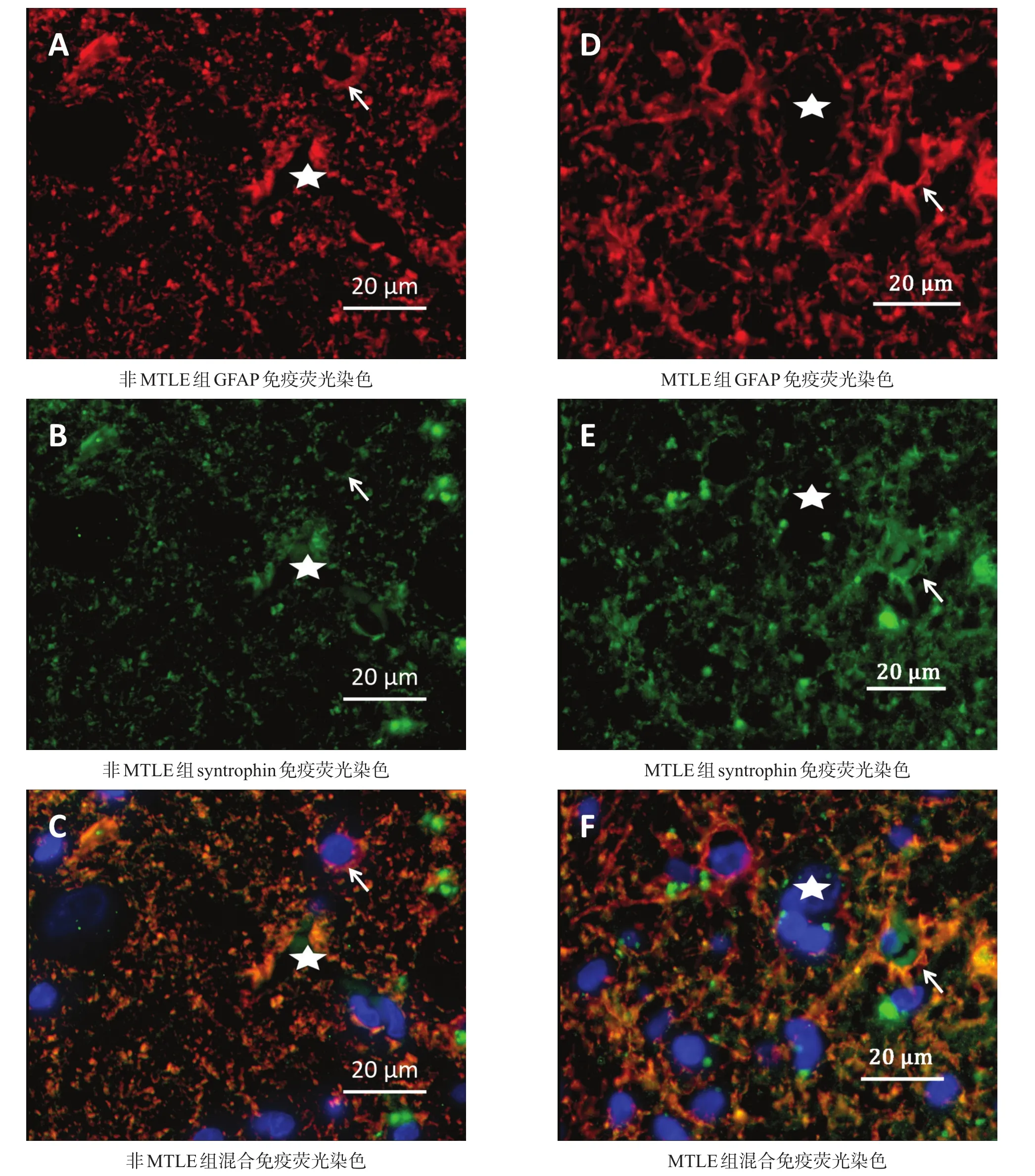
采用syntrophin与GFAP免疫荧光双重组织化学染色法观察syntrophin在星形胶质细胞上的表达。染色方法参考文献[7]。0.01 mol/L柠檬酸盐缓冲液(pH=6.0)微波热修复,山羊血清封闭,滴加syntrophin抗体(1∶100)及GFAP抗体(1∶400)。4℃孵育过夜。滴加罗丹明标记的山羊抗兔荧光二抗(1∶200)及Alexa Fluor 488标记的山羊抗小鼠荧光二抗(1∶200),室温孵育1 h,4',6-二脒基-2-苯基吲哚(4',6-diamidino-2-phenylindole,DAPI)室温孵育5 min,PBS洗涤,抗荧光淬灭封片剂封片。正置荧光显微镜观察、拍照。阴性对照用PBS代替一抗,其余步骤相同。
1.3.3免疫荧光组织化学染色
滴加syntrophin一抗(1∶100),4℃孵育过夜;滴加Alexa Fluor 488标记的山羊抗小鼠二抗(1∶200),余同免疫荧光双重组织化学染色。
应用Image J图像分析软件对syntrophin免疫荧光结果进行分析。每个标本取1张切片,镜下(400×)随机选取CA4区5个视野,图片黑白反向处理,计算5个视野中平均光密度值(average optical density,AOD)。
1.4统计学分析
采用SPSS 17.0软件进行统计分析。对年龄、病程及syntrophin表达量以(±s)表示,进行t检验。对性别以例数表示,进行χ2检验。显著性水平α=0.05。
2 结果
2.1分组结果
共观察17例结构比较完整的海马组织标本。依据HE染色及GFAP、NeuN免疫组织化学染色(图1),MTLE组13例,非MTLE组4例。两组间性别、年龄及病程均无显著性差异(P>0.05)。见表1。

表1 两组一般资料比较
2.2免疫荧光双重染色
非MTLE组syntrophin和GFAP在星形胶质细胞足突及胞膜上共表达,MTLE组syntrophin和GFAP在血管周围星形胶质细胞足突上表达减少,在星形胶质细胞胞体上表达增多(图2)。
2.3免疫荧光组织化学染色
MTLE组syntrophin阳性染色明显增强(图3)。图像分析显示,MTLE组syntrophin蛋白表达量显著增加(t=5.421,P=0.0004)。

图1 海马组织HE染色、GFAP和NeuN免疫组化染色(bar=20 μm)

图2 海马组织syntrophin与GFAP免疫双重荧光染色(bar=20 μm)

图3 海马组织syntrophin免疫荧光染色及统计结果
3 讨论
目前国内外对syntrophin及DGC在癫痫疾病中的研究都十分有限。本研究显示,syntrophin在MTLE患者海马区明显增多,且在星形胶质细胞上极性分布异常,即血管周围星形胶质细胞足突处分布减少且星形胶质细胞胞体分布增加,提示syntrophin蛋白在MTLE的发生过程中起重要作用。
MTLE的发生归因于神经元异常放电,但临床发现MTLE患者普遍对抗癫痫药物耐药,且组织学观察到神经元显著丢失及星形胶质细胞大量增生。
星形胶质细胞在癫痫中的作用逐渐受到重视[9-10]。研究发现,星形胶质细胞通过调节细胞外K+及神经递质的浓度参与癫痫的发生,这一调节作用主要依赖星形胶质细胞上的一些胞膜蛋白[11]。
DGC是一组跨膜蛋白复合物,主要由抗肌萎缩蛋白(dystrophin)、肌营养不良糖蛋白(dystroglycans)、肌聚糖蛋白(sarcoglycans)和syntrophin组成,在肌肉中主要起维持肌纤维结构和钙稳态的作用[12]。研究发现,DGC在中枢神经系统也起着重要作用[13-14]。早期研究发现,DGC在Duchenne型肌营养不良症(Duchenne muscular dystrophy,DMD)、Becker肌营养不良症(Becker muscular dystrophy,BMD)等肌肉相关疾病中存在异常[15-16];同时,许多DMD患者伴有认知障碍、学习障碍及神经精神异常等[17-18]。
已有研究证实,脑内DGC主要分布在神经元突触后膜及星形胶质细胞上,主要起连接细胞外基质和细胞内骨架蛋白的作用[19]。神经元中DGC主要由β-dystrobrevin、α/β-dystroglycan、syntrophin、dystrophin和sarcoglycans组成,星形胶质细胞上DGC主要由 α-dystrobrevin、α/β-dystroglycan、syntrophin和DP71组成[20]。
DGC中的主要成分syntrophin作为接头蛋白,由α、β1、β2、γ1、γ2五种亚基构成,每个亚基含有PDZ结构域及C端区域两个同源结构域,不同的亚基连接不同的蛋白[21]。在星形胶质细胞中,syntrophin主要以α及γ亚基的形式存在,将AQP4、Kir4.1锚定到细胞膜上。AQP4是中枢神经系统分布最广泛的水通道蛋白,起着跨膜转运水分子以维持细胞内外水代谢平衡的作用[22]。Kir4.1在中枢神经系统中主要起调节神经元膜电位和星形胶质细胞的K+缓冲作用,维持细胞内外K+平衡[23]。
本研究显示,海马组织中syntrophin在星形胶质细胞胞膜及足突处表达,但是MTLE患者中syntrophin该处表达明显减少,而在胞体处明显增加,整体表达量增加。此种分布及表达变化与我们以往对AQP4和Kir4.1的研究相同[4]。
动物实验证实,syntrophin敲除小鼠通过影响细胞外水分子和K+的平衡,参与疾病发生[24]。Heuser等[25]在MTLE中发现DGC中dystrophin和syntrophin的异常,且与Kir4.1丢失密切相关。我们推测,syntrophin在星形胶质细胞上的再分布与整体表达的增加,导致AQP4和Kir4.1再分布,加剧星形胶质细胞肿胀、细胞外间隙容积减少及细胞外K+浓度增加,参与癫痫发生。后续研究我们将进一步探究MTLE中syntrophin与AQP4/Kir4.1变化的关系。
综上所述,本研究发现,海马组织星形胶质细胞中syntrophin在细胞膜及足突处表达,MTLE患者syntrophin在血管周围星形胶质细胞足突处表达明显减少,胞体处表达增加,整体表达量增加。syntrophin可能通过改变锚定在其上的AQP4及Kir4.1的分布及表达,参与癫痫发作。本研究为星形胶质细胞参与MTLE的可能分子机制提供了新的线索及思路。
[参考文献]
[1]Joe MK,Kee C,Tomarev SI.Myocilin interacts with syntrophins and is member of dystrophin-associated protein complex[J].J Biol Chem,2012,287(16):13216-13227.
[2]Connors NC,Adams ME,Froehner SC,et al.The potassium channel Kir4.1 associates with the dystrophin-glycoprotein complex via alpha-syntrophin in glia[J].J Biol Chem,2004, 279(27):28387-28392.
[3]Guadagno E,Moukhles H.Laminin-induced aggregation of the inwardly rectifying potassium channel,Kir4.1,and the water-permeable channel,AQP4,via a dystroglycan-containing complex in astrocytes[J].Glia,2004,47(2):138-149.
[4]徐仟,孙振荣,李桂林,等.人颞叶内侧癫痫海马组织星形胶质细胞水通道蛋白4和内向整流性钾离子通道4.1的再分布[J].中国康复理论与实践,2012,18(3):215-218.
[5]Amiry-Moghaddam M,Williamson A,Palomba M,et al.Delayed K+clearance associated with aquaporin-4 mislocalization:Phenotypic defects in brains of α-syntrophin-null mice[J].Proc Natl Acad Sci U S A,2003,100(23): 13615-13620.
[6]Sinha S,Danish SF.History and technical approaches and considerations for ablative surgery for epilepsy[J].Neurosurg Clin NAm,2016,27(1):27-36.
[7]吴敏,方庆,师忠芳,等.亚甲基蓝对大鼠局灶性脑缺血再灌注血脑屏障的保护作用[J].中国康复理论与实践,2016,22(2): 125-131.
[8]Blumcke I,Thom M,Aronica E,et al.International consensus classification of hippocampal sclerosis in temporal lobe epilepsy:a Task Force report from the ILAE Commission on Diagnostic Methods[J].Epilepsia,2013,54(7):1315-1329.
[9]Steinhauser C,Grunnet M,Carmignoto G.Crucial role of astrocytes in temporal lobe epilepsy[J].Neuroscience,2016,323: 157-169.
[10]Allen NJ,Barres BA.Glia-more than just brain glue[J].Nature,2009,457(7230):675-677.
[11]Coulter DA,Steinhauser C.Role of astrocytes in epilepsy[J]. Cold Spring Harb Perspect Med,2015,5(3):a022434.
[12]Gumerson JD,Michele DE.The dystrophin-glycoprotein complex in the prevention of muscle damage[J].J Biomed Biotechnol,2011,2011:210797.
[13]Waite A,Brown SC,Blake DJ.The dystrophin-glycoprotein complex in brain development and disease[J].Trends Neurosci,2012,35(8):487-496.
[14]Haenggi T,Fritschy JM.Role of dystrophin and utrophin for assembly and function of the dystrophin glycoprotein complex in non-muscle tissue[J].Cell Mol Life Sci,2006,63(14): 1614-1631.
[15]赵蕾,胡超平,王艺,等.Duchenne型肌营养不良症患儿肌膜抗肌萎缩蛋白-糖蛋白复合物表达研究[J].中国现代神经疾病杂志,2015,15(6):448-452.
[16]van den Bergen JC,Wokke BH,Hulsker MA,et al.Studying the role of dystrophin-associated proteins in influencing Becker muscular dystrophy disease severity[J].Neuromuscul Disord,2015,25(3):231-237.
[17]付雅,吴士文.Duchenne型肌营养不良认知障碍机制研究进展[J].中国康复理论与实践,2014,20(5):451-454.
[18]D'Angelo MG,Lorusso ML,Civati F,et al.Neurocognitive profiles in Duchenne muscular dystrophy and gene mutation site[J].Pediatr Neurol,2011,45(5):292-299.
[19]Anastasi G,Tomasello F,DiMauro D,et al.Expression of sarcoglycans in the human cerebral cortex:an immunohistochenical and molecular study[J].Cells Tissues Organs,2012,196 (5):470-480.
[20]Blake DJ,Hawkes R,Benson MA,et al.Different dystrophin-like complexes are expressed in neurons and glia[J].J Cell Biol,1999,147(3):645-658.
[21]Pilgram GS,Potikanond S,Baines RA,et al.The roles of the dystrophin-associated glycoprotein complex at the synapse[J]. Mol Neurobiol,2010,41(1):1-21.
[22]Nagelhus EA,Ottersen OP.Physiological roles of aquaporin-4 in brain[J].Physiol Rev,2013,93(4):1543-1562.
[23]Nwaobi SE,Cuddapah VA,Patterson KC,et al.The role of glial-specific Kir4.1 in normal and pathological states of the CNS[J].Acta Neuropathol,2016,132(1):1-21.
[24]Dmytrenko L,Cicanic M,Anderova M,et al.The impact of alpha-syntrophin deletion on the changes in tissue structure and extracellular diffusion associated with cell swelling under physiological and pathological conditions[J].PLoS One,2013,8 (7):e68044.
[25]Heuser K,Eid T,Lauritzen F,et al.Loss of perivascular Kir4.1 potassium channels in the sclerotic hippocampus of patients with mesial temporal lobe epilepsy[J].J Neuropathol Exp Neurol,2012,71(9):814-825.
Distribution ofAstrocytic Syntrophin in Hippocampus from Human Mesial Temporal Lobe Epilepsy
WANG Xiao-xuan1,SUN Zhen-rong2,WU Min1,SHI Zhong-fang1,YAN Xu1,XU Li-xin1,DONG Li-ping1,YANG Shao-hua3,YUAN Fang1
1.Department of Pathophysiology,Beijing Neurosurgical Institute,Beijing Tiantan Hospital,Capital Medical University,Beijing 100050,China;2.Department of Neurosurgery,Beijing Tiantan Hospital,Capital Medical University, Beijing 100050,China;3.Department of Pharmacology and Neuroscience,University of North Texas Health Science Center,Fort Worth,Texas 76107,USA
YUAN Fang and SUN Zhen-rong.E-mail:florayuan@vip.sina.com(YUAN Fang),sunzr1961@ 163.com(SUN Zhen-rong)
ObjectiveTo investigate the expression changes of astrocytic syntrophin in hippocampus from human mesial temporal lobe epilepsy(MTLE).MethodsFrom April,2015 to July,2016,17 cases of hippocampus,collected from temporal lobectomy,were divided into MTLE group(n=13)and non-MTLE group(n=4)according to hematoxylin and eosin staining,glial fibrillary acidic protein and neuronal nuclei immunohistochemical staining.Immunofluorescence double labeling and immunofluorescence histochemistry were used to observe the expression of syntrophin.ResultsThe proliferation of astrocytes increased and neurons reduced in the hippocampus of MTLE group.Syntrophin was found in the membrane and foot processes of astrocyte,that was enriched along perivascular astrocyte end-feet domain in non-MTLE group,but lost in MTLE group.While the whole expression of syntrophin was more in MTLE group than in non-MTLE group (t=5.421,P<0.001).ConclusionThe distribution of syntrophin in hippocampus astrocytes may be related to the development of MTLE.
mesial temporal lobe epilepsy;hippocampus;astrocyte;syntrophin;dystrophin-associated glycoprotein complex
R742.1
A
1006-9771(2017)03-0292-06
2016-12-21
2017-01-13)
北京市自然科学基金项目(No.7152027)。
1.首都医科大学北京市神经外科研究所病理生理室,首都医科大学附属北京天坛医院,北京市100050;2.首都医科大学附属北京天坛医院神经外科,北京市100050;3.北德克萨斯大学医学中心神经与药理系,美国德克萨斯州沃思堡76107。作者简介:王晓璇(1993-),女,汉族,山东聊城市人,硕士研究生,主要研究方向:难治性癫痫的发病机制研究。通讯作者:袁芳、孙振荣。E-mail:florayuan@vip.sina.com(袁芳)、sunzr1961@163.com(孙振荣)。
10.3969/j.issn.1006-9771.2017.03.009
