Better preoperative planning improves liver resection outcomes
2017-02-24
Richmond, USA
Better preoperative planning improves liver resection outcomes
Trevor W Reichman
Richmond, USA
Since the advent of liver resection as a treatment option for benign and malignant liver diseases, liver resections have continued to become safer with most centers reporting low morbidity and mortality.[1,2]Gradual improvements in outcomes over the last decade have largely been due to improvement in surgical techniques including the more routine use of laparoscopy and advances in perioperative care.[3]Improvements in abdominal imaging have also lead to better patient selection and improved surgical planning. Despite these advances, there is still a large group of patients that are deemed “technically unresectable” due to anatomical restrictions or incompatible liver volumes. As a community, even in this day and age, we are also still far from achieving zero morbidity and mortality for patients.[4]
Many studies have demonstrated the importance of maintaining an adequate residual liver volume in order to avoid liver failure and also postoperative complications.[5,6]Residual or functional liver remnant calculated using 3D modeling has been shown to accurately predict the functional liver remnant in patients undergoing liver resection.[7]3D imaging has the potential added advantage of providing not only the functional liver remnant but also is able to provide the spatial relationship of the tumor to critical liver structures that is often not appreciated in standard 2D imaging. 3D imaging is also useful for education of residents, fellows, and other medical professionals.[8]Despite these advantages, 3D reconstructions are not routinely used in clinical practice for the planning of liver resections.
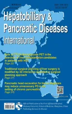
The paper[9]by Dr. Jia-Hong Dong’s group takes surgical planning beyond routine volumetrics. In this paper, the authors reviewed their experience where surgical plans were devised by two independent teams using conventional 2D imaging and compared it side-byside to 3D interactive quantitative surgical planning. For patients requiring a complex liver resection, the surgical plan was “optimized” using 3D imaging for 49 patients. More remarkably, 15 patients that were deemed unresectable by conventional 2D imaging were deemed resectable based on 3D imaging and went on to receive potentially curative surgery.
The use of 3D modeling is only the tip of the iceberg as this technology has been developed in parallel with navigation equipment for the liver. For complex neurosurgical procedures, the Stealth system combines imaging and intraoperative navigation and has largely become standard of care. Similar technology has been translated into hepatobiliary surgery and has recently been shown to be helpful for more challenging liver resections.[10]It was also found to be useful for ablations especially when the tumor was not detected via ultrasound.[11]Like 3D imaging, this technology has not become mainstream and its use is limited to only a few hospitals worldwide.
As stated by the authors, there are some limitations to the study by Dr. Dong’s group. One of the biggest issues was the study design and the inherent bias that was unavoidable. In this study, the chief surgeon always selected the operation and in all cases it was the plan devised by 3D imagery. There was also no feasible way to test the outcomes that were devised by the group that only used 2D imaging. Despite these limitations, the results of routinely utilizing 3D imagery for complex liver resection are still very promising.
Volumetrics and 3D reconstruction are essential for preoperative planning in living donor liver transplantation and afford optimal safety to both the donor and therecipient of the liver graft. Although likely not necessary for all patients undergoing liver resection for benign or malignant disease, patients that have borderline resectable disease clearly appear to benefit from this technology. There is no doubt that there is increased cost and time involved with 3D analysis, but the benefit of lives extended or saved from definitive surgical resection should outweigh these negative factors. Performing safer liver resections will likely also shorten the length of hospital stay, decrease postoperative complications, and lower mortality. Shouldn’t all patients being evaluated for a complex liver resection be afforded this expertise and technology?
Contributors:RTW wrote the whole manuscript and is the guarantor.
Funding:None.
Ethical approval:Not needed.
Competing interest:No benefits in any form have been received or will be received from a commercial party related directly or indirectly to the subject of this article.
1 Cescon M, Vetrone G, Grazi GL, Ramacciato G, Ercolani G, Ravaioli M, et al. Trends in perioperative outcome after hepatic resection: analysis of 1500 consecutive unselected cases over 20 years. Ann Surg 2009;249:995-1002.
2 Jarnagin WR, Gonen M, Fong Y, DeMatteo RP, Ben-Porat L, Little S, et al. Improvement in perioperative outcome after hepatic resection: analysis of 1803 consecutive cases over the past decade. Ann Surg 2002;236:397-407.
3 Ciria R, Cherqui D, Geller DA, Briceno J, Wakabayashi G. Comparative short-term benefits of laparoscopic liver resection: 9000 cases and climbing. Ann Surg 2016;263:761-777.
4 Dokmak S, Ftériche FS, Borscheid R, Cauchy F, Farges O, Belghiti J. 2012 Liver resections in the 21st century: we are far from zero mortality. HPB (Oxford) 2013;15:908-915.
5 Reichman TW, Sandroussi C, Azouz SM, Adcock L, Cattral MS, McGilvray ID, et al. Living donor hepatectomy: the importance of the residual liver volume. Liver Transpl 2011;17:1404-1411.
6 Shoup M, Gonen M, D’Angelica M, Jarnagin WR, DeMatteo RP, Schwartz LH, et al. Volumetric analysis predicts hepatic dysfunction in patients undergoing major liver resection. J Gastrointest Surg 2003;7:325-330.
7 DuBray BJ Jr, Levy RV, Balachandran P, Conzen KD, Upadhya GA, Anderson CD, et al. Novel three-dimensional imaging technique improves the accuracy of hepatic volumetric assessment. HPB (Oxford) 2011;13:670-674.
8 The Toronto Video Atlas of Surgery. Available from: http://pie. med.utoronto.ca/TVASurg.
9 Wang XD, Wang HG, Shi J, Duan WD, Luo Y, Ji WB, et al. Traditional surgical planning of liver surgery is modified by 3D interactive quantitative surgical planning approach: a singlecenter experience with 305 patients. Hepatobiliary Pancreat Dis Int 2017;16:271-278.
10 Cash DM, Miga MI, Glasgow SC, Dawant BM, Clements LW, Cao Z, et al. Concepts and preliminary data toward the realization of image-guided liver surgery. J Gastrointest Surg 2007;11:844-859.
11 Kingham TP, Scherer MA, Neese BW, Clements LW, Stefansic JD, Jarnagin WR. Image-guided liver surgery: intraoperative projection of computed tomography images utilizing tracked ultrasound. HPB (Oxford) 2012;14:594-603.
May 4, 2017
Accepted after revision May 12, 2017
Author Affiliations: Division of Transplantation, Department of Surgery, Virginia Commonwealth University, Richmond, VA, USA (Reichman TW)
Trevor W Reichman, MD, PhD, Associate Professor of Surgery, Division of Transplantation, Department of Surgery, Virginia Commonwealth University, PO Box 980057, Richmond, VA 23298, USA (Tel: +1-804-828-2461; Fax: +1-804-828-4858; Email: trevor.reichman@ vcuhealth.org)
© 2017, Hepatobiliary Pancreat Dis Int. All rights reserved.
10.1016/S1499-3872(17)60024-9
Published online May 23, 2017.
