aVR导联ST段抬高结合肌钙蛋白I预测非ST段抬高型急性冠状动脉综合征患者预后
2016-12-17张晓晖
张晓晖 曾 伟
泰山医学院附属医院心电图室,山东泰安 271000
aVR导联ST段抬高结合肌钙蛋白I预测非ST段抬高型急性冠状动脉综合征患者预后
张晓晖曾 伟
泰山医学院附属医院心电图室,山东泰安 271000
目的 探讨心电图aVR导联ST段抬高结合肌钙蛋白Ⅰ(cTnI)在预测非ST段抬高型急性冠状动脉综合征(NSTE-ACS)患者预后中的价值。 方法 选择本院自2014年1月~2015年1月收集205例NSTEACS患者,对所有患者均测定cTnⅠ,测量心电图aVR导联ST段抬高程度并行冠脉造影,并根据结果分别行冠脉介入治疗或药物保守治疗,出院后随访6个月。205例患者分成四组:A组,cTnI阴性结合aVR-ST不抬高组(100例);B组,cTnI阴性结合aVR-ST抬高组(31例);C组,cTnI阳性结合aVR-ST不抬高组(43例);D组,cTnI阳性结合aVR-ST抬高组(31例)。观察分析四组间左主干病变或三支冠脉病变发生率及不良心血管事件(包括死亡、心肌梗死、再梗、血运重建)的发生率,P<0.05为差异有统计学意义。结果 (1)左主干病变或三支冠脉病变发生率:D组显著高于A组(χ2=78.9973,P=0.0000)、B组(χ2=13.8091,P=0.0002)及C组(χ2=44.7846,P=0.0000);B组显著高于A组(χ2=19.7072,P=0.000)及C组(χ2=10.8407,P=0.0009);A组与C组间无统计学差异(χ2=0.0173,P=0.8952)。(2)不良心血管事件的发生率:D组显著高于A组(χ2=89.4940,P=0.0003)、B组(χ2=11.0878,P=0.0009)及C组(χ2=38.5719,P=0.0005);B组显著高于A组(χ2=29.8000,P=0.0005)及C组(χ2=9.5431,P=0.0020);A组与C组间无统计学差异(χ2=2.0582,P=0.1514)。 结论 联合心电图aVR导联ST段抬高和肌钙蛋白I阳性对预测NSTE-ACS患者的预后有重要的临床价值,值得心血管临床给予其更高程度的关注。
aVR导联ST段抬高;肌钙蛋白I;非ST段抬高型急性冠状动脉综合征;预后
非ST段抬高型急性冠状动脉综合征(non-ST-segment elevation acute coronary syndrome,NSTEACS)是由不稳定性动脉粥样硬化斑块不完全阻塞相关冠状动脉导致的急性心脏疾病,且以血小板为主的“白血栓”,是临床上最常见的冠心病类型之一。NSTE-ACS患者的冠状动脉粥样硬化斑块具有不稳定性,临床上不良心血管事件发生的危险较高。早期识别此类患者,有助于准确判断预后,在临床治疗方案的制定,减低致残率、致死率方面意义重大。据报道,在判断NSTE-ACS患者的预后方面,相对于其余导联ST段压低而言,aVR导联ST段抬高有更高的预测价值[1-2],另有文献显示,不管NSTE-ACS患者的近期或远期预后,肌钙蛋白I都与之密切相关[3-4],但目前少有研究联合二者用于NSTE-ACS患者早期危险分层,本研究旨在探讨二者的结合在预测NSTE-ACS患者预后中的价值。
1 资料与方法
1.1一般资料
本院自2014年1月~2015年1月收集的205例NSTE-ACS患者,对所有患者均测定cTnI,测量心电图aVR导联ST段抬高程度并行冠脉造影,并根据结果分别行冠脉介入治疗或药物保守治疗,出院后随访6个月。其中男134例,女71例,平均年龄(66.0±10.0)岁。诊断均符合中华医学会心血管病学分会关于NSTE-ACS诊断标准。入选标准:(1)急性非ST段抬高型心肌梗死;(2)不稳定性心绞痛,包括初发型、恶化劳累型、静息型及梗死后心绞痛。排除标准:(1)变异型心绞痛;(2)近6个月内行PCI治疗者;(3)曾行冠脉旁路移植术;(4)肾功能衰竭患者;(5)肿瘤患者;(6)血液病患者;(7)脓毒血症和感染性休克等其他非冠脉疾病。
1.2实验室检测与心电图描记
所有患者采血化验cTnI,参考值0~0.4ng/mL,排除检查标本所致肌钙蛋白假阳性,以指标高于参考值者为阳性。所有患者记录标准12导联心电图,测量ST段的方法:以TP段位为基线,把QRS波群起点当作基点,将J点后80ms作为ST段测量点,进行测量,以任何心电图导联ST段偏离基线≥0.5mm认为出现ST段变化,连续测量6个ST段,取其平均值作为测量数据,记录aVR导联ST段抬高值。
1.3治疗与随访
所有患者住院期间给予双重抗血小板、低分子肝素钙及他汀类药物,根据病情给予控制心率、扩张冠脉、改善心功能等药物。均行选择性冠状动脉造影术,冠脉造影结果确定分组,左冠状动脉主干狭窄程度≥50%者为左主干病变,右冠状动脉、左冠状动脉前降支、左冠状动脉回旋支狭窄程度均≥50%者为三支病变。随访6个月,观察患者非致死性心肌梗死(包括再梗)、心力衰竭、心血管死亡以及血运重建等不良心血管事件的发生率。
1.4统计学方法
应用SPSS17.0软件进行数据分析,计数资料以频数或率表示,计量资料以表示,计量资料采用t检验;计数资料采用χ2检验。P<0.05为差异有统计学意义。
2 结果
2.1aVR-ST不抬高组和aVR-ST抬高组两组间比较
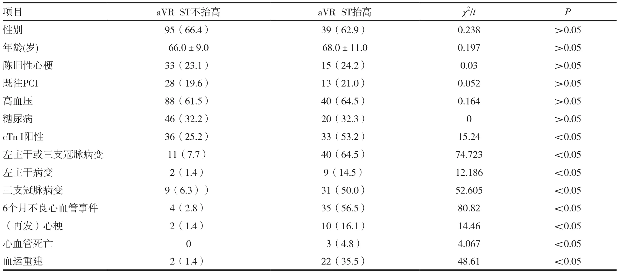
两组的性别、年龄、陈旧心梗、既往PCI、高血压及糖尿病构成无统计学差异。aVR-ST抬高组患者的cTnI阳性率、左主干或三支冠脉病变率及6个月内不良心血管事件发生率均高于aVR-ST不抬高组。差异有统计学意义(P<0.05)。见表1。
2.2cTnI阴性组和cTnI阳性组临床资料比较
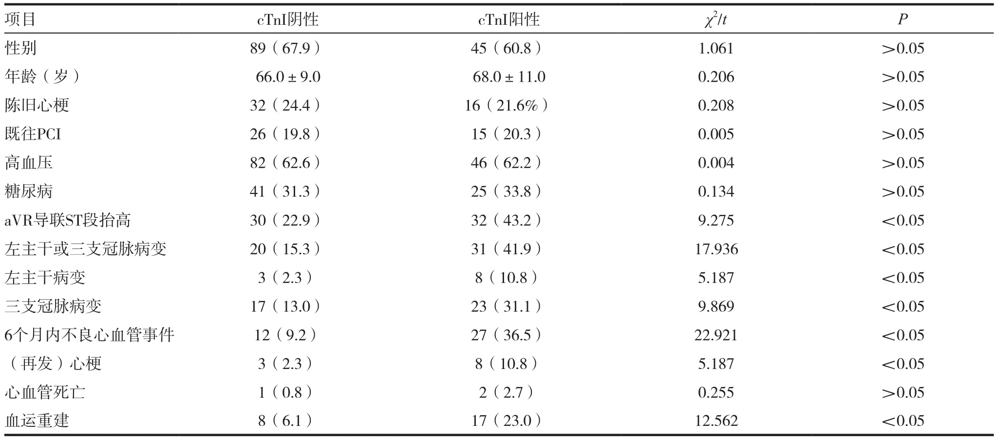
以cTnI是否升高分为cTnI阴性组(cTnI<0.4ng/mL)与cTnI阳性组(cTnI>0.4ng/mL)。两组的性别、年龄、陈旧心梗、既往PCI、高血压及糖尿病构成无统计学差异。cTnI阳性组患者的aVR导联ST段抬高率、左主干或三支冠脉病变率以及6个月内不良心血管事件发生率均高于cTnI阴性组,差异有统计学意义。见表2。
2.3aVR导联ST段变化与cTn I结合,各组不良心血管事件发生比较
将aVR导联ST段变化与cTnI结合,将患者分为4组。A组:aVR-ST不抬高与cTnI阴性,B组:aVRST抬高与cTnI阴性,C组:aVR-ST不抬高与cTnI阳性,D组:aVR-ST抬高与cTnI阳性。①两两比较 四组患者的左主干或三支冠脉病变率,以P<0.05为差异有统计学意义。A组与B组比较,χ2=19.7072,P=0.000;A组与C组比较,χ2=0.0173,P=0.8952;B组 与 C组 比 较,χ2=10.8407,P=0.0009;D组与A组比较,χ2=78.9973,P=0.0000;D组与B组比较,χ2=13.8091,P=0.0002;D组与C组比较,χ2=44.7846,P=0.0000。结果提示D组患者的左主干或三支冠脉病变率最高,B组次之,A组与C组间差异无统计学意义。②两两比较 4组患者6个月内不良心血管事件发生率,以P<0.0071为差异有统计学意义。A组与B组比较,χ2=29.80000,P=0.0005;A组与C组比较,χ2=2.0582,P=0.1514;B组与C组比较,χ2=9.5431,P=0.0020;D组与A组 比 较,χ2=89.4940,P=0.0003;D组 与 B组比较,χ2=11.0878,P=0.0009;D组与C组比较,χ2=38.5719,P=0.0005。结果提示A组与C组间差异无统计学意义,D组不良心血管事件的发生率高于其他三组。见表3。

表1 aVR-ST不抬高组和aVR-ST抬高组临床资料

表2 cTnI阴性组和cTnI阳性组临床资料比较
3 讨论
NSTE-ACS包括不稳定性心绞痛和非ST段抬高型心肌梗死(NSTEMI),并发症多、病死率高,为指导临床早期干预,Braunwald等[5]主张对NSTEACS患者进行危险分层。NSTE-ACS时,冠脉常常存在不完全阻塞,多数患者常有短暂性ST段压低或T波低平、倒置,但部分患者虽有严重狭窄,也可无心电图改变。尽管如此,体表心电图检查还是早期诊断NSTE-ACS简单有效的非创伤性检查。Barrabes等[1]研究发现,ST段上抬≥0.5mm时,aVR导联ST段抬高的程度与患者的临床预后有显著相关性。Filip等[6,12-13]研究证实aVR导联ST段抬高及其程度可以作为NSTE-ACS患者30天死亡率的独立预测因素。Kosuge等[7-8]认为aVR导联ST段抬高是对NSTE-ACS患者是否存在三支或左主干病变及其短期预后的判断的有效指标。已有研究证实,多支冠脉病变以及左主干病变患者的aVR导联ST段抬高的发生率明显高于不抬高组,并且在患者血运重建治疗情况上统计学差异,这都提示aVR导联ST段的抬高对预测NSTE-ACS患者的预后有重要价值。

表3 aVR导联ST段变化与cTn I结合,各组不良心血管事件比较[n(%)]
肌钙蛋白(troponin,Tn)是横纹肌收缩的重要调解蛋白,由TnI、TnT和TnC三个亚单位组成,TnI又分为心肌亚型(cTnI)、快骨骼肌亚型(fTnI)和慢骨骼肌亚型(sTnI)3种亚型。cTnI在基因上有特异的氨基酸排列,仅存在于心肌内,但在个体发育的各个阶段都不在骨骼肌中表达[9],因而具有高度的心肌特异性。cTnI的特异性及时间窗较肌红蛋白或CK-MB都短,对30d以内的短期预后及1年以内的长期预后有预测价值。NSTE-ACS患者CK-MB正常但cTnI增高的,其死亡风险增高。而且随着cTnI值增高, NSTE-ACS患者死亡风险增大。Apple等[10,14]指出cTnI升高可作为独立指标用以预报NSTE-ACS患者心脏事件的危险性,可以用于判断NSTE-ACS患者的预后。Merrill等[11]研究也证实,cTnI水平的测定可作为ACS患者危险分层中的重要指标。本研究显示cTnI阳性的NSTE-ACS患者其左主干或三支冠脉病变率以及6个月内不良心血管事件发生率均明显高于cTnI阴性组,提示cTnI水平的测定可用于NSTEACS患者危险分层,这与上述其他研究结果一致。
而且本研究对NSTE-ACS患者的心电图aVR导联ST段的偏移情况及cTnI水平进行不同组合,随访6个月后,结果显示,aVR导联ST段抬高合并cTnI阳性时,不管左主干病变、三支冠状动脉血管病变的发生率,还是不良心血管事件(包括心肌梗死、再梗塞、死亡、行血运重建等)的发生率均明显高于其他组合。综上所述,cTnI阳性及aVR导联ST段抬高联合判断对预测NSTE-ACS患者的风险及预后有重要的临床价值,值得我们重视。
[1]Barrabes JA,Figueras J,Moure C,et al.Prognostic value of lead aVR in patients with a first non ST segment elevation acute myocardial infarction[J].Circulation,2003,108(7):814-819.
[2]Kosuge M,Kimura K,Ishikawa T,et al.Predictors of left main or three vessel disease in patients who have acute coronary syndromes with non ST-segment elevation[J].Am J Cardio,2005,95(11):1366-1369.
[3]Ottani F,Galvani M,Nicolini FA,et al.Elevated cardiac troponin levels predict the risk of adverse outcome in patients with acute coronary syndromes[J].Am Heart J,2000,140(6):917-927.
[4]Antman EM,Tanasijevic MJ,Thompson B,et al.Cardiac specific troponin I levels to predict the risk of mortality in patients with acute coronary syndromes[J].N Engl J Med,1996,335(5):1342-1349.
[5]Braunwald E,Antman EM,Beaskey JM,et al.ACC/ AHA guidelines for the management of patients with unstable angina and non-ST-segment elevation myocardial infarction executive summany and recommendations[J]. Circulation,2000,102(10):1193-1209.
[6]Filip M,Szymański,Marcin Grabowski,et al.Admission ST-segment elevation in lead aVR as the factor improving complex risk stratification in acute coronary syndromes[J]. Am J Emer Med,2008,26(4):408-412.
[7]Kosuge M,Kimura K,Ishikawa T,et al.Combined prognostic utility of ST segment in lead aVR and troponin T on admission in non-ST-segment elevation acute coronary synodromes[J].Am J Cardiol,2006,97(3):334-339.
[8]Kosuge M,Ebina T,Hibi K,et al.ST-segment elevation resolution in lead aVR:A strong predictor of adverse outcome in patients with non-ST-segment elevation acute coronary syndromes[J].Circ J,2008,72(7):1047-1053.
[9]Bodor GS,Porterfield D,Voss EM,et al.Cardiac troponin-I is not expressed in fetal and healthy or diseased adult human skeletal muscle tissue[J].Clin Chem,1995,41(12):1710-1715.
[10]Apple FS,Smith SW,Pearce LA,et al.Use of the Centaur TnI Ultra assay for detection of myocardial infarction and adverse events in patients presenting with symptoms suggestive of acute coronary syndrome[J].Clin Chem,2008,54(4):723-728.
[11]Merrill B,Calvin JE,Klein LW, et al.Supplemental value of troponin I combined with a clinical risk model to predict in-hospital events in intermediate and high-risk patients with acute coronary syndromes[J].Invasive Cardiol,2002,14(10):603-608.
[12]Kosuge M,Ebina T,Hibi K,et al.ST-segment elevation resolution in lead aVR:a strong predictor of adverse outcomes in patients with non-ST-segment elevation acute coronary syndrome[J].Circulation Journal Official Journal of the Japanese Circulation Society,2008,72(7):1047-1053.
[13]Taglieri N,Marzocchi A,Saia F,et al.Short- and Long-Term Prognostic Significance of ST-Segment Elevation in Lead aVR in Patients With Non-ST-Segment Elevation Acute Coronary Syndrome[J].American Journal of Cardiology,2011,108(1):21-28.
[14]Morrow DA,Cannon CP,Rifai N,et al.Ability of minor elevations of troponins I and T to predict benefit from an early invasive strategy in patients with unstable angina and non-ST elevation myocardial infarction: results from a randomized trial[J].Jama the Journal of the American Medical Association,2001,286(19):2405-2412.
aVR lead ST segment elevation combined with cardiac troponin I in prediction of prognosis of patients with Non-ST-segment elevation acute coronary syndrome
ZHANG XiaohuiZENG Wei
Electrocardiographic Room,Affiliated Hospital ofTaishan Medical College,Tai'an 271000,China
Objective To investigate the value of electrocardiogram aVR lead ST segment elevation combined with cardiac troponin I in prediction of prognosis of patients with Non-ST-segment elevation acute coronary syndrome. Methods 205 cases of patients with Non-ST-segment elevation acute coronary syndrome from January 2014 to January 2015 in our hospital were included in our study.All patients were detected cTnl,detected ST segment elevation of aVR lead in electrocardiogram,and carried on coronary angiography.According to the results,all of them were followed up for 6 months after discharge.They were divided into 4 groups:group A that was cTnl negative combined with AVR-ST non- elevation group (100 cases),group B that was cTnl negative combined with AVRST elevation group(31 cases),group C that was cTnl positive combined with AVR-ST non- elevation group (43 cases),group D that was cTnl positive combined with AVR-ST elevation group (31 cases).Incidence of left main lesion or three coronary artery disease and incidence of adverse cardiovascular events (including death, myocardial infarction,reinfarction,revascularization) of the four groups were observed.There were statistical significance (P<0.05). Results (1)Incidence of left main lesion or three coronary artery disease:group D was significantly higher than group A (χ2=78.9973,P=0.0000),group B (χ2=13.8091,P=0.0002) and group C (χ2=44.7846,P=0.0000).Group B was significantly higher than group A (χ2=19.7072,P=0.000) and goup C (χ2=10.8407,P=0.0009).There was no statistical significance between group A and group C (χ2=0.0173,P=0.8952).(2)Incidence of adverse cardiovascular events:group D was significantly higher than group A (χ2=89.4940,P=0.0003),group B (χ2=11.0878,P=0.0009) and group C(χ2=38.5719,P=0.0005).Group B was sinificantly higher than group A (χ2=29.8000,P=0.0005) and group C (χ2=9.5431,P=0.0020).There was no statistical significance between group A and group C (χ2=2.0582,P=0.1514). Conclusion The value of electrocardiogram aVR lead ST segment elevation combined with cardiac troponin I inprediction of prognosis of patients with Non-ST-segment elevation acute coronary syndrome is great.It deserves a higher degree of attention to cardiovascular clinical.
aVR lead ST segment elevation;Troponin I;Non-ST-segment elevation acute coronary syndrome;Prognosis
R541.4
A
2095-0616(2016)18-11-05
(2016-07-22)
