Central neuromechanisms underlying control of intragastric pressure through acupuncture at Zusanli (ST36) in rats: the upper cervical cord is the key link between the ascending and descending pathways
2016-12-02ChunyanYongShuChenHengChenXiaoChuChaoZhangChengTanLanYeJiangshanLi
Chun-yan Yong, Shu Chen, Heng Chen, Xiao Chu, Chao Zhang, Cheng Tan, Lan Ye, Jiang-shan Li,
1 Department of Integrative Medicine, Xiangya Hospital of Central South University, Changsha, Hunan Province, China
2 School of Acupuncture and Massage, Hunan University of Chinese Medicine, Changsha, Hunan Province, China
RESEARCH ARTICLE
Central neuromechanisms underlying control of intragastric pressure through acupuncture at Zusanli (ST36) in rats: the upper cervical cord is the key link between the ascending and descending pathways
Chun-yan Yong1, Shu Chen2, Heng Chen2, Xiao Chu2, Chao Zhang2, Cheng Tan2, Lan Ye2, Jiang-shan Li2,*
1 Department of Integrative Medicine, Xiangya Hospital of Central South University, Changsha, Hunan Province, China
2 School of Acupuncture and Massage, Hunan University of Chinese Medicine, Changsha, Hunan Province, China
Graphical Abstract
orcid: 0000-0001-5482-306X (Jiang-shan Li)
Accepted: 2015-12-22
Sensory inputs stimulated by Zusanli (ST36) acupuncture in the abdomen are known to converge in the upper cervical cord. However, it is unclear whether these inputs are subsequently conveyed to the hypothalamic paraventricular nucleus and what kind of afferent fibers are involved. We focused on the upper cervical cord, where afferent inputs converge, and detected c-fos expression in oxytocinergic neurons. We found that Zusanli acupuncture therapy effectively elevated intragastric pressure, but inhibited expression of c-fos in oxytocinergic neurons of the paraventricular nucleus in upper cervical cord injured rats. These Zusanli acupuncture effects remained even after complete dorsal cord transection. However, after complete transection of the spinal cord or dorsolateral funiculus, the effects were significantly attenuated and even disappeared. These findings suggest that the paraventricular nucleus is responsible for pooling and integrating signals from the Zusanli acupuncture and sensory information from the intragastric pressure variation, thereby contributing to the regulation of intragastric pressure. The upper cervical cord serves as the key link between ascending and descending pathways, which conveys afferent inputs to the paraventricular nucleus through the dorsolateral funiculus.
nerve regeneration; traditional Chinese medicine; acupuncture; Zusanli (ST36); upper cervical cord; dorsolateral funiculus; dorsal cord; hypothalamic paraventricular nucleus; intragastric pressure; immunohistochemical double staining; nervous pathway; neural regeneration
Introduction
Zusanli (ST36), the He-Sea point of the stomach meridian, is located on the anterior aspect of the lower leg below Dubi (ST35) and one finger-breadth (middle finger) from the anterior crest of the tibia. Studies have shown that acupuncture at Zusanli influences brain activities and improves gastric mobility (Chen et al., 2013), gastric secretion (Yang et al., 2013), and gastric electrical activity (Yang et al., 2014). As previously reported (Chen et al., 2014), acupuncture at Zusanli significantly increases discharge frequency in paraventricular nucleus (PVN) neurons of the hypothalamus in gastric distension rats. It has been suggested that acupuncture plays a role in PVN neuron activation, and that the PVN neurons are activated by physiological changes due to gastric distension and stress from acupuncture (Chen et al., 2014). However, very little is known about the type of PVN neurons that are involved in this regulatory process. There are abundant oxytocinergic neurons and high-density oxytocin (OT) receptors in the PVN (Gerald et al., 2001), and oxytocinergic neurons can significantly protect gastric mucosa against ischemia-reperfusion injury by reducing gastric juice output and acidity, and play a role in mechanisms within the stomach (Zhang et al., 2008). Oxytocinergic neurons are thought to be involved in acupuncture signal transductions that regulate gastric function. However, further studies are needed to determine the effect of acupuncture at Zusanli on activation of PVN oxytocinergic neurons and the specific signal transduction pathways that are involved.
Afferent fibers from the visceral organs, as well as afferent nerves from corresponding puncture sites, converge into spinothalamic tract neurons via the same posterior root, thereby generating acupuncture signals for visceral regulation (Wessel and Lai, 1997). However, maintenance of spinal cord structural integrity is crucial for efficacy of acupuncture input and information. The spinal cord is a complex neural structure that comprises 31 spinal nerve segments (vertical linkage) and upstream and downstream fibers (horizontal linkage). The upper cervical cord (C1—3segments) is responsible for the communication of nerve centers with spinal nerve segments and plays an important role in transmission and modulation of visceral information (Qin et al., 2004). In a gastric distension experiment, half of the neural signals from the stomach were conveyed into the C1—2segments via the spinal nerves, indicating that the upper cervical cord is involved in integration of afferent information from the stomach (Qin et al., 2003). Additionally, acupuncture at Zusanli has been shown to significantly increase the number of c-fos-positive cells in the C1segment (Wang et al., 2010). Taken together, these studies have shown that acupuncture information and sensory inputs from the stomach converge in the upper spinal cord (C1—3).
In this study, micromanipulation techniques were employed for sham surgery on the upper spinal cord, as well as experimental dorsal cord transection, dorsolateral funiculus transection, and spinal cord transection in rats. Using the upper spinal cord as an entry point, we investigated the signaling pathway by which acupuncture at Zusanli regulated gastric function. We analyzed the vertical and horizontal linkages of the upper spinal cord to determine whether acupuncture information is conveyed from the upper spinal cord into the PVN, and what type of afferent fibers are involved, as well as the effects of acupuncture at Zusanli on c-fos expression in PVN oxytocinergic neurons and intragastric pressure.
Materials and Methods
Experimental animals
Forty healthy, adult, Sprague-Dawley rats (half male and female), 8—10 weeks of age, weighing 250—350 g, were provided by the Animal Laboratory of Hunan University of Chinese Medicine in China (license No. SCXK (Xiang) 2011-0003). Animals were housed at a temperature of 25 ± 2°C and a humidity of 60%, with a 12-hour light/dark cycle, and allowed free access to water and food. Then, all rats were fasted for 12 hours prior to gastric pressure measurement with free access to water, and only fed 5% glucose saline at a ratio of 1:1 by oral gavage. During the experiment, the disposal of animals conformed to relevant ethical standards of Ethical Issues in Animal Experimentation issued in 2009.
Modeling and grouping
A total of 40 rats with an equal number of males and females were randomly assigned to five groups (n = 8 per group): (1) sham surgery (sham), in which the dura was opened with no treatments; (2) sham surgery + acupuncture (S + A), in which the dura was opened with acupuncture interventions; (3) dorsal cord transection + acupuncture (DCT + A), in which the rats were given acupuncture treatment following dorsal cord transection; (4) dorsolateral funiculus transection + acupuncture (DLFT + A), in which rats were given acupuncture treatment after dorsolateral funiculus transection; (5) spinal cord transection + acupuncture (SCT + A), in which rats were given acupuncture treatment after spinal cord transection.
Following anesthesia with intraperitoneal injection of 20% urethane (50 mg/kg, Shanghai Experiment Reagent Co., Ltd., Shanghai, China), rats from each group were fixed in a prone position for cervical skin preparation. Under sterile conditions, a 2-cm-long longitudinal incision was made along the spinous process, with the C2spinous process as the center, for blunt dissection of the bilateral paraspinal muscles until the C1—3spinous processes were completely exposed. In the DCT + A and DLFT + A groups, bilateral paraspinal muscles were separated using a microscopic distractor (Martin Alber GmbH & Co., Baden-Württemberg, Germany) following exposure of the dorsal funiculus or dorsolateral funiculus, respectively. The C1—3spinous processes were then selected using microscopic bone rongeur forceps (GIMMI GmbH, Baden-Württemberg, Germany) to expose the lamina. Finally, the lamina was removed from back to front to completely expose the spinal cord under a 10× surgical microscope, followed by a 0.5-cm-long longitudinal cut to the dura mater.
In the S and S + A groups, subsequent experiments were performed after opening the dura. In the DCT + A and

Figure 1 Effects of acupuncture at Zusanli (ST36) on c-fos-positive cell number in PVN oxytocinergic neurons of rats with upper cervical cord injury.
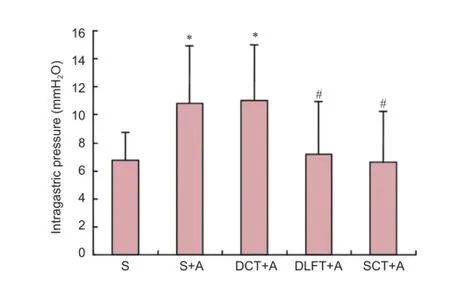
Figure 3 Acupuncture at Zusanli (ST36) controlled intragastric pressure in rats with upper cervical cord injury.
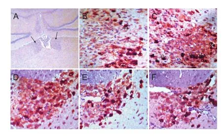
Figure 2 Effects of acupuncture at Zusanli (ST36) on c-fos expression in PVN oxytocinergicneurons of rats with upper cervical cord injury.
DLFT + A groups, the bilateral dorsal funiculus or dorsolateral funiculus was gently disconnected. To ensure complete transection of the dorsal funiculus or dorsolateral funiculus, the broken ends were repeatedly cut 3—4 times. In the SCT + A group, the spinal cord was completely resected at the C3segment, and this transection process was repeated 3—4 times (Yin et al., 2008). In all groups, the broken ends were pressed gently using gauze to stop bleeding, rinsed with normal saline, and covered with a gelatin sponge. The surrounding muscles on both ends were sutured to fill in any defects due to the laminectomy, followed by closing the fascia and skin layer by layer.
Gastric balloon insertion
After model establishment, rats from each group were bound to a board in a supine position. The hair on the abdomen was removed, and the abdomen was opened longitudinally along the midline underneath the xiphoid process to expose the gastric body. A small incision was made in the duodenum, about 0.5 cm to the pylorus, to avoid blood vessels. A balloon, which was attached to a polyethylene tube, was inserted into the antrum and moderately inflated. The other end of the polyethylene tube was pulled out from the body, followed by closing the abdominal wound. A pressure transducer, which was connected to the polyethylene tube (using a 12# needle and a T-branch pipe), was connected to a BL-420F bioinformatics recording system (TechMan Soft, Chengdu, China). After the waveform was stabilized in the sham group, average gastric pressure was recorded within 2 minutes. In the other groups, the variation in average amplitude of intragastric pressure within a unit interval was calculated based on a 2-minute recording of gastric pressures.
Acupuncture treatment
Zusanli is a bilateral acupoint about 5 mm below the fibular head and posterolateral to the knee (Li et al., 2007). Inthis study, an attachment point was selected 2 mm below the acupoint. With exception to the sham group, a sterile acupuncture needle (0.3 mm × 13.0 mm, Suzhou Medical Appliance Factory, Jiangsu, China) was inserted into the Zusanli acupoint of the right limb at a depth of 3—5 mm. Electrical stimulation was performed for 20 minutes using a G-6805 electro-acupuncture device (Shanghai Medical Electronic Instrument Factory, Shanghai, China). The anode was connected to the acupoint, and the cathode was connected to the attachment point. Electrical stimulation consisted of dilatation waves: 4-Hz sparse wave; 20-Hz dense wave; 0.5-ms pulse density; 2—4 V output voltage). The electrical stimulation was considered successful when the limbs exhibited a mild trembling.
Specimen preparation and immunohistochemical staining Researchers were not aware of study allocation. Two hours after acupuncture, rats in all the groups, except for the sham group, were transcardially perfused using a BT-200B constant pump (Shanghai Jieweifu Medical Co., Ltd., Shanghai, China; 200 mL normal saline perfused at a speed of 10 mL/min followed by 4% paraformaldehyde perfusion for 40 minutes), and then brain tissue was resected. The cerebral cortex was removed, and the hippocampus and hypothalamus were reserved. In the sham group, brain tissues were collected at 2 hours after surgery. All brain samples were fixed in 4% paraformaldehyde for 24 hours at 4°C, and the samples were then dehydrated in 30% sucrose/PBS, followed by paraffin embedding, and sliced into 5-µm-thick serial sections. Subsequently, the sections were deparaffinized and rehydrated, incubated in 3% H2O2at room temperature for 5—10 minutes, heated to boiling in a microwave with citrate buffer, and then cooled to room temperature. The sections were washed with phosphate-buffered saline (PBS) (2 minutes × 3 times) between each step. Afterwards, the sections were incubated with a mixture of primary antibodies (mouse anti-rat OT monoclonal antibody and rabbit anti-rat c-fos polyclonal antibody at a working concentration of 1:100; Abcam, USA), at 37°C for 2 hours, followed by horseradish peroxidase-labeled goat anti-mouse IgG and alkaline phosphatase-labeled goat anti-rabbit IgG (1:100; ZSGS-BIO, Beijing, China) at 37°C for 10—30 minutes, and horseradish peroxidase-labeled streptavidin working solution at 37°C for 30 minutes. The sections were also rinsed in PBS (2 minutes × 3 times) between each step. After developing the staining with diaminobenzidine tetrahydrochloride, the sections were rinsed in PBS (2 minutes × 2 times) and incubated in a mixture (ZSGS-BIO) of 4A (diluent), 4B (GBL activator), and 4C (GBL receptor substrate). After rinsing with tap water, the sections were counterstained with hematoxylin-eosin, washed with tap water, dehydrated for 5—10 minutes, cleared for 5—10 minutes, and mounted with neutral resin. The seconds were then covered with a coverslip and allowed to dry. The sections were analyzed using a 40× optical microscope (LEICA Instrument Co., Braunschweig, Germany). Five sections from each rat were randomly selected to quantify OT/c-fos co-labeled cells using a MIAS medical image analysis system (Beijing Beihang Tianhua Modern Scien-Tech Co., Ltd., Beijing, China).
Statistical analysis
Measurement data are expressed as the mean ± SD and were analyzed using SPSS17.0 statistical software (SPSS, Chicago, IL, USA). Intergroup comparison was done using one-way analysis of variance and the least significant difference test. A level of significance was selected at bilateral α = 0.05.
Results
c-fos expression in PVN oxytocinergic neurons following acupuncture at Zusanli in rats with upper cervical cord injury
Compared with the sham group, the number of OT/c-fos co-labeled cells significantly decreased in the S + A and DCT + A groups (P < 0.05), but there was no significant difference between these two groups (P > 0.05). Acupuncture at Zusanli inhibited proliferation of PVN oxytocinergic neurons, and this inhibitor effect remained after dorsal cord transection, indicating that dorsal cord transection had little effect on regulation of PVN oxytocinergic neurons following acupuncture at Zusanli. Compared with the sham group, the number of PVN OT/c-fos co-labeled cells remained unchanged in the DLFT + A and SCT + A groups (P > 0.05). However, compared with the S + A group, the number of PVN OT/c-fos co-labeled cells significantly increased in these two groups (P < 0.05). Additionally, compared with the DCT + A group, the number of PVN OT/c-fos co-labeled cells significantly increased in these two groups (P < 0.05; Figure 1).
Three types of positive cells were identified by immunohistochemical staining under an optical microscope: (1) c-fos-positive cells with brown-stained nuclei that were mostly round or oval; (2) OT-positive cells with a red cytoplasm, and nuclei were not stained; (3) OT/c-fos co-labeled cells with a brown nuclei and red cytoplasm. Double-labeled cells were evenly distributed throughout the PVN. The cells were mostly oval-shaped or spindle- and multi-polar shaped (Figure 2).
Variation of intragastric pressure in upper cervical cord injury rats after acupuncture at Zusanli
Compared with the sham group, intragastric pressure significantly increased in the S + A and DCT + A groups (P <0.05), but no significant difference was found between these two groups (P > 0.05). Acupuncture at Zusanli effectively increased intragastric pressure in rats undergoing dorsal cord transection, indicating that dorsal cord transection had little effect on regulation of intragastric pressure following acupuncture at Zusanli. Compared with the sham group, there was no significant change in intragastric pressure in the DLFT + A and SCT + A groups (P > 0.05). Compared with the S + A and DCT + A groups, intragastric pressure significantly decreased in the DLFT + A and SCT + A groups (P <0.05; Figure 3).
Discussion
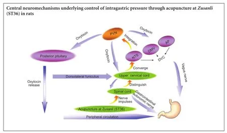
Traditional acupuncture was used to regulate gastric function and focused mainly on the peripheral nervous system rather than the central nervous system. Additionally, little has been reported on the association between ascending or descending conduction bundles with gastric function regulation. Therefore, we explored the effect of acupuncture at Zusanli on regulation of intragastric pressure after upper cervical cord injury or transection of conduction bundles, analyzing the two following aspects: (1) activation of oxytocinergic neurons in the PVN (nerve center) and (2) variation of intragastric pressure (effector). With the intermediate link (upper cervical cord) as the entry point, this study combined morphological changes of the central nervous system and visceral physiological responses, thereby providing novel insight into meridians (acupoints)-viscera research.
Results from this study showed that acupuncture at Zusanli effectively reduced the number of OT/c-fos co-labeled cells and increased intragastric pressure, although these regulatory effects were significantly decreased after dorsolateral funiculus or spinal cord transection. These results suggested that the dorsolateral funiculus is the main conduction bundle responsible for acupuncture signal transduction into the PVN. Additionally, deep sensitivity of the lower limbs almost disappears in patients suffering from dorsal cord injuries. However, these patients have no superficial sensory disorders, although there are normal acupuncture sensations in the lesioned area (National Neurosurgery Acupuncture Anesthesia Cooperation, 1986). Our results confirmed that the ascending neural pathways along the dorsal cord have little effect on acupuncture signal transduction. As reported by Silva et al. (2010), electroacupuncture-induced analgesia in the rat tail-flick test disappeared after neural block of the bilateral dorsolateral funiculus. This suggests that the dorsolateral funiculus is one of the main neural tracks for acupuncture signal transduction into the PVN.
In this study, the PVN was involved in acupuncture signal transduction, and acupuncture at Zusanli increased intragastric pressure by activating oxytocinergic neurons in the PVN in rats without upper cervical cord injuries. Flanagan et al. (1992) reported that gastric motility was inhibited by OT microinjection into the dorsal motor nucleus of the vagus (DMV) or PVN, but this inhibition was blocked by vagotomy or microinjection of an OT receptor antagonist into the PVN. Their findings suggest that PVN-released oxytocin exerts a tonic inhibitory effect on gastric function, which is consistent with our findings. Currently, this inhibitory effect is thought to be due to increased vagal neuronal excitability in the brainstem (Llewellyn-Smith et al., 2012).
The vagal preganglionic fibers innervating the stomach originate from the dorsal vagal complex in the brainstem and are regarded as one of the nerve centers contributing to regulation of gastric function using acupuncture at Zusanli (Liu et al., 2004). These fibers are composed of the DMV, nucleus tractus solitarii (NTS), and area postrema (AP). The NTS and AP receive inputs from the stomach, which are subsequently conveyed with acupuncture information via the upper cervical cord in the NTS (Li et al., 2009). There is direct contact between the NTS and PVN via some neurotransmitters (Praful et al., 2012). Therefore, signals from the NTS are projected directly to the PVN, which inhibits oxytocinergic neurons in the PVN. Projections from PVN-produced OT go through the following pathways: (1) some projections pass through the PVN-dorsal venous complex (DVC)-vagus pathway, which suggests that a large amount of DMV and PVN neurons have a single synaptic contact (Benarroch et al., 2005); the DMV, along with the nucleus ambiguous of the medulla oblongata retrograde parasympathetic innervation, controls contractions of gastric smooth muscle (Yuki et al., 2013). (2) Some PVN oxytocinergic neurons directly project to the spinal cord (Martínez-Lorenzana et al., 2007) and act on the gastrointestinal vegetative nerves, which influences the gastrointestinal system. (3) Some of the PVN-released OT is released via the posterior pituitary into the blood and acts on the gastrointestinal system through the peripheral circulation and nerve systems (Zhou et al., 2013). Consequently, acupuncture at Zusanli inhibits OT release from the PVN, which reduces PVN OT projections to the DVC, as well as decreases the number of vagal motor neurons in the brainstem. Conversely, there is decreased OT release from the PVN, which directly projects to the spinal cord, and OT levels are also reduced in the peripheral circulation. This results in decreased gastrointestinal inhibitory information and agitated intragastric pressure. If the spinal cord or dorsolateral funiculus is completely transected, the ascending signal pathway for transferring acupuncture information is interrupted, and the OT responses to the acupuncture at Zusanli are weakened and even disappear. Correspondingly, OT projections to different parts of the brain increase, resulting in an inhibitory effect on intragastric pressure.
In summary, the PVN serves as a command center for pooling and integrating information from acupuncture at Zusanli, and the PVN regulates intragastric pressure by modulating OT descending projections. Moreover, the upper cervical cord serves as the key link between ascending and descending pathways and exerts its transduction role via the dorsolateral funiculus. Owing to the limitations of our techniques and conditions, in this study, we only explored the relationship of the PVN, upper cervical cord, and stomach with Zusanli acupuncture in this study. Further investigations on the regulation of gastric function by Zusanli acupuncture through the PVN and upper cervical cord are warranted.
Acknowledgments: We are very grateful to Professor Jiangshan Li, Professor Zhi-gao Li and all the staff from the laboratory of Hunan University of Chinese Medicine in China for their support.
Author contributions: CYY and JSL designed the study, collected data, and reviewed the paper. CYY, CZ, CT, LY conducted the experiment. HC and XC assisted in the implementation of the experiment. CYY and SC were responsible for statistical analysis. All authors approved the final version of this paper for publication.
Conflicts of interest: None declared.
Plagiarism check: This paper was screened twice using Cross-Check to verify originality before publication.
Peer review: This paper was double-blinded and stringently reviewed by international expert reviewers.
References
Benarroch EE (2005) Paraventricular nucleus, stress response, and cardiovascular disease. Clin Auton Res 15:254-263.
Chen S, Yong CY, Chen H, Chu X, Zhang C, Tan C, Ye L, Li JS (2014) Response of gastric-related neurons in the hypothalamic paraventricular nucleus to acupuncture at Neiguan and Zusanli in a rat model of gastric distension. Zhongguo Zuzhi Gongcheng Yanjiu 18:675-680.
Chen Y, Xu JJ, Liu S, Hou XH (2013) Electroacupuncture at ST36 increases contraction of the gastric antrum and improves the SCF/c-kit pathway in diabetic rats. Am J Chin Med 41:1233.
Condés-Lara M, Martínez-Lorenzana G, Rojas-Piloni G, Rodríguez-Jiménez J (2007) Branched oxytocinergic innervations from the paraventricular hypothalamic nuclei to superficial layers in the spinal cord. Brain Res 1160:2029.
Flanagan LM, Olson BR, Sved AF, Verbalis JG, Stricker EM (1992) Gastric motility in conscious rats given oxytocin and an oxytocin antagonist centrally. Brain Res 578:256-260.
Gerald G, Falk F (2001) The oxytocin receptor system: structure,function,and regulation. Physiol Rev 81:629-683.
Li JS, Yan J, He JF (2009) The effect of acupuncture on gastric pressure and c-fos express in nucleus of the solitary tract of rats gastric distention model. Zhongguo Xiandai Yisheng 45:62-64.
Li ZR (2007) Experimental Acupuncture Science. Beijing: China Press of Traditional Chinese Medicine.
Liu JH, Yan J, Yi SX, Chang XR, Lin YP, Hu JM (2004) Effects of electroacupuncture on gastric myoelectric activity and substance P in the dorsal vagal complex of rats. Neurosci Lett 356:99-102.
Llewellyn-Smith IJ, Kellett DO, Jordan D, Browning KN, Travagli RA (2012) Oxytocin-immunoreactive innervation of identified neurons in the rat dorsal vagal complex. Neurogastroenterol Motil 24:136-146.
Miyano Y, Sakata I, Kuroda K, Aizawa S, Tanaka T, Jogahara T, Kurotani R, Sakai T (2013) The role of the vagus nerve in the migrating motor complex and ghrelin- and motilin-induced gastric contraction in suncus. PLoS One 8:1-7.
Qin C, Chandler MJ, Jou CJ (2004) Responses and afferent path-ways of C1-C2spinal neurons to cervical and thoracic esophageal stimulation in rats. J Neurophysiol 91:2227-2235.
Qin C, Chandler MJ, Miller KE, Foreman RD (2003) Responses and afferent pathways of C(1)-C(2)spinal neurons to gastric distension in rats. Auton Neurosci 104:128-136.
National Neurosurgery Acupuncture Anesthesia Cooperation (1986) Application and research of acupuncture anesthesia in brain surgery. Acupunct Anesthesia Res Med 663:670.
Silva ML, Silva JR, Prado WA (2010) The integrity of the anterior pretectal nucleus and dorsolateral funiculus is necessary for electroacupuncture-induced analgesia in the rat tail-flick test. Eur J Pain 14:249-254.
Singru PS, Wittmann G, Farkas E, Zséli G, Fekete C, Lechan RM (2012) Refeeding-activated glutamatergic neurons in the hypothalamic paraventricular nucleus (PVN) mediate effects of melanocortin signaling in the nucleus tractus solitarius (NTS). Endocrinology 153:3804-3814.
Wang HJ, Ji LJ, Yan LP, Wang B, Zhang XY, Zhang TS, Jin XF (2010) Effects on the expression of c-fos protein at medulla oblongata and Hypothalamus of the rats with acute gastric mucosal lesions treated by “Wei bing fang” points with electro-acupuncture. Yixue Xinxi 23:153-154.
Wessel MU, Lai J (1997) Mechanism of visceral referral pain: uterine inflammation in the adult virgin rat results in neurogic plasma extravasation in the skin. Pain 73:309.
Yang Q, Xie YD, Zhang MX, Huang B, Zhang C, Li HY, Zhang R, Qin M, Huang YX, Wang JJ (2014) Effect of electroacupuncture stimulation at Zusanli acupoint (ST36) on gastric motility: possible through PKC and MAPK signal transduction pathways. BMC Complement Altern Med 14:137.
Yang ZB, Wang CG, Gong A, Xie YF, Liu Q, Yang Q (2013) Regulation of moxibustion for expression of gastric mucosa cell-related marker protein in rats with acute gastriculcer. Zhongguo Zhen Jiu 33:1017-1021.
Yin WH, Jin DD, Lu KW (2008) Preliminary research on the standard rat model of transsection spinal injury. Zhongguo Linchuang Jiepouxue Zazhi 26:647-651.
Zhang J, Liu S, Tang M, Chen JD (2008) Optimal locations and parameters of gastric electrical stimulation in altering ghrelin and oxytocin in the hypothalamus of rats. Neurosci Res 62:262-269.
Zhou HC, Zhou ZM, Hai X, Xu M, Chen Z, Dong R (2013) The effects of mild stress to gastrointestinal motility and oxytocin in hypothalamic paraventricular nucleus of rats. Zhonghua Xingwei Yixue yu Naokexue Zazhi 22:788-790.
Copyedited by Cooper C, Wysong S, Wang J, Wang L, Li CH, Song LP, Zhao M
10.4103/1673-5374.184497
How to cite this article: Yong CY, Chen S, Chen H, Chu X, Zhang C, Tan C, Ye L, Li JS (2016) Central neuromechanisms underlying control of intragastric pressure through acupuncture at Zusanli (ST36) in rats∶ the upper cervical cord is the key link between the ascending and descending pathways. Neural Regen Res 11(6)∶971-976.
Funding: This study was supported by the National Natural Science Foundation of China, No. 81273677; and a grant from the Postgraduate Innovation Topics of Hunan University of Chinese Medicine in China in 2014, No. 2014CX01.
*Correspondence to: Jiang-shan Li, M.D., ljs8268@126.com.
杂志排行
中国神经再生研究(英文版)的其它文章
- Bone marrow mesenchymal stem cell therapy in ischemic stroke: mechanisms of action and treatment optimization strategies
- Optic radiation injury in a patient with intraventricular hemorrhage: a diffusion tensor tractography study
- Synergetic effects of ciliary neurotrophic factor and olfactory ensheathing cells on optic nerve reparation (complete translation)
- miR-148b-3p promotes migration of Schwann cells by targeting cullin-associated and neddylationdissociated 1
- Transplantation of human adipose tissue-derived stem cells for repair of injured spiral ganglion neurons in deaf guinea pigs
- Indirubin-3′-monoxime suppresses amyloid-betainduced apoptosis by inhibiting tau hyperphosphorylation