Neurotrophic factors: promising candidates in tissue regeneration
2016-12-02NanXiao
PERSPECTIVE
Neurotrophic factors: promising candidates in tissue regeneration
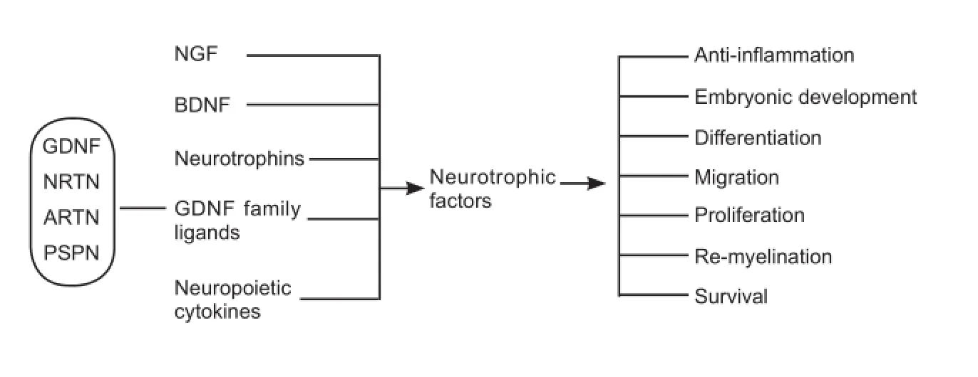
Neurotrophic factors, also referred as neurotrophins, are growth factors originally identified in the nervous system. As indicated by the name, neurotrophic factors are essential for the survival and development of neurons. Nerve growth factor (NGF) is the first identified neurotrophic factor, and is necessary for the development of sensory neurons in the dorsal root ganglia, and cholinergic neurons (Levi-Montalcini and Hamburger, 1951). Brain derived neurotrophic factor (BDNF) is required for survival of sensory neuron in the dorsal root ganglia, hippocampus and cerebral cortex, but not for motor neurons (Conover et al., 1995). On the other hand, Neurotrophin-3 (NT-3) is critical for the survival and proliferation of both sensory neurons, and motor neurons (Woolley et al., 2005). Glial cell derived neurotrophic factors (GDNF) family of ligands (GFLs) include GDNF, neurturin (NRTN), artemin (ARTN) and persephin (PSPN). GFLs enhance the survival of dopaminergic neurons in the midbrain (Lin et al., 1993), as well as the survival of enteric neurons in the gastrointestinal tract (Sanchez et al., 1996). Neuropoietic cytokines, such as ciliary neurotrophic factor (CNTF) and leukemia inhibitory factor (LIF) are also considered members of neurotrophic factor family, and promote survival of multiple types of neurons and glial cells (Stolp, 2013).
There are numerous reports on the role of neurotrophic factors in healthy and damaged neurons. Neurotrophic factors promote the development, survival, proliferation, and differentiation of healthy neurons. They also exhibit a role in anti-inflammation, anti-apoptosis, re-myelination and axon regeneration, thereby facilitating neuronal tissue regeneration.
More recent discoveries indicate that neurotrophic factors are expressed in non-neuronal tissues, and may play a role in the tissue repair as well. NGF, BDNF, and NT3 have been noted to promote proliferation, vascularization, and neural differentiation in mesenchymal cells such as bone marrow stem cells (BMSCs) and fibroblasts. NT3 and GDNF are reported to promote ovarian follicle differentiation (Nilsson et al., 2009) and spermatogenesis respectively (Meng et al., 2000). GDNF is also critical in the ureteric bud origination during kidney development (Sanchez et al., 1996). In addition, increased level of NGF is linked with improved regeneration of cardiomyocytes (Caporali et al., 2008) and pancreatic islet (Hata et al., 2015)(Figure 1).
In a recent study, we reported that GDNF is enriched in the salivary gland stem cells (SSCs), but not in the acinar cell. More interestingly, the expression of GDNF in the SSCs is elevated post radiation to the head and neck region in both mice and humans. This neurotrophic factor is able to increase the proliferation of SSCs in a dose dependent manner in vitro. We also found that one time delivery of GDNF to the salivary gland either before or post radiation to the head and neck region of the mice would significantly increase the number of surviving SSCs, improve the morphology of the salivary gland and rescue the saliva secretion. The data suggested that GDNF protects the salivary gland from radiation induced damage by promoting salivary gland stem cell regeneration and proliferation in vivo (Xiao et al., 2014).
Besides the healthy tissues, neurotrophic factors are also reported to regulate tumor cell growth, invasion, metastasis along nerve, and resistance to therapy. BDNF is found to enhance the proliferation of malignant gliomas, breast cancer and lung cancer. GDNF pathways have been associated with growth and metastasis of neuroblastoma, glioma, breast cancer, small cell lung cancer, thyroid cancer, pancreatic cancer, colon cancer and testicular cancer. It would be necessary to test whether GDNF promotes head and cancer (HNC) survival, proliferation and migration, before applying it to the post radiation HNC patients.
The mechanism through which neurotrophic factors mediate tissue regeneration and tumor cell behaviorstill needs further investigation. NGF, BDNF and NTs (Neurotrophins) bind to the neurotrophin receptor p75 at low affinity. The binding between NGF, BDNF, NTs and the receptor tyrosine kinases (Trk) are stronger and more specific. NGF specifically binds to TrkA while BDNF preferentially binds to TrkB. NT-3 preferentially binds to TrkC, but can also activate TrkA and TrkB, while NT-4/5 preferentially binds to TrkB. The PI3K/AKT, MEK/ERK are reported to be downstream of the neurotrophin activation (Skaper, 2012). GDNF is the most well studied member of the GFL family. It preferentially binds to the receptor GFRα1, which then activates the receptor tyrosine kinase RET or the neural cell adhesion molecule (NCAM) (Zhou et al., 2003). The PI3K/AKT, MEK/ ERK, SRC/c-Jun kinase (JNK), FYN/focal adhesion kinase(FAK) pathways have all been reported to be downstream of the GDNF signal. In our study, elevated GDNF were found co-localized with NCAM and phosphorylated FAK signal in the SSC of post radiation HNC patients. Although phosphorylated AKT and phosphorylated ERK level both increased in the post radiation salivary gland, the signal was neither enriched in SSCs nor overlay with GDNF. The results indicate that GDNF mainly work through the NCAM and FAK pathway in promoting salivary gland regeneration (Xiao et al., 2014).

Figure 1 Schematic map of neurotrophic factors and their functions.
These results indicate that neurotrophic factors could be promising drug candidates for tissue regeneration in the future. Neurotrophic factors and their modulators are tested in clinical trials already for treating neurodegenerative diseases, such as Alzheimer's disease (NCT01163825, NCT00876863, NCT02271750) and Parkinson's disease (NCT00985517, NCT01621581), progressive supranuclear palsy (NCT00005903), traumatic brain injury (NCT01212679, NCT02276079), and cerebral radiation necrosis (NCT02032147). Neurotrophic factors are delivered to patients through direct injection, infusion pumps, encapsulated particles, virus mediated infection, etc. A clinical study on the safety and efficacy of a recombinant human NGF eye drop solution is ongoing recruiting patients with persistent epithelial defect of cornea or keratitis of the cornea (NCT01756456, NCT01411657). Neuropoietic cytokine CNTF has been shown in several clinical models to enhance survival and regeneration of the retinal ganglion neurons (NCT01408472). There are controversies about applying the neurotrophic factors. Amgen withheld the clinical trial of GDNF on patients with Parkinson's in 2004 concerning the efficacy and safety issues. With the increasing evidences indicating the drug delivery method may play critical role in the effect, MedGenesis Therapeutix Inc. reopened the trial using convection enhanced delivery method to deliver GDNF.
It is promising to see more bedside studies on the potential therapeutic effect of neurotrophic factors, as more and more benchside studies demonstrate that neurotrophic factors play important roles in regulating stem cell behavior and promotes tissue regeneration.
Nan Xiao*
Department of Biomedical Sciences, Arthur A. Dugoni School of Dentistry, University of the Pacific, San Francisco, CA, USA
*Correspondence to: Nan Xiao, Ph.D., nxiao@pacific.edu.
Accepted: 2016-03-10
orcid: 0000-0001-7264-3046 (Nan Xiao)
Caporali A, Sala-Newby GB, Meloni M, Graiani G, Pani E, Cristofaro B, Newby AC, Madeddu P, Emanueli C (2008) Identification of the prosurvival activity of nerve growth factor on cardiac myocytes. Cell Death Differ 15:299-311.
Conover JC, Erickson JT, Katz DM, Bianchi LM, Poueymirou WT, Mc-Clain J, Pan L, Helgren M, Ip NY, Boland P, Friedman B, Wiegand S, Vejsada R, Kato AC, Dechiara TM, Yancopoulos GD (1995) Neuronal deficits, not involving motor neurons, in mice lacking BDNF and/or NT4. Nature 375:235-238.
Hata T, Sakata N, Yoshimatsu G, Tsuchiya H, Fukase M, Ishida M, Aoki T, Katayose Y, Egawa S, Unno M (2015) Nerve growth factor improves survival and function of transplanted islets via trka-mediated beta cell proliferation and revascularization. Transplantation 99:1132-1143.
Levi-Montalcini R, Hamburger V (1951) Selective growth stimulating effects of mouse sarcoma on the sensory and sympathetic nervous system of the chick embryo. J Exp Zool 116:321-361.
Lin LF, Doherty DH, Lile JD, Bektesh S, Collins F (1993) GDNF: a glial cell line-derived neurotrophic factor for midbrain dopaminergic neurons. Science 260:1130-1132.
Meng X, Lindahl M, Hyvonen ME, Parvinen M, de Rooij DG, Hess MW, Raatikainen-Ahokas A, Sainio K, Rauvala H, Lakso M, Pichel JG, Westphal H, Saarma M, Sariola H (2000) Regulation of cell fate decision of undifferentiated spermatogonia by GDNF. Science 287:1489-1493.
Nilsson E, Dole G, Skinner MK (2009) Neurotrophin NT3 promotes ovarian primordial to primary follicle transition. Reproduction 138:697-707.
Sanchez MP, Silos-Santiago I, Frisen J, He B, Lira SA, Barbacid M (1996) Renal agenesis and the absence of enteric neurons in mice lacking GDNF. Nature 382:70-73.
Skaper SD (2012) The neurotrophin family of neurotrophic factors: an overview. Methods Mol Biol 846:1-12.
Stolp HB (2013) Neuropoietic cytokines in normal brain development and neurodevelopmental disorders. Mol Cell Neurosci 53:63-68.
Woolley AG, Sheard PW, Duxson MJ (2005) Neurotrophin-3 null mutant mice display a postnatal motor neuropathy. Eur J Neurosci 21:2100-2110.
Xiao N, Lin Y, Cao H, Sirjani D, Giaccia AJ, Koong AC, Kong CS, Diehn M, Le QT (2014) Neurotrophic factor GDNF promotes survival of salivary stem cells. J Clin Invest 124:3364-3377.
Zhou FQ, Zhong J, Snider WD (2003) Extracellular crosstalk: when GDNF meets N-CAM. Cell 113:814-815.
10.4103/1673-5374.182696 http∶//www.nrronline.org/
How to cite this article: Xiao N (2016) Neurotrophic factors: promising candidates in tissue regeneration. Neural Regen Res 11(5):735-736.
杂志排行
中国神经再生研究(英文版)的其它文章
- Recovery of injured fornical crura following neurosurgical operation of a brain tumor: a case report
- Gender difference in the neuroprotective effect of rat bone marrow mesenchymal cells against hypoxiainduced apoptosis of retinal ganglion cells
- Vitamin B complex and vitamin B12levels after peripheral nerve injury
- Methylprednisolone microsphere sustained-release membrane inhibits scar formation at the site of peripheral nerve lesion
- A self-made, low-cost infrared system for evaluating the sciatic functional index in mice
- Methylprednisolone exerts neuroprotective effects by regulating autophagy and apoptosis
