重视肱骨近端骨折手术中修复重建技术的合理应用
2016-03-15郭庆山
郭庆山
·专家论坛·
重视肱骨近端骨折手术中修复重建技术的合理应用
郭庆山
【摘要】肱骨近端骨折常见,具有老年骨质疏松骨折的共同特点,即并发症率及死亡率高、手术难度大等。治疗中应基于患者特性、骨折特点和医生特长,采取个性化的治疗方案;在术中还应重视该类患者普遍存在的骨质疏松、骨缺损、肩袖损伤等,综合运用一些特殊修复与重建技巧,如间接复位技术、内侧柱重建加强技术、肩袖修复技术等,以期恢复最佳的肩关节功能。
【关键词】肱骨近端骨折; 修复; 重建
作者单位: 400042 重庆,第三军医大学大坪医院野战外科研究所全军战创伤中心创伤外科
肱骨近端骨折常见,占所有骨折的4%~5%,男女比例3∶7。70%以上的患者年龄>60岁,在老年骨质疏松骨折中占第三位[1],前两位是股骨粗隆间骨折、桡骨远端骨折。2013年一项针对高龄(平均83岁)患者进行的死亡率调查表明,伤后30、90d,肱骨近端骨折死亡率分别为3.6%、4.1%,接近于股骨近端骨折(分别是3.6%、8.1%)[2]。肱骨近端骨折同样具有老年骨质疏松骨折的高并发症、高死亡率风险,治疗中应关注治疗方式和手术指征,以及骨折本身普遍存在的骨质疏松、骨缺损、肩袖损伤等,在术中运用一些特殊技术,这均直接关系到治疗成败,值得高度重视。
1肱骨近端骨折非手术与手术治疗指征
虽然肱骨近端骨折常见,但关于治疗的随机对照研究(RCTs)却很少。关于手术还是非手术治疗,目前仍没有标准及普遍接受的判断指标。可接受的观点是应基于患者特性、骨折特点和医生特长,采取个性化的方案。肱骨近端骨折80%~90%移位不明显,非手术治疗可达较好结果,10%~20%需行外科手术,包括内固定术、半肩置换术等[3-4]。非手术治疗是更具成本效益的方法,适于无移位或轻度移位骨折,在老年、低需求、明显内侧粉碎、太过复杂的骨折也可考虑非手术治疗。骨折块移位严重时非手术治疗效果差,手术治疗适用于年轻、高需求和多发、复杂骨折等情况,治疗目标是解剖复位和稳定固定。老年患者依据损伤的严重程度初期使用假体也是可供选择的方案。
2骨质疏松外科处理技术
骨质疏松骨折是一个尚未解决的问题,锁定钢板已经彻底改变了骨质疏松性骨折的治疗,但并非没有并发症[5-6]。常规用非可吸收粗线缝合肩袖以补充内固定、内侧柱支撑螺钉[7-8]等固定策略的应用,也未达成真正的共识,故目前仍然没有最佳的治疗方法。以下简要介绍几种治疗技术。
2.1间接复位技术骨质疏松的三、四部分骨折复位困难,应用直接复位技术常需要广泛剥离软组织,甚至切开关节囊,如此加重了血供破坏,也容易导致骨折块进一步碎裂,故间接复位技术很重要,最常用的是缝线牵引复位或牵引联合钢板提拉复位技术。
不论骨折如何粉碎,肩袖总是附着于结节骨块上,用非吸收缝线标记肩袖,通过牵引肩袖缝线可对骨折碎片进行间接操作。通过纵向牵引上肢使肱骨干内收或外展、旋转或偏侧,结合直接撬拨复位技术,同时牵拉缝合线完成间接复位。注意缝线应置于腱-骨交界处以防切出,尤其是老年患者。
在某些头下型骨折,胸肌的牵拉常导致骨干内侧移位。可在肱骨头和大体复位后的骨干外侧安置钢板,骨折远端第一孔钻入3.5mm皮质骨螺钉,通过拧紧螺钉牵拉骨干外移贴向钢板达到间接精确复位。注意螺钉拧紧复位后可造成钢板轻度上移,预先安置钢板时应稍偏下方。
2.2内侧柱重建技术近年来的研究均强调稳定内侧柱的重要性,内侧皮质足够时内侧铰链易于复位,可对抗内翻崩溃、钢板失败、螺钉切出等机械并发症。
2.2.1内柱需要加强和重建的指征目前尚无确切的标准,需要考虑多种因素,如骨折类型、粉碎程度、复位质量、患者相关因素等,且术中经常会改变重建加强策略[9]。Tingart等[10]认为肱骨干骨皮质厚度是预测骨矿物质密度和内固定是否成功的相对可靠和可重复的指标,建议通过测量复合骨皮质厚度(combined cortical thickness,CCT),评估骨质疏松程度,评价内侧皮质状态。复合骨皮质厚度是肱骨干的两个平面上内侧和外侧皮质厚度的平均值(调整X线片放大倍数后,图1)。如果内侧皮质完整,皮质骨厚度>4mm,不常规加强;内侧皮质完整,皮质骨厚度<4mm,常用可注射骨水泥或骨颗粒填塞肱骨头的空腔。若内侧皮质不完整,不管皮质厚度,强烈建议用植骨、髓内同种异体腓骨、肱骨距特殊螺钉等技术来稳定内侧柱。

图1平面1是肱骨干近端皮质骨内边界开始变平行处;平面2位于平面1远端20mm,平面(黄线)厚度(红线)
2.2.2推荐的几个重建措施髓内钢板:如果骨折粉碎、明显骨质疏松甚至肱骨距缺损时,不易将肱骨头稳定于骨干上,有人设计髓内钢板维持该复位[11],该钢板安置过程中会进一步造成骨折间隙空腔形成,其应用非常少见。
髓内结构性植骨下内侧支撑技术:在内侧柱严重粉碎的骨折,可在髓腔内植入大骨块,通过结构性支撑恢复内侧肱骨距。Walch等[12]于1996年介绍了髓内腓骨支撑技术,并得到较多作者的支持[13-15],认为钢板加腓骨的生物力学明显优于单纯锁定钢板。最近,Neviaser等[16]回顾了38例腓骨移植患者75周的随访资料,无软骨下螺钉穿出或切割,仅1例内翻,1例头坏死,肩关节功能良好。也有作者利用髂骨块结构植骨[17]用于Neer四部分外展嵌插骨折,随访38.8个月,骨折愈合良好,无肱骨头坏死。
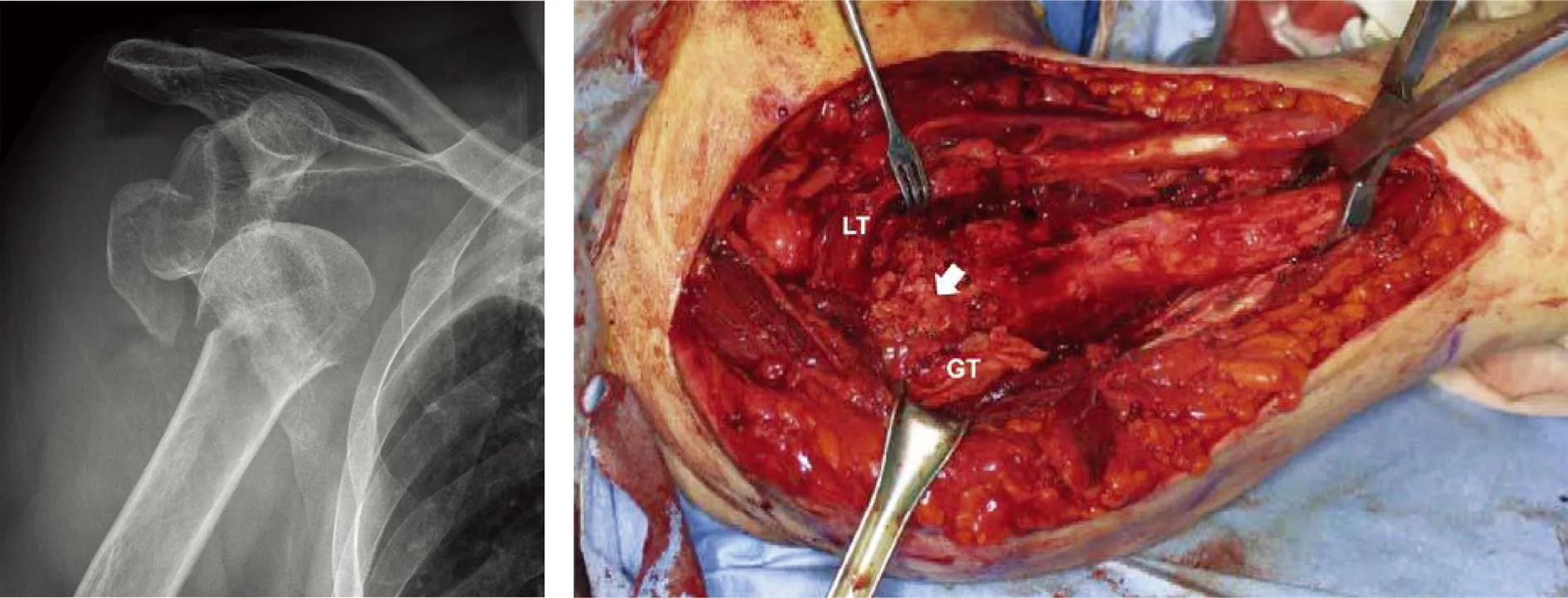
锁定钢板加打压植骨技术:骨质疏松患者往往有明显骨缺损,打压植骨也是一个常用的内侧柱加强技术。其操作要点:复位并临时维持头、干骨块位置,通过外侧骨窗(粉碎骨折始终存在)植入骨粒并嵌紧,将头包裹并稳定在骨干上,然后复位大小结节拴紧缝合于锁定板上(图2)。Kim等[18]评价了该技术对21例四部分骨折患者的效果,所有病例无头坏死,骨折均愈合,无钢板失败和内翻畸形,术后颈干角维持良好,Neer评分显示功能恢复佳,认为该方法可以获得良好的结果。

ab
图2a.患者女性,82岁,四部分骨折脱位; b.通过骨折窗植入颗粒骨(箭头)。GT:大结节,LT:小结节
磷酸氢钙或硫酸盐骨水泥填充技术:将骨水泥注入肱骨头加强钢板固定,可明显减少骨块间运动、增加抗扭刚度。Robinson等[19-20]用骨水泥注入肱骨头或干骺端加强钢板固定,复位保持良好。但因是非随机对照,关于其优缺点尚无法定论。
3肩袖缝合技术
虽然有作者认为全层肩袖撕裂可以行非手术治疗,并进行了临床研究,如Nanda等[21]非手术治疗85例,包括43例全层撕裂者(60岁以上者多见),认为肩关节功能与肩袖损伤无明确相关性。但目前可接受的观点是,肩袖撕裂明显影响功能,需要进一步的修复。常用技术如下。
3.1钻孔缝合技术经骨缝合张力带固定,可用于移位小的骨折,肩袖结构经常完整。Park等[22]缝合固定二或三部分骨折,Flatow等[23]固定大结节骨折,骨愈合和功能均良好。虽然Panagopoulos等[24]也用于外展嵌插型四部分骨折,并认为效果优良,但对高度不稳定四部分骨折、严重粉碎骨折,多数作者不推荐该方法。钻孔缝合技术也是肩关节置换的常规技术。
3.2吊索缝合技术钻孔缝合技术需要在骨干上钻孔,缝线常被锋利的骨孔切断,骨质疏松时缝线常会切出,导致术中或术后结节移位。吊索缝合技术为一个新的大结节固定技术,在骨干上固定索环,缝线边缘不会被锐利骨孔切割。Pijls等[25]将该技术与常规钻孔技术进行了比较,认为常规钻孔缝合复位维持差,大结节出现骨吸收现象,比吊索缝合差。吊索缝合技术骨不连发生率为3%,常规技术11%。具体方法是将3号非可吸收缝线末端折叠,头端并拢绕过骨干后自尾端环中穿出做成吊索,朝向大结节方向在骨干内外侧各置2根。结节上钻孔,内侧吊索纵向置入大结节,外侧吊索纵向置入小结节,收紧打结使结节复位,内外侧线尾再水平打结加强固定。
3.3热气球技术Park等[26]2006年报道了热气球技术,即将髓内钉、张力带和锁定缝合技术联合应用。在老年骨质疏松患者显示固定效果优良,术后肩关节功能良好。具体做法是先复位头、干骨块后髓内钉固定,提供旋转稳定和支撑作用;再于结节间水平缝合将大小结节拼合在一起增加结节间稳定性,称为锁定缝合;最后于大结节和骨干间行垂直缝合,通过髓内钉支撑的反作用力形成张力带作用,防止肱骨头内翻(图3)。如此类似热气球的结构,提供旋转稳定和支撑的交锁钉是热空气,锁定缝合是水平受力带,张力带缝合是垂直受力带,受力带分担气球膨胀的压力。

ab
图3热气球技术。a.锁定缝合,岗下肌和肩胛下肌间用5号爱惜帮编织缝合; b.张力带缝合,在肱骨头、冈上肌、冈下肌、肩胛下肌与骨干间形成纵向张力
以上仅介绍了几种常用的手术技术,更多的修复与重建技巧仍然需要进一步研究和总结提高。在骨质疏松性肱骨近端骨折的临床治疗中,应基于患者特性、骨折特点和医生特长,采取个性化的治疗方案。需手术者应进行良好的术前设计,综合利用间接复位技术、内侧柱重建加强技术以及肩袖修复技术等,以利于骨折的稳定和愈合,利于关节功能的恢复。
参考文献:
[1] Baron JA,Barrett JA,Karagas MR.The epidemiology of peripheral fractures[J].Bone,1996,18(3S):S209-213.
[2] Liem IS,Kammerlander C,Raas C,et al.Is there a difference in timing and cause of death after fractures in the elderly[J].Clin Orthop Relat Res,2013,471(9):2846-2851.
[3] Neer CS,Mcllveen SJ.Recent results and technique of prosthetic replacement for four-part proximal humeral fractures[J].Orthop Trans,1986,10:475.
[4] Clavert P,Adam P,Bevort A,et al.Pitfalls and complications with locking plate for proximal humerus fracture[J].J Shoulder Elbow Surg,2010,19(4):489-494.
[5] Sproul RC,Iyengar JJ,Devcic Z,et al.A systematic review of locking plate fixation of proximal humerus fractures[J].Injury,2011,42(4):408-413.
[6] Brorson S,Rasmussen JV,Frich LH,et al.Benefits and harms of locking plate osteosynthesis in intraarticular (OTA Type C) fractures of the proximal humerus: a systematic review[J].Injury,2012,43(7):999-1005.
[7] Badman BL,Mighell M.Fixed-angle locked plating of two-, three-, and four-part proximal humerus fractures[J].J Am Acad Orthop Surg,2008,16(5):294-302.
[8] Zhang L,Zheng J,Wang W,et al.The clinical benefit of medial support screws in locking plating of proximal humerus fractures: a prospective randomized study[J].Int Orthop,2011,35(11):1655-1661.
[9] Namdari S,Voleti PB,Mehta S.Evaluation of the osteoporotic proximal humeral fracture and strategies for structural augmentation during surgical treatment[J].J Shoulder Elbow Surg,2012,21(12):1787-1795.
[10] Tingart MJ,Apreleva M,von Stechow D,et al.The cortical thickness of the proximal humeral diaphysis predicts bone mineral density of the proximal humerus[J].J Bone Joint Surg(Br),2003,85(4):611-617.
[11] Sperling JW,Cuomo F,Hill JD,et al.The difficult proximal humerus fracture: tips and techniques to avoid complications and improve results[J].Instr Course Lect,2007,56:45-57.
[12] Walch G,Badet R,Nove-Josserand L,et al.Nonunions of the surgical neck of the humerus: surgical treatment with an intramedullary bone peg, internal fixation, and cancellous bone grafting[J].J Shoulder Elbow Surg,1996,5(3):161-168.
[13] Bae JH,Oh JK,Chon CS,et al.The biomechanical performance of locking plate fixation with intramedullary fibular strut graft augmentation in the treatment of unstable fractures of the proximal humerus[J].J Bone Joint Surg(Br),2011,93(7):937-941.
[14] Chow RM,Begum F,Beaupre LA,et al.Proximal humeral fracture fixation: locking plate construct ± intramedullary fibular allograft[J].J Shoulder Elbow Surg,2012,21(7):894-901.
[15] Osterhoff G,Baumgartner D,Favre P,et al.Medial support by fibula bone graft in angular stable plate fixation of proximal humeral fractures: an in vitro study with synthetic bone[J].J Shoulder Elbow Surg,2011,20(5):740-746.
[16] Neviaser AS,Hettrich CM,Beamer BS,et al.Endosteal strut augment reduces complications associated with proximal humeral locking plates[J].Clin Orthop Relat Res,2011,469(12):3300-3306.
[17] Atalar AC,Demirhan M,Uysal M,et al.Treatment of Neer type 4 impacted valgus fractures of the proximal humerus with open reduction,elevation,and grafting[J].Acta Orthop Traumatol Turc,2007,41(2):113-119.
[18] Kim SH,Lee YH,Chung SW,et al.Outcomes for four-part proximal humerus fractures treated with a locking compression plate and an autologous iliac bone impaction graft[J].Injury,2012,43(10):1724-1731.
[19] Robinson CM,Page RS.Severely impacted valgus proximal humeral fractures. Results of operative treatment[J].J Bone Joint Surg(Am),2003,85-A(9):1647-1655.
[20] Lee CW,Shin SJ.Prognostic factors for unstable proximal humeral fractures treated with locking-plate fixation[J].J Shoulder Elbow Surg,2009,18(1):83-88.
[21] Nanda R,Goodchild L,Gamble A,et al.Does the presence of a full-thickness rotator cuff tear influence outcome after proximal humeral fractures[J].J Trauma,2007,62(6):1436-1439.
[22] Park MC,Murthi AM,Roth NS,et al.Two-part and three-part fractures of the proximal humerus treated with suture fixation[J].J Orthop Trauma,2003,17(5):319-325.
[23] Flatow EL,Cuomo F,Maday MG,et al.Open reduction and internal fixation of two-part displaced fractures of the greater tuberosity of the proximal part of the humerus[J].J Bone Joint Surg(Am),1991,73(8):1213-1218.
[24] Panagopoulos AM,Dimakopoulos P,Tyllianakis M,et al.Valgus impacted proximal humeral fractures and their blood supply after transosseous suturing[J].Int Orthop,2004,28(6):333-337.
[25] Pijls BG,Werner PH,Eggen PJ.Alternative humeral tubercle fixation in shoulder hemiarthroplasty for fractures of the proximal humerus[J].J Shoulder Elbow Surg,2010,19(2):282-289.
[26] Park JY,An JW,Oh JH.Open intramedullary nailing with tension band and locking sutures for proximal humeral fracture: hot air balloon technique[J].J Shoulder Elbow Surg,2006,15(5):594-601.
(本文编辑: 黄小英)
Emphasizing the rational use of repair and reconstruction techniques in treating proximal humeral fractures
GUOQing-shan
(Trauma Center,State Key Laboratory of Trauma,Burns and Combined Injury,Institute of Surgery Research,
Daping Hospital,Third Military Medical University,Chongqing400042,China)
【Abstract】Proximal humeral fractures are common,which have common features of osteoporotic fractures,such as high complication rate, high mortality and great difficulty in operation. Its treatment should be based on patient-pecific,fracture-specific and physician-specific features,and should apply personalized treatment programs;attention should be paid to the prevalence of osteoporosis,bone defects,rotator cuff injury in these patients during the operating process;some special repair and reconstruction techniques,such as indirect reduction techniques,medial column reconstruction strengthen technique,rotator cuff repair technology, should be comprehensively used so as to restore optimal shoulder function.
【Key words】proximal humeral fractures; repair; reconstruction
(收稿日期:2015-11-10; 修回日期: 2015-12-20)
基金项目:国家科技支撑计划(2012BAI11B01);国家科技惠民计划(2013GS500101)
【中图分类号】R 683.41
【文献标识码】A【DOI】 10.3969/j.issn.1009-4237.2016.02.001
文章编号:1009-4237(2016)02-0065-04
