动脉硬化无创检测研究进展
2015-06-01孙欣李广义李远洋邱天爽
孙欣,李广义,李远洋,邱天爽
1. 山东大学附属省立医院,山东 济南250021;2. 大连理工大学 电信学部生物医学工程系,辽宁 大连 116024
动脉硬化无创检测研究进展
孙欣1,李广义1,李远洋1,邱天爽2
1. 山东大学附属省立医院,山东 济南250021;2. 大连理工大学 电信学部生物医学工程系,辽宁 大连 116024
动脉硬化度的无创检测对于心血管疾病的“三早”预防具有重要意义。本文介绍了踝臂指数、脉搏波传播速度等目前用于动脉硬化无创检测的主要指标及其相关技术、装置,对近年来动脉弹性功能检测领域的研究进展进行了阐述。
动脉硬化;无创检测;踝臂指数;脉搏波传播速度;超声检查;脉搏波波形分析;AASI
心血管疾病是全球的头号死因,其高发病率、高致残率和高死亡率也已经成为我国的重大公共卫生问题。动脉硬化是心血管疾病的重要病理基础,是心血管系统功能退行的重要原因,也是其主要结果和体现。动脉硬化度的检测对于心血管疾病的早期预防和病理研究等方面具有重要的作用。本文将就动脉硬化无创检测的相关指标、检测技术进行综述,并对近年来该领域的研究进展进行阐述。
1 踝臂指数
踝臂指数(Ankle-Brachial Index,ABI)于20世纪50年代由Winsor[1]提出,后经Yao等[2]的发展,目前已成为诊断外周动脉疾病(Peripheral Arterial Disease,PAD)尤其是下肢动脉硬化闭塞症的重要指标[3-4]。随着研究的深入,人们还发现踝臂指数对于评价心血管系统风险具有较好的效果[5-6],有独立预测心血管潜在危险的临床价值。ABI通常定义为人体下肢踝部收缩压与上肢肱动脉收缩压之比,即

其中SPa表示下肢踝部收缩压,SPb表示上臂肱动脉收缩压。人体两侧每侧各能得到一个ABI值。根据ACCF/AHA的推荐[7],当某侧的ABI值< 0.90时,则该侧下肢可能出现动脉狭窄或闭塞的情况。
近年来,运动后ABI(Post-exercise ABI)逐渐成为研究关注点之一。与静息状态下的ABI相比,运动后测得的ABI能够提供更多的诊断和预后信息[8-10],特别是对于那些静息ABI值正常的患者。目前,运动后ABI的测量仍存在一些争议,例如不同操作者间测量结果的差异[11]以及对PAD的诊断价值等[12],这些问题都有待进一步流行病学研究和测量技术改进。
2 脉搏波传播速度
脉搏波传播速度(Pulse Wave Velocity,PWV)是指脉搏波在人体动脉树中传播时从一点传播到另一点的速度,即

其中D表示脉搏波的传播距离,PTT表示脉搏波传播时间(Pulse Transit Time,PTT)。根据Moens-Korteweg 公式:

其中E表示动脉管壁的杨氏模量,h为管壁的厚度,R为动脉舒张末期的半径,ρ表示血液密度,脉搏波传播速度随着动脉管壁杨氏模量的增加而逐渐增大,因此可以通过测定PWV来了解人体动脉的硬化程度。
测量不同节段动脉的脉搏波传播速度可以得到不同部位动脉的硬化程度。根据动脉部位的不同,PWV可分为颈动脉-股动脉PWV(carotid-femoral PWV, cfPWV)、肱动脉-桡动脉PWV(brachial-radial PWV,brPWV)、臂踝PWV(brachial-anklePWV,baPWV)等。在许多研究中,人们还往往关注脉搏波从心脏出发传播到动脉某处的传播速度,例如心脏-桡动脉PWV(heart-radial PWV,hrPWV)、心脏-踝部PWV(heart-ankle PWV,haPW)等等。在各节段动脉中,主动脉在人体动脉系统中起着重要的缓冲作用,主动脉的硬化(常以cfPWV表征)往往预示着心血管疾病的发生。研究表明,cfPWV对于心血管系统的危险具有重要的预测作用[13-15]。cfPWV 常被认为是动脉硬化无创测量的“金标准”[16]。
近年来,baPWV的测量在动脉硬化检测以及心血管疾病早期检测领域的应用日趋广泛。baPWV是指脉搏波在上臂和脚踝部之间的传播速度。研究表明,baPWV与主动脉PWV具有很好的相关性[17-18],与多项心血管疾病的危险因子如年龄[19]、高血压[20]存在相关性,在评估动脉硬化和心血管风险方面具有巨大潜力[21-23]。
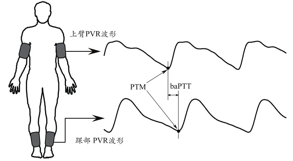
如图1所示,baPWV测量时将4个袖带分别绑缚与人体的双侧上臂和踝部,为袖带充气至某一较低压力值,然后恒压以采集、记录四肢的脉搏波。这种采集脉搏波的方法常称为脉搏波容积描记(Pulse Volume Recording,PVR)。根据人体同侧臂部和踝部的脉搏波,可计算出臂踝脉搏波传播时间(baPTT);再使用(2)式即可得到baPWV,其中臂踝之间的脉搏波传播距离由经验公式得到[24]。另外,利用四肢袖带测量四肢血压(一般采用示波法)还可得到受试者的ABI。显而易见,除具有重要临床意义,baPWV测量的优势还在于简便快捷,同时还能测量受试者的踝臂指数ABI,极为适合用于心血管疾病的早期筛查。相关的商用产品已经出现,较有代表性的产品有日本欧姆龙公司的BP-203RPE系列动脉硬化诊断装置以及我国汇医融工公司的CVDF系列心血管系统状态监测仪。

虽然上述商用产品得到了广泛应用,目前baPWV测量技术仍存在需要进一步深入探讨的问题。计算时使用的臂踝脉搏波传播距离由经验公式得到,而该经验公式是由亚洲人数据得到的,因此增加了此项技术向全球范围推广应用的阻力。另外,脉搏波时标(Pulse Timing Mark,PTM)也有待进一步统一。PTM是在脉搏波波形上定义的一个可识别的表征脉搏波到达时间的标记点(图1)。研究表明[25],使用脉搏波上不同的特征点作为PTM会得到不同的baPWV测量结果;尽管该研究推荐使用INS点作为PTM,但仍需要更大规模数据的验证并形成统一的专家共识。
3 超声技术的应用
超声检查既可以获取动脉血管形态学指标,也可以获取动脉功能指标。在形态学方面,临床上早已开始使用超声技术检测人体颈动脉内中膜厚度(Intima-Media Thickness,IMT)[26],以评估动脉硬化程度。形态学的改变往往滞后于功能衰退,因此对于动脉功能的检测更有利于心血管疾病的提早预防。在动脉功能检测方面,利用超声技术可以测算诸如杨氏弹性模量、顺应性等直接表征动脉管壁弹性功能的物理量[27],还可以通过观测动脉反应性充血(也称作流致扩张,Flow-Mediated Dilation,FMD)来评估动脉的内皮功能[28]。
近年来,超声技术在动脉弹性功能检测方面的应用出现了许多新的进展。几种高时间分辨率(高帧率)超声技术逐渐用于测量局部、瞬时PWV,这些技术包括超快成像技术[29]、PWI技术[30]以及空间混叠成像技术[31]等。先前用于心脏超声成像的斑点跟踪技术在动脉功能评估领域也得到了应用,主要用于测量动脉舒缩过程中的应变曲线[32-33],进而评价动脉的硬化程度。超声剪切波弹性成像技术也从之前的肝脏等大器官应用进入到动脉检测领域,目前相关研究主要集中在颈动脉弹性功能检测[34]和斑块特性检测[35]方面。
4 其他指标和检测技术
早在1967年,Goldwyn和Watt[36]依据单弹性腔理论对外周动脉压力波形进行了分析研究,推算了受检者动脉的顺应性。后来,Cohn等[37]在此基础上提出使用大动脉弹性指数C1和小动脉弹性指数C2表征人体动脉的顺应性。该项技术采集人体桡动脉脉搏波,在双弹性腔模型基础上分析桡动脉脉搏波舒张期的衰减波形而得出上述大小动脉弹性指数,对于评估动脉弹性具有一定价值[38-39]。
与上述舒张期波形分析不同,作为另一项脉搏波波形分析(Pulse Wave Analysis,PWA)技术,增强指数(Augmentation Index,AI)则是由分析中心动脉脉搏波收缩期波形而得到的。脉搏波由心脏产生并由主动脉向外周传播,当脉搏波遇到血管分叉、管径变化等情况时会产生反射,从外周反射回来波又重新回到主动脉处叠加在主动脉的前向波的主峰上。AI定义为中心动脉处脉压减去前向波主峰波幅再除以脉压,结果用百分比表示。测量AI需通过转换函数[40]将从外周动脉(如桡动脉、颈动脉)采集到的脉搏波波形转换成中心动脉处的波形,然后再进行分析计算。虽然受到年龄、血压、心率等因素的影响[41],但由于其在评估动脉弹性功能特别是获取中心动脉相关信息方面具有重要作用,AI一直以来都备受关注。
脉压(Pulse Pressure,PP)是一个较早用于评价动脉硬化度的传统指标,其定义为收缩压与舒张压之差。动脉硬化是导致脉压增大的重要因素之一,脉压的增大反过来又会加速动脉硬化,二者相互影响形成恶性循环。脉压测量方便,在深入检查条件不允许的情况下,可使用脉压来评估患者的动脉硬化度和心血管风险[42]。动态血压监护技术及设备为脉压的长时监测提供了可行的技术手段。在此基础上,动脉硬化指数(Ambulatory Arterial Stiffness Index,AASI)被提出来用于表征动脉的整体硬化程度[43]。已有的研究证据提示,AASI对于心血管系统风险具有预测作用[44-45]。目前,许多研究人员对AASI仍抱有浓厚的兴趣,不仅因为AASI可通过动态血压监测设备进行测量,数据较易获取,更重要的原因是AASI来自于长时监测的数据,研究人员更希望通过对长时间监测数据的分析来进一步挖掘出AASI更丰富的临床价值。这也从侧面提示我们,动脉硬化无创检测领域已经出现一个新的发展方向——动脉功能的动态长时监测。
[1]W insor T.Influence of Arterial Disease on the Systolic Blood Pressure Gradients of the Extrem ity[J].Am J Med Sci,1950,220(2):117-126..
[2]Yao ST,Hobbs JT,Irvine WT.Ankle systolic pressure measurements in arterial disease affecting the lower extremities[J].Br J Surg,1969, 56(9):676-679.
[3]Kurtoglu M,Dolay K,Karamustafaoglu B,et al.The role of the ankle brachial pressure index in the diagnosis of peripheral arterial injury[J].Ulus Travma Acil Cerrahi Derg,2009,15(5):448-452.
[4]Stott P.Detecting peripheral arterial disease using the anklebrachial index[J].Int J Clin Pract,2009,63(1):2-3.
[5]Fow kes FG,M urray GD,Butcher I,et al.Ankle brachial index combined with Fram ingham Risk Score to predict cardiovascular events and mortality:a meta-analysis[J].Jama,2008,300(2):197-208.
[6]Velescu A,Clara A,Penafiel J,et al.Adding low ankle brachial index to classical risk factors improves the prediction of major cardiovascular events.The REGICOR study[J].Atherosclerosis, 2015,241(2):357-363.
[7]Rooke TW,Hirsch AT,M isra S,et al.2011 ACCF/AHA Focused Update of the Guideline for the M anagement of patients w ith peripheral artery disease(Updating the 2005 Guideline):a report of the American College of Cardiology Foundation/American Heart Association Task Force on practice guidelines[J].Circulation,2011, 124(18):2020-2045.
[8]Sato S,Masam i K,O tsuki S,et al.Post-exercise ankle-brachial pressure index demonstrates altered endothelial function in the elderly[J].Jpn Clin Med,2011,2:21-24.
[9]de Liefde II,Klein J,Bax JJ,et al.Exercise ankle brachial index adds important prognostic information on long-term out-come only in patients with a normal resting ankle brachial index[J].Atherosclerosis,2011,216(2):365-369.
[10]Hammad TA,Strefling JA,Zellers PR,et al.The Effect of Post-Exercise Ankle-Brachial Index on Low er Extrem ity Revascularization[J].JACC Cardiovasc Interv,2015,8(9):1238-1244.
[11]van Langen H,van Gurp J,Rubbens L.Interobserver variability of ankle-brachial index measurements at rest and post exercise in patients w ith interm ittent claudication[J].Vasc Med,2009, 14(3):221-226.
[12]Gouveri E,Papanas N,Marakom ichelakis G,et al.Post-exercise ankle-brachial index is not an indispensable tool for the detection of peripheral arterial disease in an epidem iological survey. A post-hoc analysis of the Athens Study[J].Int Angiol,2013, 32(5):518-525.
[13]Sutton-Tyrrell K,Najjar SS,Boudreau RM,et al.Elevated aortic pulse wave velocity,a marker of arterial stiffness,predicts cardiovascular events in well-functioning older adults[J].Circulati on,2005,111(25):3384-3390.
[14]Meaume S,Benetos A,Henry OF,et al.Aortic pulse wave velocity predicts cardiovascular mortality in subjects >70 years of age[J].Arterioscler Thromb Vasc Biol,2001,21(12):2046-2050.
[15]Laurent S,Katsahian S,Fassot C,et al.Aortic stiffness is an independent predictor of fatal stroke in essential hypertension[J].Stroke,2003,34(5):1203-1206.
[16]Laurent S,Cockcroft J,Van Bortel L,et al.Expert consensus document on arterial stiffness:methodological issues and clinical applications[J].Eur Heart J,2006,27(21):2588-2605.
[17]Tanaka H,Munakata M,Kawano Y,et al.Comparison between carotid-femoral and brachial-ankle pulse wave velocity as measures of arterial stiffness[J].J Hypertens,2009,27(10):2022-2027.
[18]Yamashina A,Tomiyama H,Takeda K,et al.Validity, reproducibility, and clinical significance o f noninvasive brachial-ank le pulse wave velocity measurement[J].Hypertens Res,2002, 25(3):359-364.
[19]Tom iyama H,Yamashina A,Arai T,et al.Influences of age and gender on results of noninvasive brachial-ankle pulse wave velocity measurement-a survey of 12517 subjects[J].Atherosclerosis,2003,166(2):303-309.
[20]Chung CM,Cheng HW,Chang JJ,et al.Relationship between resistant hypertension and arterial stiffness assessed by brachialankle pulse wave velocity in the older patient[J].Clin IntervAging,2014,9:1495-1502.
[21]Sheng CS,Li Y,Li LH,et al.Brachial-ankle pulse wave velocity as a predictor of mortality in elderly Chinese[J].Hypertension,2014,64(5):1124-1130.
[22]Han JY,Choi DH,Choi SW,et al.Predictive value of brachialankle pulse wave velocity for cardiovascular events[J].Am J Med Sci,2013,346(2):92-97.
[23]Yamashina A,Tom iyama H,Arai T,et al.Brachial-ankle pulse wave velocity as a marker of atherosclerotic vascular damage and cardiovascular risk[J].Hypertens Res,2003,26(8):615-622.
[24]Yam ashina A,Tom iyam a H,Takeda K,et a l.Validity, reproducibility, and clinical significance of noninvasive brachialankle pulse w ave velocity measurement[J].Hypertens Res, 2002,25(3):359-364.
[25]Sun X,Li K,Ren H,et al.Influence of timing algorithm on brachial -ankle pulse wave velocity measurement[J].Biomed Mater Eng,2014,24(1):255-261.
[26]O'Leary DH,Polak JF,Kronmal RA,et al.Carotid-artery intima and media thickness as a risk factor for myocardial infarction and stroke in older adults.Cardiovascular Health Study Collaborative Research Group[J].N Engl J Med,1999,340(1):14-22.
[27]Brands PJ,Hoeks AP,W illigers J,et al.An integrated system for the non-invasive assessment of vessel wall and hemodynamic properties of large arteries by means of ultrasound[J].Eur J Ultrasound, 1999,9(3):257-266.
[28]Celermajer DS,Sorensen KE,Gooch VM,et al.Non-invasive detection of endothelial dysfunction in children and adults at risk of atherosclerosis[J].Lancet,1992,340(8828):1111-1115.
[29]Couade M,Pernot M,Messas E,et al.U ltrafast imaging of the arterial pulse wave[J].Irbm,2011,32(2):106-108.
[30]Vappou J,Luo J,Okajima K,et al.Aortic pulse wave velocity measured by pulse wave imaging(PW I):A com parison w ith applanation tonometry[J].Artery Res,2011,1,5(2):65-71.
[31]Nagaoka R,M asuno G,Kobayashi K,et al.M easurement of regional pulse-wave velocity using spatial compound imaging of the common carotid artery in vivo[J].U ltrasonics,2015, 55:92-103.
[32]Saito M,Okayama H,Inoue K,et al.Carotid arterial circum ferential strain by two-dimensional speckle tracking:a novel parameter of arterial elasticity[J].Hypertens Res,2012,35(9):897-902.
[33]Yang EY,Brunner G,Dokainish H,et al.Application of speckletracking in the evaluation of carotid artery function in subjects w ith hypertension and diabetes[J].J Am Soc Echocardiogr, 2013,26(8):901-909.e1.
[34]Couade M,Pernot M,Prada C,et al.QUANTITATIVE ASSESSMENT OF ARTER IAL WALL BIOMECHAN ICAL PROPERTIES USING SHEAR WAVE IMAGING[J].Ultrasound Med Biol,2010, 36(10):1662-1676.
[35]Ramnarine KV,Garrard JW,Kanber B,et al.Shear wave elastography imaging of carotid plaques:feasible, reproducible and of clinical potential[J].Cardiovasc Ultrasound,2014,12(1):49.
[36]Goldwyn RM,Watt TB.Arterial Pressure Pulse Contour Analysis Via a Mathematical M odel for the Clinical Quantification of Human Vascular Properties[J].IEEE Trans Biomed Eng, 1967,14(1):11-17.
[37]Cohn JN,Finkelstein S,M cveigh G,et al.Noninvasive pulse wave analysis for the early detection of vascular disease[J].Hype rtension,1995,26(3):503-508.
[38]Manning TS,Shykoff B E,Izzo J L,Jr.Validity and reliability of diastolic pulse contour analysis(w indkessel model)in humans[J].Hypertension,2002,39(5):963-968.
[39]Duprez DA,Jacobs DR,Jr.,Lutsey PL,et al.Association of small artery elasticity w ith incident cardiovascular disease in older adults:the multi-ethnic study of atherosclerosis[J].Am J Epidemiol,2011,174(5):528-536.
[40]Pauca AL,O'Rourke MF,Kon ND.Prospective evaluation of a method for estimating ascending aortic pressure from the radial artery pressure waveform[J].Hypertension,2001,38(4):932-937.
[41]Stoner L,Faulkner J,Lowe A,et al.Should the augmentation index be normalized to heart rate?[J].J Atheroscler Thromb,2014, 21(1):11-16.
[42]N ijdam ME,Plantinga Y,Hulsen HT,et al.Pulse pressure am p lification and risk o f cardiovascu lar disease[J].Am J Hypertens,2008,21(4):388-392.
[43]Li Y,W ang J G,Dolan E,et al.Ambulatory arterial stiffness index derived from 24-hour am bulatory blood pressure monitoring[J].Hypertension,2006,47(3):359-364.
[44]Aznaouridis K,Vlachopoulos C,Protogerou A,et al.Ambulatory systolic-diastolic pressure regression index as a predictor of clinical events:a meta-analysis of longitudinal studies[J].Stroke,2012,43(3):733-739.
[45]Kollias A,Stergiou GS,Dolan E,et al.Am bulatory arterial stiffness index:a systematic review and meta-analysis[J].Atheros clerosis,2012,224(2):291-301.
Progress in Arterial Stiffness Non-invasive M easurement
SUN Xin1, LI Guang-yi1, LI Yuan-yang1, QIU Tian-shuang2
1. Shandong Provincial Hospital A ffi liated to Shandong University, Shandong Jinan 250021, China;2. Department o f Biom edical Engineering, Dalian University of Technology, Liaoning Dalian 116024, China
Non-invasive detection is essential to the primary and secondary prevention of cardiovascular diseases. This paper reviewed the main indicators, related techniques and devices, and delivered a report on state-of-the-art technology in the field of arterial elastic function measurement.
arterial stiffness;non-invasive detection;ankle-brachial index;pulse wave velocity;ultrasound;pulse wave analysis;AASI
R541.5
A
10.3969/j.issn.1674-1633.2015.10.004
1674-1633(2015)10-0014-04
2015-09-21
2015年高等学校本科教学改革与教学质量工程建设项目;国家科技支撑计划项目(2012BAJ18B06)。
邱天爽,教授。
通讯作者邮箱:qiutsh@dlut.edu.cn
