Double Roots of Mandibular Premolar in Full-mouth Periapical Films
2015-02-08
1Department of Stomatology, Peking Union Medical College Hospital, Chinese Academy of Medical Sciences & Peking Union Medical College, Beijing 100730, China
2Department of Radiology, Peking University School of Stomatology, Beijing 100081, China
Double Roots of Mandibular Premolar in Full-mouth Periapical Films
Ling-jia Kong1, Kuo Wan1*, and Deng-gao Liu
1Department of Stomatology, Peking Union Medical College Hospital, Chinese Academy of Medical Sciences & Peking Union Medical College, Beijing 100730, China
2Department of Radiology, Peking University School of Stomatology, Beijing 100081, China
first mandibular premolar; second mandibular premolar; double roots; full-mouth periapical film
ObjectiveTo evaluate the incidence of two-rooted mandibular premolar morphology using full-mouth periapical film series in a Chinese population, with particular emphasis on bilateral incidence, so as to provide a clinical anatomical basis for root canal treatment in mandibular premolars.
MethodsA total of 2015 patients who underwent dental treatment and had full mouth periapical radiographs at the Peking University School of Stomatology from April 2011 to April 2012 were enrolled in this study. Three experienced dentists reviewed the patients’ periapical films and classified the root morphology of mandibular premolars bilaterally. The incidence of unilateral and bilateral double roots were recorded and calculated, including confirmed and suspected bucco-lingual root types.
ResultsIn terms of the morphology of two-rooted mandibular first premolars, of the 2015 cases with complete root formation, two-rooted first premolars were detected in 120 cases, with a total number of 159 teeth. According to the number of teeth, the overall incidence of double roots was 4.03% (159/3972). In terms of the morphology of two-rooted mandibular second premolars, of the 2015 cases with complete root formation, two-rooted second premolars were detected in 24 cases, with a total number of 33 teeth. According to the number of teeth, the overall incidence of double roots was 0.85% (33/3880).
ConclusionsThe roots of mandibular premolars display specific morphological patterns. Based on a large sample, we observed and calculated not only the occurrence rate of bucco-lingual and mesio-distal double roots in first and second mandibular premolars, but also the incidence of unilateral or bilateral double roots within the same mandible. These findings could provide useful information on the anatomical structure of mandibular premolars for endodontic, prosthodontic and surgical procedures, and could improve the quality of treatment and reduce complications.
Chin Med Sci J 2015; 30(3):174-178
MANDIBULAR premolar teeth normally have single roots, but have always been talked of having an unusual anatomy.1Slowey2suggested mandibular premolar to be the teeth which is the most difficult to successfully perform endodontic treatment. Mandibular premolars are undoubtedly an endodontic challenge, especially because the presence of extra roots or root canals may occur far more often than one can expect.3The literature is replete with reports of extra canals in mandibular premolars, but reports regarding the incidence of extra roots in these teeth are quite rare. Hoen et al4reported an incidence rate of 42% for missed roots or canals in the teeth that needed retreatment. In an assessment of the results of endodontic therapy, the failure rates for the first and second mandibular premolars were 11.45% and 4.45%, respectively, possibly due to the complex root canal anatomy of some of the studied teeth.5Therefore, it is highly important to understand root number variations to administer appropriate root canal therapy. However, the morphology of roots and root canals varies among different populations, regions and races. The aim of this study is to investigate the incidence of double roots in mandibular premolars, especially those that occur bilaterally, to provide guidance for clinical operation on these types of teeth.
MATERIALS AND METHODS
Sample collection
Data from 2015 patients with full-mouth periapical films who visited the Department of Oral Radiology at Peking University Stomatological Hospital between April 2011 and April 2012 were assessed. The inclusion criteria were as follows: 12-78 years of age, with full-mouth periapical films acquired with a bisecting angle technique to clearly reflect the morphology of every tooth. The exclusion criteria were: patients with severely damaged radiographs, residual premolar roots, missing bilateral mandibular premolars, or those without periapical films. The initial reason for taking periapical films was not considered.
Diagnostic criteria
It is difficult to observe bucco-lingual double roots because they are oriented in the same direction as the radiation source ion. Therefore, the bucco-lingual double roots might appear as overlapped. This issue can be addressed by the following methods. First, the periodontal image can help to identify overlapping roots, shown as continuous lowdensity contour lines with consistent width around the dental roots on the periapical film; therefore, the diagnosis of multiple roots can be established if there are many lines on the periodontal image. Second, comparison of different full-mouth periapical films taken in different angles can indicate the presence of bucco-lingual double roots. Three examiners, 2 endodontists and 1 dental radiologist, assessed the periapical films of the included patients for double roots, including bucco-lingual and mesio-distal double roots, in the 2015 cases of mandibular first and second premolars, focusing on the incidence of bilateral double roots. In cases of controversial, a consensus agreement was reached via discussion among the examiners.
Radiographs are not the gold standard for identifying bucco-lingual double roots; they can only help in screening. Therefore, the patients were divided into groups for confirmed and suspected bucco-lingual double roots. For mesio-distal double roots, cases where the two roots bifurcated in a mesio-distal direction from the middle, coronal, or apical third of the roots were investigated in analysis. The following criteria were used to divide the cases into confirmed and suspected groups.
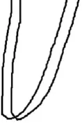
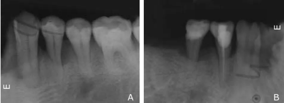
Confirmed bucco-lingual double roots: the standard for confirmed bucco-lingual double roots was the presence of four contour lines in the periodontal image intersecting at a periapical point with no overlap. Figure 1 is a schematic diagram illustrating bucco-lingual double roots, and Figures 2A and 2B show confirmed bucco-lingual double roots in mandibular first and second premolars, respectively.

Figure 1. A schematic diagram of confirmed bucco-lingual double roots.

Figure 2. Confirmed bucco-lingual double roots in the mandibular first premolar (A) and the mandibular second premolar (B).
Suspected bucco-lingual double roots: this group included the less-suspected and highly suspected cases. The less suspected cases had four contour lines in the periodontal image overlapping at a periapical point, whereas the highly suspected cases had four contour lines in the periodontal image that did not overlap at the periapical point. Figures 3A and 3B show suspected buccolingual double roots in a mandibular first premolar and a mandibular second premolar, respectively.
Confirmed non-buccolingual double roots: non-buccolingual double roots were confirmed when there were only two or three contour lines in the periodontal image in the films. Figure 4 is a schematic diagram of confirmed non-double roots. Figures 5A and 5B illustrate confirmed non-bucco-lingual double roots in a mandibular first premolar and a mandibular second premolar, respectively.
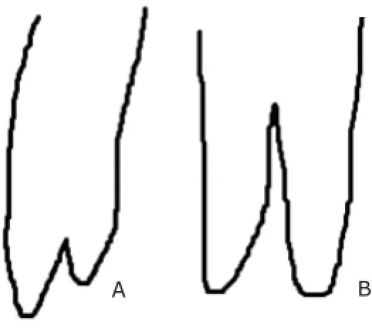
Mesio-distal double roots: mesio-distal double roots included those that bifurcate from the apical, middle, and coronal third of the roots (Fig. 6). Figures 7A and 7B show bifurcation in apical third of roots in a mandibular first premolar and a mandibular second premolar, respectively; figures 7C and 7D demonstrate bifurcation in middle third of roots in a mandibular first premolar and a mandibular second premolar, respectively.
RESULTS
Incidence of mandibular first premolar double roots
A total of 2015 patients with full-mouth periapical films were investigated. Excluding five patients with bilateral loss of mandibular first premolars, and another 48 cases for the absence of unilateral mandibular first premolars, there were 159 teeth with double roots (incidence rate: 4.03%). Overall, 39 of 3972 cases had bilateral double roots (incidence rate: 0.98%), including 6 cases with bilateral mesio-distal double roots, 28 cases with bilateral buccolingual double roots, and 5 cases with bucco-lingual double roots on one side and mesio-distal double roots on the other. Among the 81 patients with unilateral double roots, one case was excluded from the analysis due to tooth losson the other side, and the remaining 80 were with single roots on the other side.

Figure 3. Suspected bucco-lingual double roots in the mandibular first premolar (A) and the mandibular second premolar (B).

Figure 4. A schematic diagram of the confirmed non-double roots.

Figure 5. Confirmed non-bucco-lingual double roots in the mandibular first premolar (A) and the mandibular second premolar (B).

Figure 6. Schematic diagrams of the mesio-distal double roots divided from the periapical section (A), the middle section or the upper section of the roots (B).

Figure 7. Mesio-distal double roots divided from the periapical section in the mandibular first premolar (A) and the mandibular second premolar (B), and those divided from the middle section in the mandibular first premolar (C) and the mandibular second premolar (D).
Incidence of mandibular second premolar double roots
Of the 2015 patients with full-mouth periapical films, 17 were excluded for bilateral loss of mandibular second premolars, and another 116 because of the absence of unilateral mandibular second premolars. There were 33 teeth with double roots (incidence rate: 0.85%). Overall, 9 of 3880 cases had bilateral double roots (incidence rate: 0.23%), including 8 cases with bilateral buccolingual double roots, and 1 with buccolingual double root on one side and mesio-distal double root on the other. Among the 15 patients with unilateral double roots, 2 cases was excluded from the analysis due to tooth loss on the other side, and the remaining 13 cases were with single roots on the other side.
DISCUSSION
This study used full-mouth periapical films to evaluate double roots of premolars in 2015 Chinese individuals. Periapical films are very important for examining dental and periodontal tissues, including enamel, dentin, cementum, pulp cavity, pericementum, alveolar bone, and lamina dura.6Full-mouth periapical films are especially useful in that they provide information on all maxillary and mandibular teeth. Periapical radiographic techniques are divided into bisecting angle and paralleling techniques. The paralleling technique can accurately and clearly reveal the morphology and location of dental and periodontal structures. However, this technique requires the use of a film holder and a long position-indicating device, and it is quite time-consuming to acquire all films.7In contrast, no special filming device is required for the bisecting angle technique; however, its result is not always accurate enough because of deformation and distortion of the dental film due to non-vertical angulation along the X-ray midline and the long axis of the tooth. Therefore, in this study, three experienced dentists simultaneously interpreted the films to reduce the possibility of misdiagnosis. Although this method may not be completely accurate, the radiographic findings are clinically relevant.
It is the standard test to verify double roots on ex vivo teeth rather than on full-mouth periapical films. Despite this limitation, the bisecting angle technique is the most widely used for full-mouth periapical radiography in China. Even though it is less accurate than ex vivo teeth examination to use full-mouth periapical film to evaluate the number of premolar roots, it is of great advantage for the analysis of the incidence of bilateral double roots.
In this study, only 4.03% mandibular first premolars had double roots, and 0.85% mandibular second premolars had double roots, higher than previous findings, which showed that the incidence of double roots in mandibular first premolars and second premolars was 0.2%8and 0.3%9, respectively.
Five anatomical studies that included 672 teeth reported the number of roots in the mandibular first premolar.10-14The majority of the teeth in these studies (93.9%) had a single root; double roots were found in 5.65% of the teeth studied and three roots found in 0.45%.
Eight anatomic studies that included 4 436 teeth reported the number of roots in the mandibular second premolar.9,14-19The majority of the teeth in these studies (99.34%) had a single root; double roots were found in only 0.52% of the teeth studied; three-rooted (0.14%) teeth were extremely rare.
Numerous factors contribute to variations in the root and root canal studies, including ethnicity, age, sex, and unintentional bias in selection of clinical examples of teeth (specialty endodontic practice versus general dental practice) and study design (in vitro versus in vivo).20Scott et al21described the accessory root of mandibular first premolar. They observed ethnic differences in the root morphology, and reported the highest incidence (>25%) of accessory roots in the Australian and sub-Saharan African populations. The lowest incidence (0-10%) occurred in the American, Arctic, New Guinea, Jomon and Western Eurasian populations. Sert et al22also reported sex differences in canal morphology, observing higher incidence (44%) of accessory roots and canals in females as compared to males (34%).
There are no previous reports of bilateral mandibular premolar double roots; however, our results showed that a person may present with bilateral double roots in the mandibular first premolar or the mandibular second premolar. The incidence is 0.98% and 0.23%, respectively. Although the probability is very low, clinicians must consider bilateral double roots if a patient presents with mandibular premolar double roots on one side.
In conclusion, accurate pre-operative radiographs are very important. Straight and angled radiographs and the use of paralleling technique are very helpful in providing clues for the number of roots. Morphologic variations in pulpal anatomy must be always considered before starting treatment. A thorough understanding of root canal anatomy and its variations, careful interpretation of the radiograph, close clinical inspection of the floor of the pulpchamber, and proper modification of access opening are essential for a successful treatment outcome.
1. Gandhi B, Patil AC. Root canal treatment of a mandibular second premolar with three roots and canals—an anatomic variation. J Dent (Tehran) 2013; 10: 569-74.
2. Slowey RR. Root canal anatomy. Road map to successful endodontics. Dent Clin North Am 1979; 23: 555-73.
3. Vaghela DJ, Sinha AA. Endodontic management of four rooted mandibular first premolar. J Conserv Dent 2013; 16: 87-9.
4. Hoen MM, Pink FE. Contemporary endodontic retreatments: an analysis based on clinical treatment findings. J Endod 2002; 28: 834-6.
5. Shenoy A, Bolla N, Vemuri S, et al. Endodontic retreatment—unusual anatomy of a maxillary second and mandibular first premolar: report of two cases. Indian J Dent Res 2013; 24: 123-7.
6. Yu SF. Oral Histology and Pathology. Beijing: People’s Medical Publishing House, 2007.
7. Ma XC. Oral and maxillofacial medical diagnostic imaging. Beijing: People’s Medical Publishing House, 2008.
8. Cleghorn BM, Christie WH, Dong CC. The root and root canal morphology of the human mandibular first premolar: a literature review. J Endod 2007; 33: 509-16.
9. Cleghorn BM, Christie WH, Dong CC. The root and root canal morphology of the human mandibular second premolar: a literature review. J Endod 2007; 33: 1031-7.
10. Lin ZM, Fang YY, Ling JQ. Morphological characteristics of mandibular first premolars in people from Pearl River Delta Region in Guangdong province. Hua Xi Kou Qiang Yi Xue Za Zhi 2008; 26: 526-30.
11. Yang HZ, Lei WH, Yi YZ. Root canal case study of the first premolar in vitro of young people. Xinjiang Yi Xue 2006; 36: 34-5.
12. Guan QL. Investigate and measure the morphology of the first permanent premolars of Han youth and junior in Shandong region. Jinan: Shandong University, 2010.
13. Jain A, Bahuguna R. Root canal morphology of mandibular first premolar in a gujarati population—an in vitro study. Dent Res J (Isfahan) 2011;8: 118-22.
14. Xu HM, Gao XJ. Morphological characteristics of the root canal in premolars of the Chinese. Chin J Conserv Dent 2008;18: 126-9.
15. Geider P, Perrin C, Fontaine M. Endodontic anatomy of lower premolars-apropos of 669 cases. J Odontol Conserv 1989; 11-5.
16. Vertucci FJ, Gegauff A. Root canal morphology of the maxillary first premolar. J Am Dent Assoc 1979; 99: 194-8.
17. Prakash R, Nandini S, Ballal S, et al. Two-rooted mandibular second premolars: case report and survey. Indian J Dent Res 2008; 19: 70-3.
18. Zaatar E I, Al-Kandari A M, Alhomaidah S, et al. Frequency of endodontic treatment in Kuwait: radiographic evaluation of 846 endodontically treated teeth. J Endod 1997; 23: 453-6.
19. Zillich R, Dowson J. Root canal morphology of mandibular first and second premolars. Oral Surg Oral Med Oral Pathol 1973; 36:738-44.
20. Kottoor J, Albuquerque D, Velmurugan N, et al. Root anatomy and root canal configuration of human permanent mandibular premolars: a systematic review. Anat Res Int 2013; 2013: 254250.
21. Scott R, Turner C. The anthropology of modern human teeth. 2nd ed. Cambrideg: Cambrigde University Press, 2000.
22. Sert S, Bayirli GS. Evaluation of the root canal configurations of the mandibular and maxillary permanent teeth by gender in the Turkish population. J Endod 2004; 30: 391-8.
Received for publication June 23, 2014.
*Corresponding author Tel: 86-10-69155256, E-mail: wankuo@126.com
杂志排行
Chinese Medical Sciences Journal的其它文章
- Paraneoplastic Dermatomyositis Accompanying Nasopharyngeal Carcinoma
- Genetic Effects on Sensorineural Hearing Loss and Evidence-based Treatment for Sensorineural Hearing Loss
- Serum Myeloperoxidase Level in Systemic Lupus Erythematosus
- Pulmonary Carcinosarcoma with Intracardiac Extension: a Case Report
- Postpartum Atypical Hemolytic Uremic Syndrome: an Unusual and Severe Complication Associated with IgA Nephropathy
- Cerebrospinal Fluid Biomarkers in Dementia Patients with Cerebral Amyloid Angiopathy
